


Article Figures & Data
Figures
- FIGURE 1.
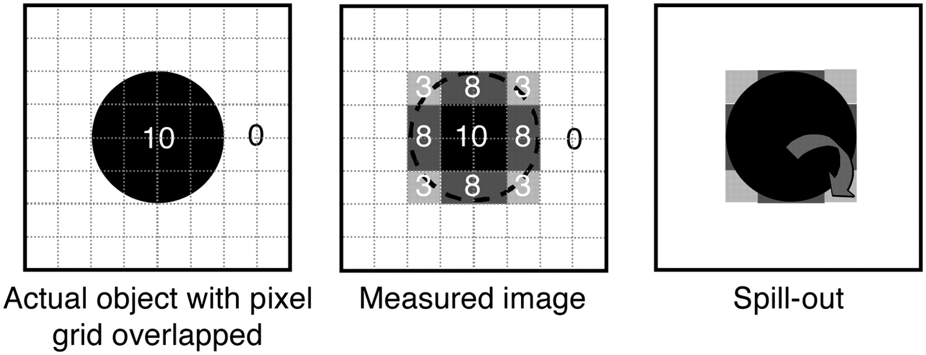
Circular source (diameter of 10 mm) of uniform activity (100 arbitrary units) in nonradioactive background yields measured image in which part of signal emanating from source is seen outside actual source. Maximum activity in measured image is reduced to 85.
- FIGURE 2.
Influence of image sampling on PVE. Pixels on edges of source include both source and background tissues. Signal intensity in these pixels is mean of signal intensities of underlying tissues. Part of signal emanating from source is seen outside actual object and therefore is described as spilling out.
- FIGURE 3.
The measured image (D) of the activity distribution (A) results from mixture of spilling out (B) and spilling in (C). Image sampling affects background activity, creating spilling in within tumor (C). Resulting image is sum of spilling in and spilling out (D).
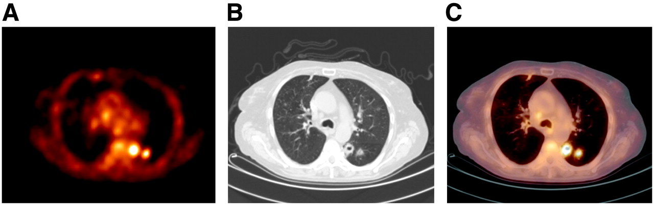
- FIGURE 4.
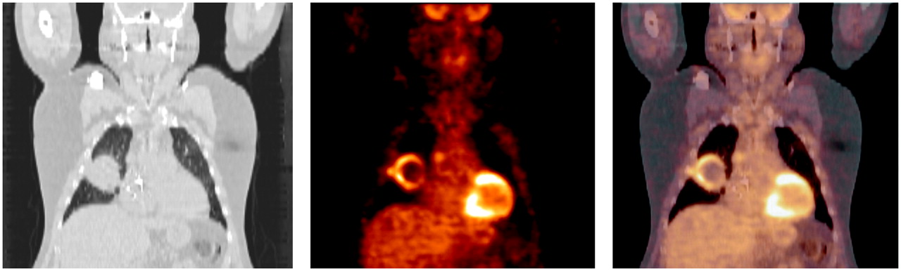
(A) PET image. (B) Corresponding CT image. (C) PET/CT image. Discrepancy between tumor contours as seen on CT and PET images is clearly visible.
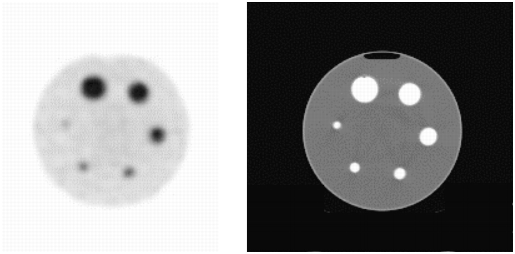
- FIGURE 5.
Transverse PET slice of 6 radioactive spheres with different diameters (10, 12, 16, 22, 28, and 34 mm) and filled with same radioactivity concentrations in uniform radioactive background (left) and corresponding CT slice (right). PVE makes apparent uptake decrease when sphere size decreases.
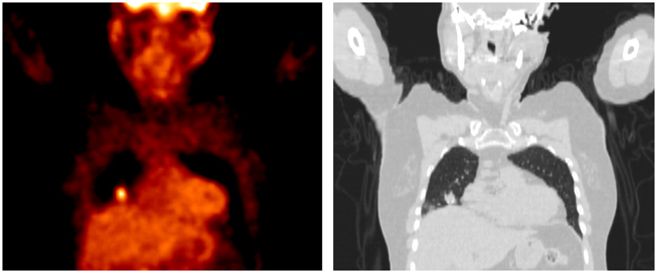
- FIGURE 6.
PET slice (left) and corresponding CT slice (right). Tumor is close to 3 types of tissues (lung, liver, and mediastinum).
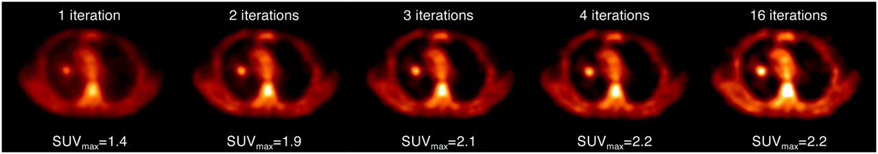
- FIGURE 7.
Transverse PET slices of same PET data reconstructed with ordered-subset expectation maximization with different numbers of iterations (8 subsets). SUVmax varies substantially at early iterations.
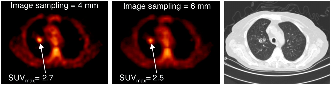
- FIGURE 8.
Transverse PET slices from same patient with image sampling at 4 and 6 mm, resulting SUVmax, and corresponding CT slice.
- FIGURE 9.
Different measurement methods yield different SUVs. SUVmax was calculated from maximum uptake in tumor. SUV75% and SUV50% were mean values in ROI corresponding to isocontours equal to 75% and 50% SUVmax, respectively. SUV15×15 was measured in fixed rectangular region of 15 × 15 mm. SUVmean was measured in manually drawn region (represented in red on CT slice [right]).
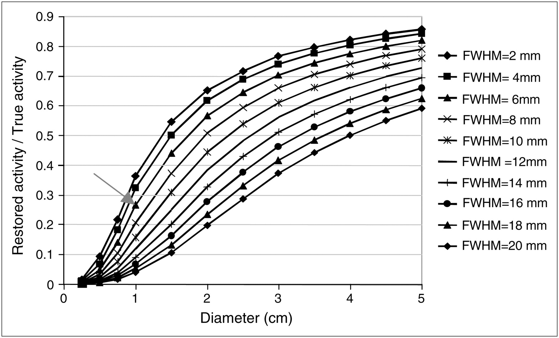
- FIGURE 10.
Restored activity measured in actual contour of spheres in cold background as function of sphere diameter and spatial resolution of imaging system.
- FIGURE 11.
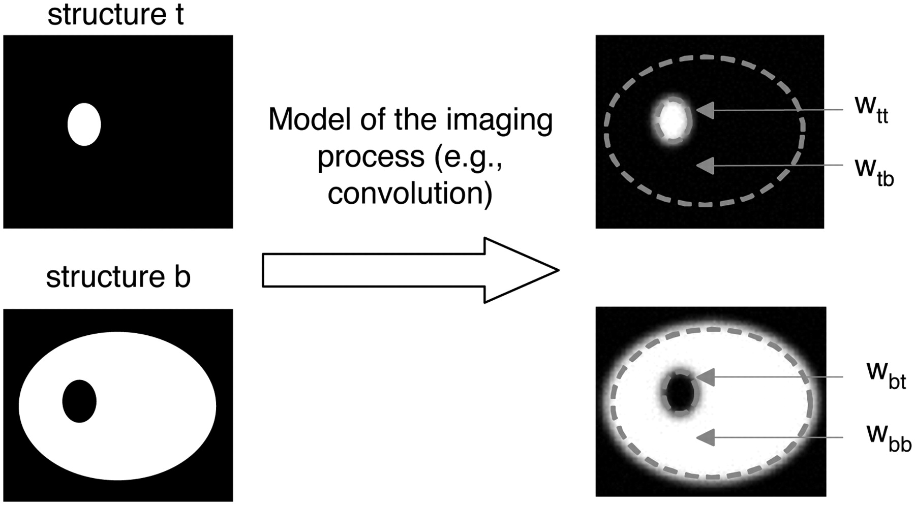
Calculation of transfer coefficients for 2 compartments (tumor [t] and background [b]). Image of each binary compartment as seen by imaging system is obtained by modeling imaging system response. Resulting image is nonbinary image from which 4 transfer coefficients can be calculated. For example, Wtt corresponds to fraction of signal emanating from tumor and detected in tumor, whereas Wtb corresponds to fraction of signal emanating from tumor and detected in background.
- FIGURE 12.
CT image (left), corresponding PET image (middle), and PET/CT image (right) of tumor with no uptake in center. Delineation of tumor from CT image would yield inappropriate definition of metabolically active part of tumor.
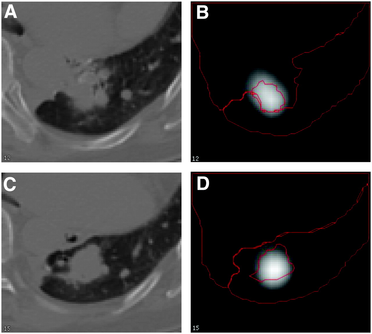
- FIGURE 13.
CT (A and C) and PET (B and D) images corresponding to 2 slices through lung tumor. Compartment contours as drawn from CT are shown in red on PET images.


Tables
Property RC GTM Deconvolution Partition based Multiresolution Fitting Anatomic maximum a posteriori Kinetic modeling Assumes tumor is spheric? No No No No No Yes No No Assumes known tumor volume? Yes Yes No Yes No Yes Yes No Assumes uniform tumor uptake? Yes Yes Not necessarily Yes No Yes No No Assumption(s) regarding tissues surrounding tumor Locally uniform, known uptake Piecewise constant, unknown uptake No Piecewise constant, known uptake Gray levels correlated with those of anatomic data Locally uniform, unknown uptake Partially known No Needs anatomic data? Not necessarily Yes, registered No Yes, registered Yes, registered No Yes Not necessarily Mode of action After reconstruction After reconstruction After reconstruction After reconstruction After reconstruction After reconstruction During reconstruction After reconstruction, time series required Type of results Tumor uptake value Tumor average uptake value Tumor average uptake value Image of tumor compartment PVE-corrected image Tumor average uptake value PVE-corrected image Kinetic parameters compensated for PVE Reference(s) for PET tumor imaging 7,17,18,22,23,50,51,53,54 30 37 38 47,50 ↵* Desirable properties are shown in bold type.


Jump to section
Related Articles
Cited By...
- Prevalence and Medium-Term Outcomes of Patients with Biopsy-Proven Intermediate- to High-Risk Prostate Adenocarcinoma with Low Intraprostatic Uptake on [68Ga]Ga-PSMA-11 PET/CT in the proPSMA Study
- MIRD Pamphlet No. 32: A MIRD Recovery Coefficient Model for Resolution Characterization and Shape-Specific Partial-Volume Correction
- Characterizing meniscal calcifications with photon counting-based dual-energy computed tomography
- Cardiac Neuroendocrine Tumor Metastases on 68Ga-DOTATATE PET/CT: Identification and Prognostic Significance
- ISIT-QA: In Silico Imaging Trial to Evaluate a Low-Count Quantitative SPECT Method Across Multiple Scanner-Collimator Configurations for 223Ra-Based Radiopharmaceutical Therapies
- Impact of PET Reconstruction on Amyloid-{beta} Quantitation in Cross-Sectional and Longitudinal Analyses
- Artificial Intelligence for PET and SPECT Image Enhancement
- Long Versus Short Axial Field of View Immuno-PET/CT: Semiquantitative Evaluation for 89Zr-Trastuzumab
- Inter-scanner A{beta}-amyloid PET harmonization using barrel phantom spatial resolution matching
- Impact of PET reconstruction on A{beta}-amyloid quantitation in cross-sectional and longitudinal analyses
- 68Ga-PSMA PET/CT-Based Atlas for Prostate Bed Recurrence After Radical Prostatectomy: Clinical Implications for Salvage Radiation Therapy Contouring Guidelines
- Hot Spot Imaging in Cardiovascular Diseases: An Information Statement from SNMMI, ASNC, and EANM
- A Guide to ComBat Harmonization of Imaging Biomarkers in Multicenter Studies
- Investigating the in vivo biodistribution of extracellular vesicles isolated from various human cell sources using positron emission tomography
- MRI-based 3D retinal shape determination
- Semiautomatic Tumor Delineation for Evaluation of 64Cu-DOTATATE PET/CT in Patients with Neuroendocrine Neoplasms: Prognostication Based on Lowest Lesion Uptake and Total Tumor Volume
- Accuracy Assessment of SUV Measurements in SPECT/CT: A Phantom Study
- Converting an Anti-Mouse CD4 Monoclonal Antibody into an scFv Positron Emission Tomography Imaging Agent for Longitudinal Monitoring of CD4+ T Cells
- Optimization of Number of Iterations as a Reconstruction Parameter in Bone SPECT Imaging Using a Novel Thoracic Spine Phantom
- Multicenter Study of Quantitative SPECT: Reproducibility of 99mTc Quantitation Using a Conjugated-Gradient Minimization Reconstruction Algorithm
- Have Volume-based Parameters of Positron Emission Tomography/Computed Tomography Prognostic Relevance for Patients With Potentially Platinum-responsive Recurrent Ovarian Cancer? A Single Center Italian Study
- Total nodule number is an independent prognostic factor in resected stage III non-small cell lung cancer: a deep learning powered study
- Semiautomatically Quantified Tumor Volume Using 68Ga-PSMA-11 PET as a Biomarker for Survival in Patients with Advanced Prostate Cancer
- Radiomics Features of 18F-fluorodeoxyglucose Positron-Emission Tomography as a Novel Prognostic Signature in Colorectal Cancer
- 18F-DCFPyL PET/CT in Patients with Subclinical Recurrence of Prostate Cancer: Effect of Lesion Size, Smoothing Filter, and Partial-Volume Correction on PROMISE Criteria
- Performance of Digital PET Compared with High-Resolution Conventional PET in Patients with Cancer
- Assessing PET Parameters in Oncologic 18F-FDG Studies
- Revisiting Weight-Normalized SUV and Lean-Body-Mass-Normalized SUV in PET Studies
- Head-to-Head Comparison of 68Ga-DOTA-JR11 and 68Ga-DOTATATE PET/CT in Patients with Metastatic, Well-Differentiated Neuroendocrine Tumors: A Prospective Study
- Standardization of Preclinical PET/CT Imaging to Improve Quantitative Accuracy, Precision, and Reproducibility: A Multicenter Study
- 18Fluorodeoxyglucose Accumulation in Arterial Tissues Determined by PET Signal Analysis
- Assessing 18F-FDG Uptake in the Sentinel Lymph Node in Breast Cancer
- The Influence of Minimal Misalignment on the Repeatability of PET Images Examined by the Repositioning of Point Sources
- Time-of-Flight Information Improved the Detectability of Subcentimeter Spheres Using a Clinical PET/CT Scanner
- Generation of Structural MR Images from Amyloid PET: Application to MR-Less Quantification
- Targeting CXCR4 (CXC Chemokine Receptor Type 4) for Molecular Imaging of Aldosterone-Producing Adenoma
- Androgen and Estrogen Receptor Imaging in Metastatic Breast Cancer Patients as a Surrogate for Tissue Biopsies
- Mapping of human brown adipose tissue in lean and obese young men
- Rigor and Reproducibility in Analysis of Vascular Calcification
- Associations Between Somatic Mutations and Metabolic Imaging Phenotypes in Non-Small Cell Lung Cancer
- Baseline Metabolic Tumor Volume Predicts Outcome in High-Tumor-Burden Follicular Lymphoma: A Pooled Analysis of Three Multicenter Studies
- Accuracy and Precision of Partial-Volume Correction in Oncological PET/CT Studies
- Partial-Volume Effect Correction Improves Quantitative Analysis of 18F-Florbetaben {beta}-Amyloid PET Scans
- An Effective Immuno-PET Imaging Method to Monitor CD8-Dependent Responses to Immunotherapy
- SUVpeak Performance in Lung Cancer: Comparison to Average SUV from the 40 Hottest Voxels
- Primary tumour standardised uptake value is prognostic in nonsmall cell lung cancer: a multivariate pooled analysis of individual data
- Targeting CD146 with a 64Cu-labeled antibody enables in vivo immunoPET imaging of high-grade gliomas
- Estimation of Tumor Volumes by 11C-MeAIB and 18F-FDG PET in an Orthotopic Glioblastoma Rat Model
- Relationship Between 18F-FDG PET/CT Scans and KRAS Mutations in Metastatic Colorectal Cancer
- Influence of Statistical Fluctuation on Reproducibility and Accuracy of SUVmax and SUVpeak: A Phantom Study
- Multimodal Partial-Volume Correction: Application to 18F-Fluoride PET/CT Bone Metastases Studies
- Immuno-PET of Murine T Cell Reconstitution Postadoptive Stem Cell Transplantation Using Anti-CD4 and Anti-CD8 Cys-Diabodies
- Localization of Hidden Insulinomas with 68Ga-DOTA-Exendin-4 PET/CT: A Pilot Study
- [18F]-Fluorodeoxyglucose Positron Emission Tomography Standardized Uptake Value as a Predictor of Adjuvant Chemotherapy Benefits in Patients With Nasopharyngeal Carcinoma
- Variability and Uncertainty of 18F-FDG PET Imaging Protocols for Assessing Inflammation in Atherosclerosis: Suggestions for Improvement
- Metabolic PET/CT-Guided Lung Lesion Biopsies: Impact on Diagnostic Accuracy and Rate of Sampling Error
- Improving the Detection of Small Lesions Using a State-of-the-Art Time-of-Flight PET/CT System and Small-Voxel Reconstructions
- Effects of Intratumoral Inflammatory Process on 18F-FDG Uptake: Pathologic and Comparative Study with 18F-Fluoro-{alpha}-Methyltyrosine PET/CT in Oral Squamous Cell Carcinoma
- Clinical translation of an ultrasmall inorganic optical-PET imaging nanoparticle probe
- A Compartmental Model of Mouse Thrombopoiesis and Erythropoiesis to Predict Bone Marrow Toxicity After Internal Irradiation
- Glypican-3-Targeted 89Zr PET Imaging of Hepatocellular Carcinoma
- Prognostic Value of Metabolic Parameters in Patients with Synchronous Colorectal Cancer Liver Metastasis Following Curative-Intent Colorectal and Hepatic Surgery
- Quantitative ImmunoPET of Prostate Cancer Xenografts with 89Zr- and 124I-Labeled Anti-PSCA A11 Minibody
- The Effect of Small Tumor Volumes on Studies of Intratumoral Heterogeneity of Tracer Uptake
- Outcome Analysis of 18F-Fluorodeoxyglucose Positron-Emission Tomography in Patients with Lung Cancer After Partial Volume Correction
- Pre-clinical Validation of Orthotopically-implanted Pulmonary Tumor by Imaging with 18F-Fluorothymidine-Positron Emission Tomography/Computed Tomography
- Variability of Total Lesion Glycolysis by 18F-FDG-Positive Tissue Thresholding in Lung Cancer
- Combination of 18F-FDG PET/CT and Diffusion-Weighted MR Imaging as a Predictor of Histologic Response to Neoadjuvant Chemotherapy: Preliminary Results in Osteosarcoma
- Early Dynamic 18F-FDG PET to Detect Hyperperfusion in Hepatocellular Carcinoma Liver Lesions
- Subcentimeter Tumor Lesion Delineation for High-Resolution 18F-FDG PET Images: Optimizing Correction for Partial-Volume Effects
- Effect of Reconstruction Parameters in High-Definition PET/CT on Assessment of Lymph Node Metastases in Head and Neck Squamous Cell Carcinoma
- MR Imaging-Based Correction for Partial Volume Effect Improves Detectability of Intractable Epileptogenic Foci on Iodine 123 Iomazenil Brain SPECT Images: An Extended Study with a Larger Sample Size
- 18F-FDG Uptake by Metastatic Axillary Lymph Nodes on Pretreatment PET/CT as a Prognostic Factor for Recurrence in Patients with Invasive Ductal Breast Cancer
- Clinical PET of Neuroendocrine Tumors Using 64Cu-DOTATATE: First-in-Humans Study
- Prognostic PET 18F-FDG Uptake Imaging Features Are Associated with Major Oncogenomic Alterations in Patients with Resected Non-Small Cell Lung Cancer
- Noise Considerations for PET Quantification Using Maximum and Peak Standardized Uptake Value
- Relationship between 18F-Fluorodeoxyglucose Accumulation and KRAS/BRAF Mutations in Colorectal Cancer
- Large Decreases in Standardized Uptake Values After Definitive Radiation Are Associated with Better Survival of Patients with Locally Advanced Non-Small Cell Lung Cancer
- Impact of Partial-Volume Effect Correction on the Predictive and Prognostic Value of Baseline 18F-FDG PET Images in Esophageal Cancer
- 18F-Fluorodeoxy-glucose Positron Emission Tomography Marks MYC-Overexpressing Human Basal-Like Breast Cancers
- Value of 4-Dimensional 18F-FDG PET/CT in the Classification of Pulmonary Lesions
- 18F-FDG PET/CT for the Prediction and Detection of Local Recurrence After Radiofrequency Ablation of Malignant Lung Lesions
- 18F-FLT PET as a Surrogate Marker of Drug Efficacy During mTOR Inhibition by Everolimus in a Preclinical Cisplatin-Resistant Ovarian Tumor Model
- Kinetic Analysis of 18F-Fluoride PET Images of Breast Cancer Bone Metastases
- SUV: From Silly Useless Value to Smart Uptake Value
- Comparative Assessment of Methods for Estimating Tumor Volume and Standardized Uptake Value in 18F-FDG PET
- In Vivo Tumor Grading of Prostate Cancer Using Quantitative 111In-Capromab Pendetide SPECT/CT
- Standards for PET Image Acquisition and Quantitative Data Analysis
- One Step Closer to Imaging Vulnerable Plaque in the Coronary Arteries