


Article Figures & Data
Figures
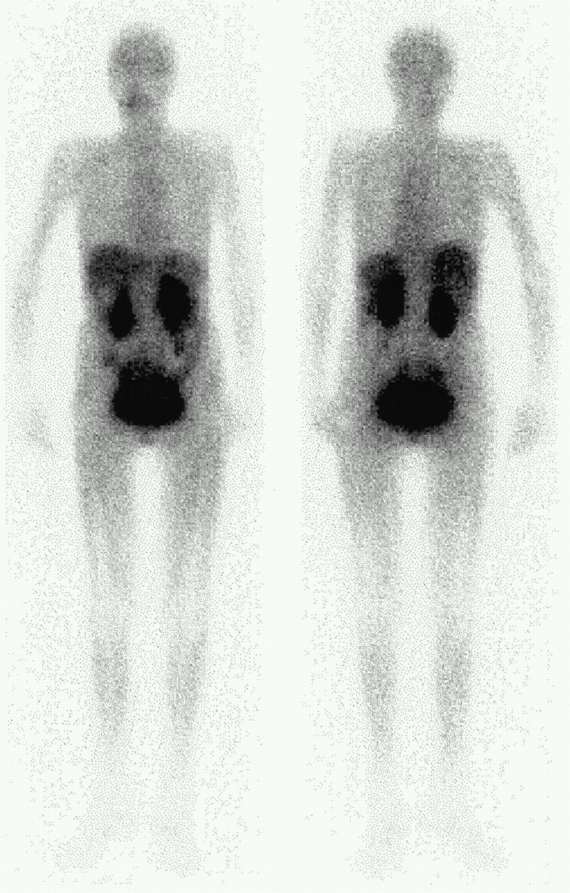
- FIGURE 1.
Anterior (left) and posterior (right) whole-body IMT scintigrams of healthy volunteer 30 min after injection show low-grade brain, liver, and spleen uptake and intense kidney and urinary system uptake.
- FIGURE 2.
Maximum-intensity projection from TYR PET study shows normal distribution in chest and upper abdomen; low uptake in bone marrow, liver, and stomach; and intense uptake in pancreas.
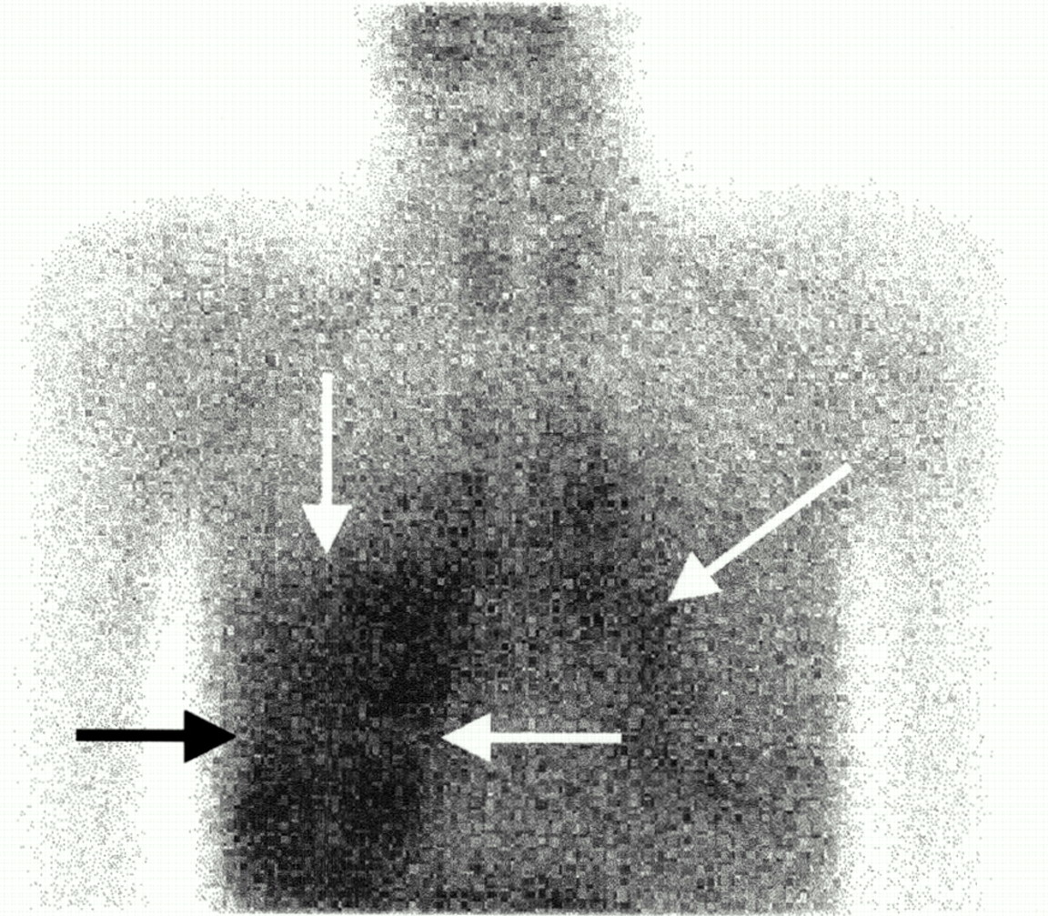
- FIGURE 3.
Planar IMT image obtained 1 wk after 60 Gy radiotherapy in patient with non–small cell lung carcinoma in right middle lobe shows nonspecific increased uptake in irradiated field (arrows).
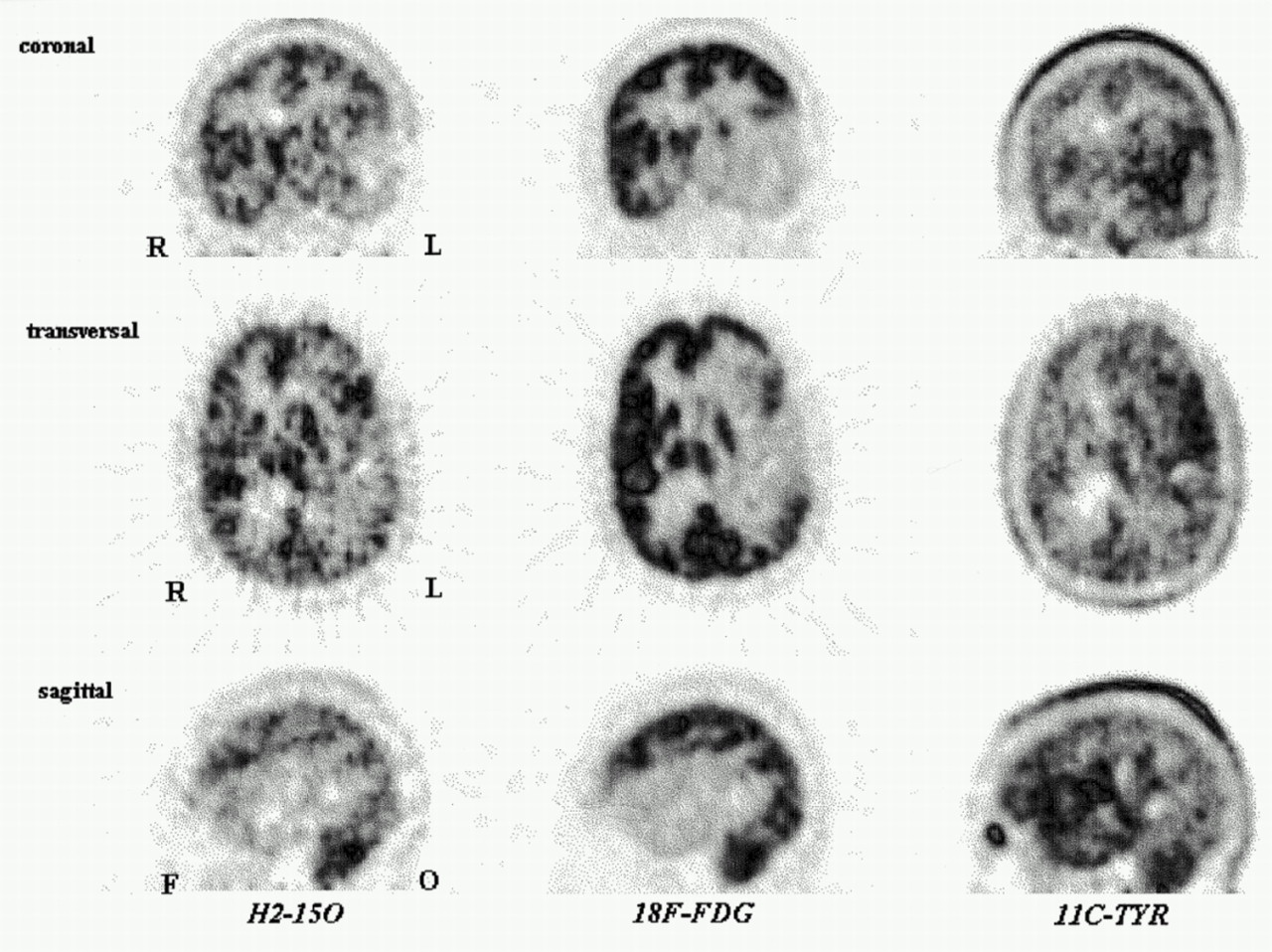
- FIGURE 4.
Coronal, transverse, and sagittal images obtained using H215O (perfusion, left column), FDG (middle column), and TYR (right column) in patient with large, low-grade astrocytoma in left temporoparietal region show that tumor is not intensely perfused and glucose metabolism is low. However, large area of irregularly increased amino acid uptake is clearly seen, and amino acid uptake is noted in lacrimal gland.
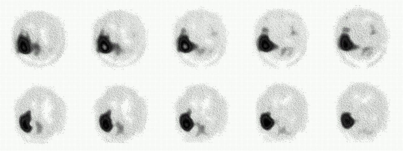
- FIGURE 5.
IMT SPECT (top row) and MET PET (bottom row) images of brain of patient with glioma show similar uptake and tumor delineation. Resolution of MET PET was converted to SPECT resolution.
- FIGURE 6.
Coronal and sagittal projections of TYR PET study of patient with large, recurrent squamous cell carcinoma of right maxillary sinus extending into skull base show irregularly increased TYR uptake in tumor (thick arrows). Because of irradiation, uptake in both parotid glands and right submandibular gland has disappeared, and uptake in left submandibular gland is visible (thin arrows).
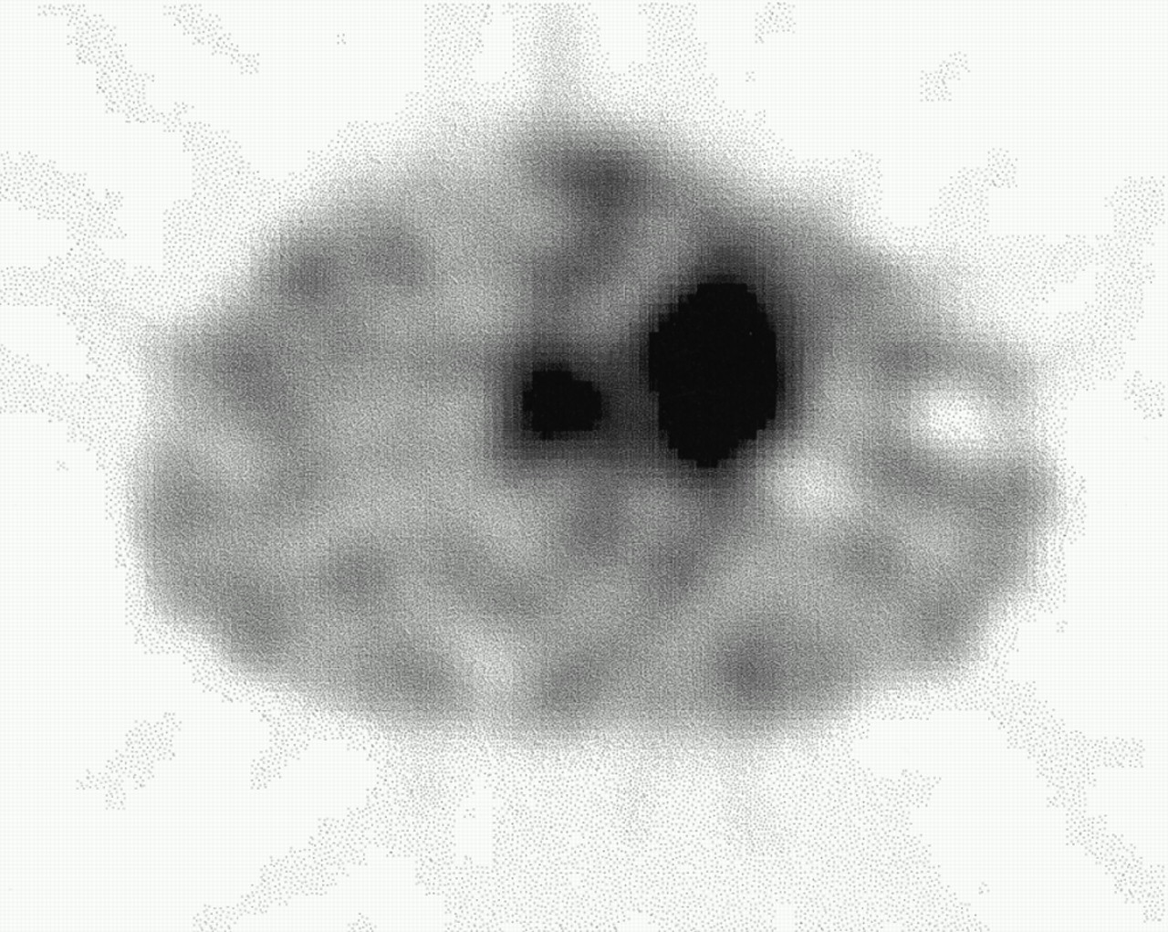
- FIGURE 7.
Coronal chest IMT SPECT section through 6-cm squamous cell carcinoma in right middle lobe of same patient as in Figure 3 shows high IMT uptake.
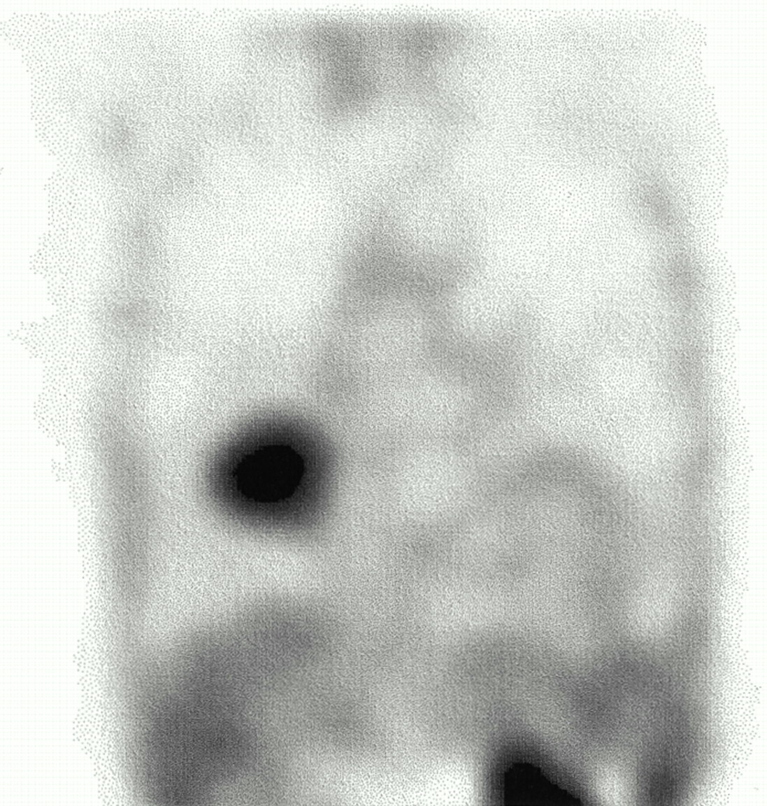
- FIGURE 8.
Transverse chest IMT SPECT image of patient with large cell carcinoma of left upper lobe and mediastinal metastasis shows avid uptake in both.
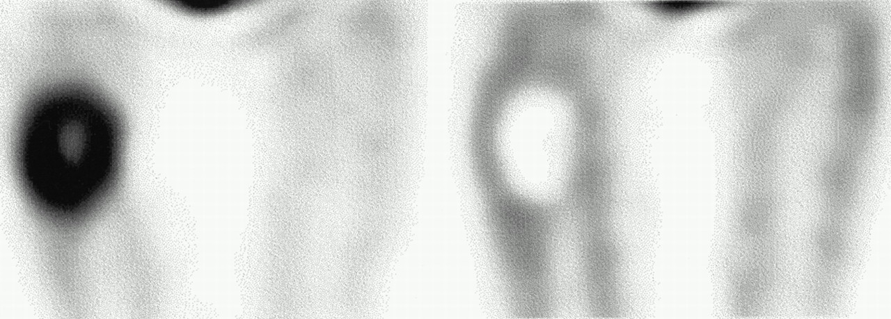
- FIGURE 9.
Coronal IMT SPECT sections through upper legs of patient with high-grade malignant fibrous histiocytoma, before (left) and after (right) regional hyperthermic cytostatic perfusion of leg, show disappearance of irregular intense IMT uptake after perfusion, in agreement with complete tumor necrosis.


Tables
Level Diagnostic test Seeks answers to whether the application … 1 Feasibility is feasible 2 Accuracy is sufficiently sensitive and specific 3 Diagnostic value performs well in relation to other tests 4 Therapeutic value results in better treatment 5 Patient and societal value results in better survival and quality of life, at acceptable cost Table is slightly adapted from (58).
Study Year No. of patients Purpose Sensitivity (%) Remarks and findings Biersack (59) 1989 10 Detection 100 First study Langen (36) 1990 32 Detection 88 Kuwert (55) 1995 53 Detection 50–82 Differentiation between high grade, low grade, and benign; specificity 83%–100% Weber (75) 1997 19 Detection 97 IMT uptake ratios superior to those of FDG PET Langen (115) 1997 14 Detection 100 Similar to MET PET Woesler (116) 1997 23 Detection 83 Differentiation between high and low grades; IMT similar to FDG PET Grosu (76) 2000 30 Detection 100 Significant impact on radiotherapy planning Guth (84) 1995 17 Evaluation 82 Recurrence detection Molenkamp (117) 1998 11 Evaluation 100 Detection of progression in low-grade childhood tumors Kuwert (118) 1998 27 Evaluation 78 Recurrence detection; specificity 100% Bader (69) 1999 30 Evaluation 75–100 Detection of recurrence, grades 2–4; superior to FDG PET Study Year No. of patients Tumor type Sensitivity (%) Remarks and findings Pruim (56) 1995 22 Primary brain 92 Specificity 67%; no correlation with grade Heesters (85) 1998 10 Primary brain — PSR within original tumor volume unchanged after radiotherapy Braams (89) 1996 11 Oral cavity 83 In nodal staging, better than MRI or CT; specificity 95% Kole (100) 1997 13 Breast cancer 100 For primary tumor; visually less uptake than for FDG in fibrocystic disease Ginkel (107) 1999 17 Sarcoma 82 Partial vs. complete remission distinguished after chemotherapy; specificity 100% Plaat (106) 1999 21 Sarcoma — Correlation of PSR with Ki-67, not with grade Kole (57) 1999 25 Sarcoma — FDG better for grading; TYR better for correlation with proliferation Kole (111) 1998 10 Nonseminoma 20 Kole (119) 1997 22 Various types 94 Chondrosarcoma not visualized Que (120) 2000 10 Cervix 80 Interfering bone marrow and intestinal uptake present PSR = protein synthesis rate.
Study Year No. of patients Tumor type Sensitivity (%) Remarks and findings Flamen (90) 1999 11 Head and neck 91 For primary tumors; ∼60% for nodal spread Jager (37) 1998 20 Various types — Feasible in breast cancer, lung cancer, sarcoma, and lymphoma Boni (109) 1997 7 Melanoma 37 For lesion detection Jager (110) 2000 22 Carcinoid 43–60 Correlation with secretory activity Jager (42) 2000 32 Sarcoma 100 88% specificity for differentiation between benign and malignant; correlation with proliferation Jager (96) 2000 17 Lung cancer 94 For primary tumors; 60% for mediastinal lesions
In this issue


Jump to section
Related Articles
Cited By...
- Diagnostic value of PET with different radiotracers and MRI for recurrent glioma: a Bayesian network meta-analysis
- Interaction of Halogenated Tyrosine/Phenylalanine Derivatives with Organic Anion Transporter 1 in the Renal Handling of Tumor Imaging Probes
- Radiosynthesis, in vitro and preliminary in vivo evaluation of the novel glutamine derived PET tracers [18F]fluorophenylglutamine and [18F]fluorobiphenylglutamine
- The L-type amino acid transporter LAT1 inhibits osteoclastogenesis and maintains bone homeostasis through the mTORC1 pathway
- Determination of an Optimal Pharmacokinetic Model of 18F-FET for Quantitative Applications in Rat Brain Tumors
- Evaluation of Prostate Cancer with Radiolabeled Amino Acid Analogs
- Anti-3-18F-FACBC (18F-Fluciclovine) PET/CT of Breast Cancer: An Exploratory Study
- Serial 18F-FET PET Imaging of Primarily 18F-FET-Negative Glioma: Does It Make Sense?
- Diagnostic and Prognostic Value of 11C-Methionine PET for Nonenhancing Gliomas
- Boramino acid as a marker for amino acid transporters
- Anti-1-Amino-3-18F-Fluorocyclobutane-1-Carboxylic Acid: Physiologic Uptake Patterns, Incidental Findings, and Variants That May Simulate Disease
- First Clinical Results of (D)-18F-Fluoromethyltyrosine (BAY 86-9596) PET/CT in Patients with Non-Small Cell Lung Cancer and Head and Neck Squamous Cell Carcinoma
- Preclinical Characterization of 5-Amino-4-Oxo-[6-11C]Hexanoic Acid as an Imaging Probe to Estimate Protoporphyrin IX Accumulation Induced by Exogenous Aminolevulinic Acid
- Comparison of the Amino Acid Tracers 18F-FET and 18F-DOPA in High-Grade Glioma Patients
- Treatment Response Evaluation Using 18F-FDOPA PET in Patients with Recurrent Malignant Glioma on Bevacizumab Therapy
- A Meta-Analysis on the Diagnostic Performance of 18F-FDG and 11C-Methionine PET for Differentiating Brain Tumors
- 11C-Methionine PET for Grading and Prognostication in Gliomas: A Comparison Study with 18F-FDG PET and Contrast Enhancement on MRI
- Comparative Evaluation of 18F-Labeled Glutamic Acid and Glutamine as Tumor Metabolic Imaging Agents
- Transport of 3-Fluoro-L-{alpha}-Methyl-Tyrosine by Tumor-Upregulated L-Type Amino Acid Transporter 1: A Cause of the Tumor Uptake in PET
- Assessment of Treatment Response in Patients with Glioblastoma Using O-(2-18F-Fluoroethyl)-L-Tyrosine PET in Comparison to MRI
- Impact of 3,4-Dihydroxy-6-18F-Fluoro-L-Phenylalanine PET/CT on Managing Patients with Brain Tumors: The Referring Physician's Perspective
- Evaluation of 4'-[Methyl-11C]Thiothymidine in a Rodent Tumor and Inflammation Model
- Specific biomarkers of receptors, pathways of inhibition and targeted therapies: clinical applications
- O-(2-18F-Fluoroethyl)-L-Tyrosine PET Predicts Failure of Antiangiogenic Treatment in Patients with Recurrent High-Grade Glioma
- Putative Transport Mechanism and Intracellular Fate of Trans-1-Amino-3-18F-Fluorocyclobutanecarboxylic Acid in Human Prostate Cancer
- S-11C-Methyl-L-Cysteine: A New Amino Acid PET Tracer for Cancer Imaging
- Correlation of 6-18F-Fluoro-L-Dopa PET Uptake with Proliferation and Tumor Grade in Newly Diagnosed and Recurrent Gliomas
- Methyl-L-11C-Methionine PET as a Diagnostic Marker for Malignant Progression in Patients with Glioma
- Efficacy of Systemic Radionuclide Therapy with p-131I-Iodo-L-Phenylalanine Combined with External Beam Photon Irradiation in Treating Malignant Gliomas
- Tumor Cell Metabolism Imaging
- 6-L-18F-Fluorodihydroxyphenylalanine PET in Neuroendocrine Tumors: Basic Aspects and Emerging Clinical Applications
- Non-islet cell tumour-induced hypoglycaemia: a review of the literature including two new cases
- L-Type Amino Acid Transporters LAT1 and LAT4 in Cancer: Uptake of 3-O-Methyl-6- 18F-Fluoro-L-Dopa in Human Adenocarcinoma and Squamous Cell Carcinoma In Vitro and In Vivo
- 18F-FDOPA Kinetics in Brain Tumors
- Clinical Applications of PET in Brain Tumors
- Biodistribution and Radiation Dosimetry of the Synthetic Nonmetabolized Amino Acid Analogue Anti-18F-FACBC in Humans
- PET/CT of Skull Base Meningiomas Using 2-18F-Fluoro-L-Tyrosine: Initial Report
- Positron Emission Tomography As an Imaging Biomarker
- 18F-FDOPA PET Imaging of Brain Tumors: Comparison Study with 18F-FDG PET and Evaluation of Diagnostic Accuracy
- 18F-FET PET Differentiation of Ring-Enhancing Brain Lesions
- Comparison of Sigma-Ligands and Metabolic PET Tracers for Differentiating Tumor from Inflammation
- In Vivo Evaluation and Dosimetry of 123I-2-Iodo-D-Phenylalanine, a New Potential Tumor-Specific Tracer for SPECT, in an R1M Rhabdomyosarcoma Athymic Mouse Model
- In Vivo Characterization of 123/125I-2-Iodo-L-Phenylalanine in an R1M Rhabdomyosarcoma Athymic Mouse Model as a Potential Tumor Tracer for SPECT
- PET with O-(2-18F-Fluoroethyl)-L-Tyrosine in Peripheral Tumors: First Clinical Results
- 3-O-Methyl-6-18F-Fluoro-L-Dopa, a New Tumor Imaging Agent: Investigation of Transport Mechanism In Vitro
- Prediction of Survival and Therapy Outcome with 11C-Tyrosine PET in Patients with Laryngeal Carcinoma
- Delineation of Brain Tumor Extent with [11C]L-Methionine Positron Emission Tomography: Local Comparison with Stereotactic Histopathology
- Molecular Transport Mechanisms of Radiolabeled Amino Acids for PET and SPECT
- Quantitation of Small-Animal 124I Activity Distributions Using a Clinical PET/CT Scanner
- Comparison of O-(2-18F-Fluoroethyl)-L-Tyrosine PET and 3-123I-Iodo-{alpha}-Methyl-L-Tyrosine SPECT in Brain Tumors
- 18F-FLT PET for Visualization of Laryngeal Cancer: Comparison with 18F-FDG PET
- Comparative Biodistribution of Iodinated Amino Acids in Rats: Selection of the Optimal Analog for Oncologic Imaging Outside the Brain
- Whole-Body Tumor Imaging Using PET and 2-18F-Fluoro-L-Tyrosine: Preliminary Evaluation and Comparison with 18F-FDG
- Early Response to Chemotherapy in Hypopharyngeal Cancer: Assessment with 11C-Methionine PET, Correlation with Morphologic Response, and Clinical Outcome
- An Artificial Amino Acid, 4-Iodo-L-meta-Tyrosine: Biodistribution and Excretion via Kidney
- L-1-11C-Tyrosine PET in Patients with Laryngeal Carcinomas: Comparison of Standardized Uptake Value and Protein Synthesis Rate
- Isoform Selectivity of 3-125I-Iodo-{alpha}-Methyl-L-Tyrosine Membrane Transport in Human L-Type Amino Acid Transporters
- Increased Tumor Uptake of 3-123I-Iodo-L-{alpha}-Methyltyrosine After Preloading with Amino Acids: An In Vivo Animal Imaging Study
- Improving Amino Acid Imaging: Hungry or Stuffed?