


Article Figures & Data
Figures
- FIGURE 1.
Summary of data: additional value of PET/CT for single-step detection of malignancy, definition of its location and extent, and improved management.
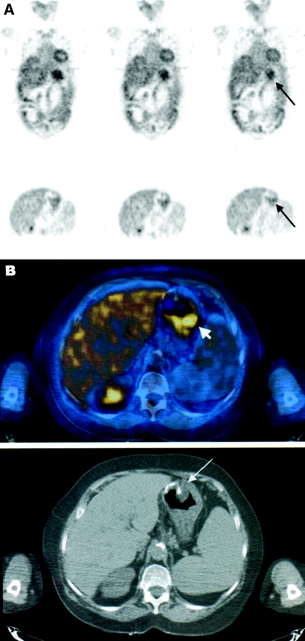
- FIGURE 2.
Precise localization of increased 18F-FDG uptake and exclusion of malignancy, after PET/CT. A 68-y-old man, 3 y after partial gastrectomy for adenocarcinoma of stomach, was referred for 18F-FDG PET/CT for further evaluation of polypoid mass in gastric stump detected on routine follow-up gastroscopy, with equivocal biopsy results. (A) 18F-FDG PET coronal images (top) and axial images (bottom) show increased 18F-FDG uptake in region of stomach (arrow). (B) Hybrid PET/CT axial image (top) precisely localizes and defines uptake as physiologic activity at gastric stump (arrowhead). Suspicious polypoid mass in anastomotic region (arrow), seen on corresponding hybrid and CT slices (bottom) obtained during same acquisition, shows no uptake of 18F-FDG. Findings on PET/CT were interpreted as physiologic 18F-FDG uptake in stomach and nonviable residual mass. Patient showed no evidence of disease for follow-up of 7 mo.
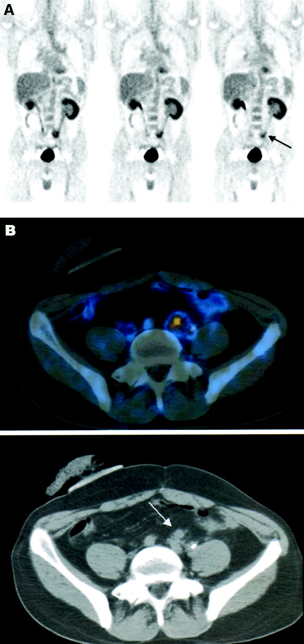
- FIGURE 3.
Precise characterization of increased 18F-FDG uptake and retrospective lesion detection on CT, after PET/CT. A 35-y-old man, 22 mo after treatment for colon cancer, with negative high-resolution contrast-enhanced CT and normal levels of serum tumor markers, was referred for 18F-FDG PET for further assessment of pelvic pain. (A) Coronal PET images show area of increased 18F-FDG uptake in left pelvic region (arrow), interpreted as equivocal for malignancy, possibly related to inflammatory changes associated with ureteral stent or to physiologic bowel uptake. (B) Hybrid PET/CT axial image (top) precisely localizes uptake to soft-tissue mass adjacent to left ureter, anterior to left iliac vessels. Mass (arrow) was detected only retrospectively on both diagnostic CT and CT component of hybrid imaging study (bottom). Patient received chemotherapy, resulting in pain relief and decrease in size of pelvic mass on follow-up CT.
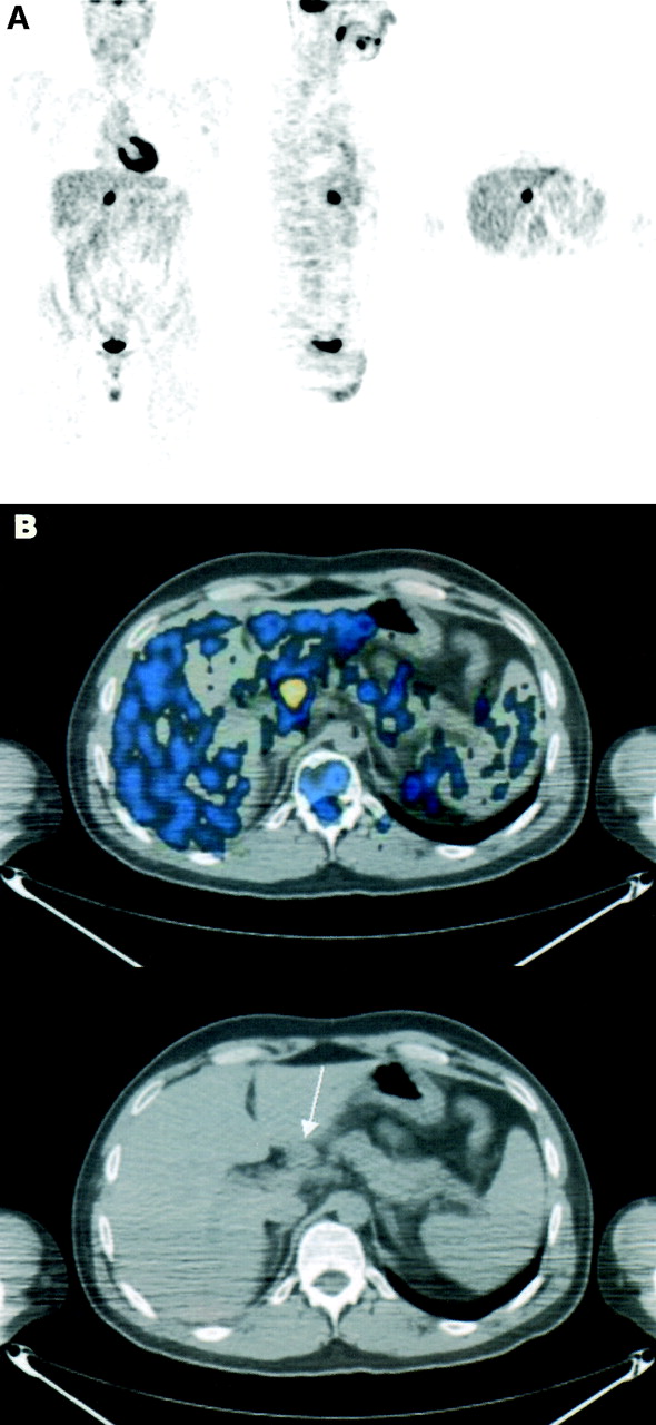
- FIGURE 4.
Precise anatomic localization of malignant 18F-FDG uptake and retrospective lesion detection on CT, after PET/CT. A 33-y-old man with Hodgkin’s disease in left cervical region was referred for 18F-FDG PET for staging. No other sites of disease were reported on CT. (A) PET images show infradiaphragmatic focus of abnormal 18F-FDG uptake in medial border of liver, consistent with either liver involvement (stage IV disease?) or nodal disease in porta hepatis (stage III disease?). (B) Hybrid PET/CT axial image (top) precisely localizes 18F-FDG uptake to adenopathy at porta hepatis, only retrospectively detected on corresponding CT image (bottom) (arrow). Patient was treated as having stage III disease and achieved complete response, showing no evidence of disease for follow-up of 12 mo.


Tables
Criteria Head and neck Chest Abdominopelvic Soft tissue and bone Total Precise lesion characterization and localization on PET after PET/CT As benign 2 34 16 8 60 As malignant 5 10 14 2 31 Precise localization of malignant lesion — 12 17 8 37 Retrospective lesion detection after PET/CT On PET 2 4 1 — 7 On CT* 6 19 21 17 63 Total† (%) 12 71 61 34 178 (32) (24) (41) (31) (30) Patient no. Diagnosis PET/CT additional value* Lesion definition after PET/CT Impact and follow-up 1 Lung cancer, treatment response a Vascular structure Exclusion of cancer, NED on 7-mo follow-up 2 Lung cancer, suspected recurrence a Vascular structure Exclusion of cancer, NED on 7-mo follow-up 3 Lung cancer, suspected recurrence a Vascular structure Exclusion of cancer, NED on 7-mo follow-up 4 Stomach cancer, suspected recurrence a Physiologic uptake in gastric lumen Exclusion of cancer, NED on 7-mo follow-up 5 Lung cancer, suspected recurrence a Vascular structure Exclusion of hilar metastasis, radiotherapy of single bone metastasis 6 Melanoma, staging b, e Thyroid lesion Guiding biopsy, benign nodule 7 Lung cancer, staging c Lesion in ischium Guiding biopsy, tendinitis 8 Breast cancer, suspected recurrence b Mediastinal LN Guiding mediastinoscopy, antracotic LN 9 Colon cancer, suspected recurrence c Lesion in colon Guiding colonoscopy and biopsy, recurrence 10 SPN, diagnosis c Lesion in colon Guiding biopsy, primary colon cancer and lung metastasis 11 Colon cancer, suspected recurrence b, e Supraclavicular LN Guiding biopsy, metastatic adenopathy 12 Renal tumor, suspected recurrence b Mass in pancreas Guiding biopsy, metastasis in pancreas 13 SPN, diagnosis d Lesion in lung Guiding curative surgery, bronchoalveolar cancer 14 Colon cancer, suspected recurrence c Abdominal mass Guiding surgery, omental metastases 15 Colon cancer, suspected recurrence c Parasplenic LN Guiding surgery, LN metastases 16 Colon cancer, suspected recurrence b, e Pelvic LN Guiding surgery, LN metastases 17 Recurrent colon cancer, restaging e Hepatic lesion Planning surgical approach, liver wedge resection 18 Colon cancer, suspected recurrence b, e Mesenteric LN Referred to chemotherapy 19 Colon cancer, suspected recurrence b, e Parailiac LN Referred to chemotherapy 20 Recurrent ovary cancer, restaging b, e Parasplenic LN Referred to chemotherapy, surgery canceled 21 Recurrent lung cancer, restaging c, e Lung lesion Referred to chemotherapy, surgery canceled 22 Lung cancer, suspected recurrence c, e Retrocrural LN Referred to chemotherapy in addition to surgery and radiotherapy 23 Lung cancer, suspected recurrence b, e Lesion in left ilium Referred to radiotherapy 24 Lymphoma, treatment response d Nasopharynx Referred to radiotherapy 25 Lung cancer, suspected recurrence e Mediastinal LN Change in radiation fields 26 Bladder cancer, suspected recurrence b, e Pelvic LN Change in radiation fields 27 Cervical cancer, staging c Retroperitoneal LN Change in radiation fields 28 Lung cancer, suspected recurrence a Vascular structure Exclusion of additional malignant site, change in radiation fields ↵* PET/CT additional value: Categories by which PET/CT induced changes in image interpretation of suspected sites were classified as follows: a = characterization as definitely benign (exclusion of malignancy); b = characterization as definitely malignant; c = precise anatomic localization of malignant site; d = retrospective detection on PET; e = retrospective detection on high-resolution contrast-enhanced CT and on CT of PET/CT.
NED = no evidence of disease; LN = lymph node.
In this issue


Jump to section
Related Articles
Cited By...
- Relationship Between 18F-FDG-PET/CT-derived Tumor Glucose Metabolic Activity, Nutritional Risk, and Survival in Patients With Soft-tissue Sarcoma
- Limiting surveillance imaging for patients with lymphoma in remission: a mixed methods study leading to a Choosing Wisely recommendation
- Performance of Digital PET Compared with High-Resolution Conventional PET in Patients with Cancer
- Novel Method to Detect and Characterize 18F-FDG Infiltration at the Injection Site: A Single-Institution Experience
- Whole-Body 18F-FDG PET/CT Is Superior to CT as First-Line Diagnostic Imaging in Patients Referred with Serious Nonspecific Symptoms or Signs of Cancer: A Randomized Prospective Study of 200 Patients
- Initial Preclinical Evaluation of 18F-Fluorodeoxysorbitol PET as a Novel Functional Renal Imaging Agent
- Functional Flow Patterns and Static Blood Pooling in Tumors Revealed by Combined Contrast-Enhanced Ultrasound and Photoacoustic Imaging
- Assessment of incidental and clinically unsuspected fluorodeoxyglucose-avid foci detected on oncological positron emission tomography/CT
- Positron emission tomography/computed tomography surveillance in patients with Hodgkin lymphoma in first remission has a low positive predictive value and high costs
- PET/CT and MRI of intra-osseous haemangioma of the tibia
- Imaging techniques in lung cancer
- Uptake of 18F-FDG in Acute Aortic Dissection: A Determinant of Unfavorable Outcome
- Prospective Evaluation of Whole-Body Cancer Screening With Multiple Modalities Including [18F]Fluorodeoxyglucose Positron Emission Tomography in a Healthy Population: A Preliminary Report
- Integrated PET/CT in the staging of nonsmall cell lung cancer: technical aspects and clinical integration
- Relationship Between Cancer Type and Impact of PET and PET/CT on Intended Management: Findings of the National Oncologic PET Registry
- PET/CT assessment of clinically unsuspected, incidental FDG-avid lesions in oncological patients
- 18F-FDG PET/CT in Evaluating Non-CNS Pediatric Malignancies
- Multimodality imaging of direct ureteric involvement in non-Hodgkin's lymphoma using PET/CT, CT urography and antegrade CT pyelography
- Value of contrast-enhanced multiphase CT in combined PET/CT protocols for oncological imaging
- Detection of extrapulmonary lesions with integrated PET/CT in the staging of lung cancer
- SPECT/Multislice Low-Dose CT: A Clinically Relevant Constituent in the Imaging Algorithm of Nononcologic Patients Referred for Bone Scintigraphy
- Screening for Cancer with PET and PET/CT: Potential and Limitations
- Early Detection of Cancer Recurrence: 18F-FDG PET/CT Can Make a Difference in Diagnosis and Patient Care
- PET-Based Treatment Planning in Radiotherapy: A New Standard?
- Clinical Significance of Small Pulmonary Nodules with Little or No 18F-FDG Uptake on PET/CT Images of Patients with Nonthoracic Malignancies
- PET/CT in Lymphoma: Prospective Study of Enhanced Full-Dose PET/CT Versus Unenhanced Low-Dose PET/CT
- Additional Value of PET/CT over PET in Assessment of Locoregional Lymph Nodes in Thoracic Esophageal Squamous Cell Cancer
- 11C-Acetate Positron Emission Tomography Imaging and Image Fusion With Computed Tomography and Magnetic Resonance Imaging in Patients With Recurrent Prostate Cancer
- Focal Thyroid Lesions Incidentally Identified by Integrated 18F-FDG PET/CT: Clinical Significance and Improved Characterization
- Whole body positron emission tomography/computed tomography (PET/CT) tumour staging with integrated PET/CT colonography: technical feasibility and first experiences in patients with colorectal cancer
- Improved Detection of Second Primary Cancer Using Integrated [18F] Fluorodeoxyglucose Positron Emission Tomography and Computed Tomography for Initial Tumor Staging
- Detection and Localization of Prostate Cancer: Correlation of 11C-Choline PET/CT with Histopathologic Step-Section Analysis
- Etiology of Solitary Extrapulmonary Positron Emission Tomography and Computed Tomography Findings in Patients With Lung Cancer
- Attenuation Correction of PET Images with Respiration-Averaged CT Images in PET/CT
- CT in PET/CT: Essential Features of Interpretation
- Imaging of Malignant Bone Involvement by Morphologic, Scintigraphic, and Hybrid Modalities
- Clinically Significant Incidental Findings on the Unenhanced CT Portion of PET/CT Studies: Frequency in 250 Patients
- Concurrent PET/CT with an Integrated Imaging System: Intersociety Dialogue from the Joint Working Group of the American College of Radiology, the Society of Nuclear Medicine, and the Society of Computed Body Tomography and Magnetic Resonance
- PET/CT Detection of Unexpected Gastrointestinal Foci of 18F-FDG Uptake: Incidence, Localization Patterns, and Clinical Significance
- Progress and Promise of FDG-PET Imaging for Cancer Patient Management and Oncologic Drug Development
- Is 18F-FDG PET/CT Useful for Imaging and Management of Patients with Suspected Occult Recurrence of Cancer?
- Evaluation of 18F-FDG Uptake and Arterial Wall Calcifications Using 18F-FDG PET/CT
- PET/CT Using 18F-FDG in Suspected Lung Cancer Recurrence: Diagnostic Value and Impact on Patient Management
- Assessment of Malignant Skeletal Disease: Initial Experience with 18F-Fluoride PET/CT and Comparison Between 18F-Fluoride PET and 18F-Fluoride PET/CT
- PET/CT Image Navigation and Communication
- Why Most PET of Lung and Head-and-Neck Cancer Will Be PET/CT
- PET/CT Today and Tomorrow
- Software Approach to Merging Molecular with Anatomic Information
- To Enhance or Not to Enhance? 18F-FDG and CT Contrast Agents in Dual-Modality 18F-FDG PET/CT
- Why Nearly All PET of Abdominal and Pelvic Cancers Will Be Performed as PET/CT
- Acquisition Protocol Considerations for Combined PET/CT Imaging