


Article Figures & Data
Figures
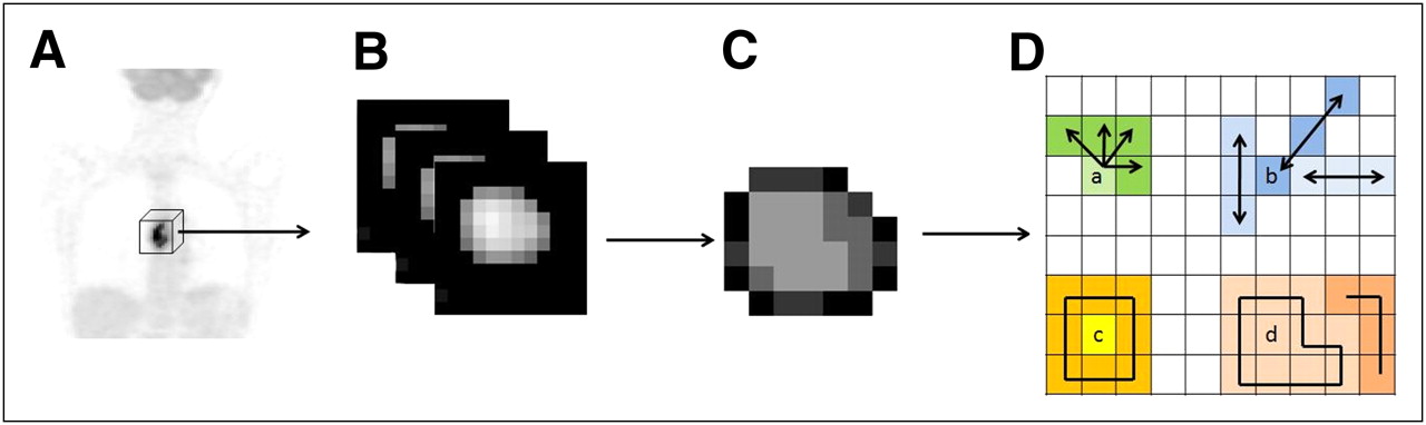
- FIGURE 1.
Whole-body 18F-FDG PET scan (A), tumor segmentation (B), and voxel-intensity resampling (C) allowing extraction of different features (D) by analysis of consecutive voxels in a direction (for cooccurrence matrices) (a), alignment of voxels with same intensity (b), difference between voxels and their neighbors (c), and zones of voxels with same intensity (d).
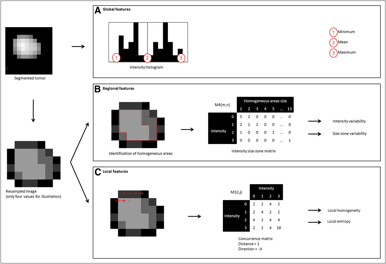
- FIGURE 2.
Examples of features extracted from tumor resampled on 4 values: 3 global features computed using intensity histogram, 2 regional features computed using M4 matrix, and 2 local features computed using M1 texture matrices.
- FIGURE 3.
Box-plot representation of parameters’ values in function of patient response (0, NR; 1, PR; and 2, CR) for SUVmax (P = 0.106) (A), SUVpeak (P = 0.045) (B), local entropy (P = 0.0006) (C), and regional intensity variability (P = 0.0002) (D).
- FIGURE 4.
Example of different extracted features and associated values for tumors of CRs, PRs, and NRs (results are normalized in [0–1] interval using range of observed values for local and regional parameters).
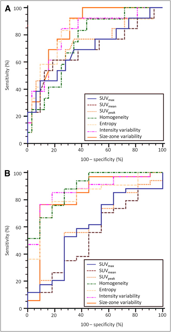
- FIGURE 5.
ROC curves for SUVmax, SUVmean, SUVpeak, local homogeneity, uniform tumor areas, intensity variability, and size-zone variability for identification of CRs (A) and PRs or CRs (B).


Tables
Characteristic No. of patients Sex Male 35 (85) Female 6 (15) Primary site Upper esophagus 10 (24) Middle esophagus 15 (37) Lower esophagus 16 (39) Tumor cell type Squamous cell carcinoma 31 (76) Adenocarcinoma 10 (24) Histologic grade Well differentiated 12 (29) Moderately differentiated 11 (27) Poorly differentiated 3 (7) Unknown 15 (37) TNM stage T1 6 (15) T2 7 (17) T3 21 (51) T4 7 (17) N0 16 (39) N1 25 (61) M0 24 (59) M1 17 (41) AJCC stage I 4 (10) IIa 6 (15) IIb 5 (12) III 12 (29) IVa 4 (10) IVb 10 (24) RECIST CR 9 (22) PR 21 (51) Stable disease (NR) 7 (17) Progressive disease (NR) 4 (10) Data in parentheses are percentages.
Type Feature Scale Features based on intensity histogram Minimum intensity Global Maximum intensity Mean intensity Variance SD Skewness Kurtosis Features based on voxel-alignment matrix (M2) Short run emphasis Regional Long run emphasis Intensity variability Run-length variability Run percentage Low-intensity run emphasis High-intensity run emphasis Low-intensity short-run emphasis High-intensity short-run emphasis Low-intensity long-run emphasis High-intensity long-run emphasis Features based on intensity–size–zone matrix (M4) Short-zone emphasis Regional Large-zone emphasis Intensity variability Size-zone variability Zone percentage Low-intensity zone emphasis High-intensity zone emphasis Low-intensity short-zone emphasis High-intensity short-zone emphasis Low-intensity large-zone emphasis High-intensity large-zone emphasis Features based on cooccurrence matrices (M1) Second angular moment Local Contrast (inertia) Entropy Correlation Homogeneity Dissimilarity Features based on neighborhood intensity-difference matrix (M3) Coarseness Local Contrast Busyness - TABLE 3
Sensitivity and Specificity (Along with Corresponding 95% Confidence Intervals) of 3 SUV-Based Measurements, 2 Cooccurrence Features, and 2 Size-Zone Features
Comparison Parameters Sensitivity (%) 95% confidence interval (%) Specificity (%) 95% confidence interval (%) NR vs. PR + CR SUVmax 53 35.1–70.2 73 39.0–94.0 SUVmean 71 52.5–84.9 45 16.7–76.6 SUVpeak 56 37.9–72.8 73 39.0–94.0 Local homogeneity 88 71.8–96.6 73 39.0–94.0 Local entropy 79 61.1–91.0 82 48.2–97.7 Size-zone 76 58.8–89.8 91 58.7–99.8 Intensity variability 76 58.7–89.3 91 58.7–99.8 NR + PR vs. CR SUVmax 46 19.2–74.9 91 75.0–98.0 SUVmean 62 31.6–86.1 81 63.6–92.8 SUVpeak 62 31.6–86.1 81 63.6–92.8 Local homogeneity 92 61.5–99.8 56 37.7–73.6 Local entropy 92 61.5–99.8 69 50.0–83.9 Size-zone 92 64.0–99.8 69 50.0–83.9 Intensity variability 85 54.6–98.1 75 56.6–88.5 Data in top part of table are evaluation of parameters to distinguish PR or CR; data on bottom part of table are evaluation of parameters to differentiate CRs.


Jump to section
Related Articles
Cited By...
- Computed tomography radiomics-based cross-sectional detection of mandibular osteoradionecrosis in head and neck cancer survivors
- Repeatability of 18F-FDG PET Radiomic Features in Cervical Cancer
- The contribution of evolutionary game theory to understanding and treating cancer
- Texture Feature Comparison Between Step-and-Shoot and Continuous-Bed-Motion 18F-FDG PET
- Radiomics: Data Are Also Images
- Optimized Feature Extraction for Radiomics Analysis of 18F-FDG PET Imaging
- Pretreatment 18F-FDG Uptake Heterogeneity Predicts Treatment Outcome of First-Line Chemotherapy in Patients with Metastatic Triple-Negative Breast Cancer
- Noninvasive Imaging of Drug-Induced Liver Injury with 18F-DFA PET
- Metabolic heterogeneity on baseline 18FDG-PET/CT scan is a predictor of outcome in primary mediastinal B-cell lymphoma
- Comprehensive Computed Tomography Radiomics Analysis of Lung Adenocarcinoma for Prognostication
- Assessing the Clinical Utility of Computed Tomography-Based Radiomics
- Glioma Survival Prediction with Combined Analysis of In Vivo 11C-MET PET Features, Ex Vivo Features, and Patient Features by Supervised Machine Learning
- Responsible Radiomics Research for Faster Clinical Translation
- Tumor Metabolic Features Identified by 18F-FDG PET Correlate with Gene Networks of Immune Cell Microenvironment in Head and Neck Cancer
- Serum levels of chemical elements in esophageal squamous cell carcinoma in Anyang, China: a case-control study based on machine learning methods
- Differentiation of Enhancing Glioma and Primary Central Nervous System Lymphoma by Texture-Based Machine Learning
- Predicting Response to Neoadjuvant Chemoradiotherapy in Esophageal Cancer with Textural Features Derived from Pretreatment 18F-FDG PET/CT Imaging
- Understanding Changes in Tumor Texture Indices in PET: A Comparison Between Visual Assessment and Index Values in Simulated and Patient Data
- Comparison of Tumor Uptake Heterogeneity Characterization Between Static and Parametric 18F-FDG PET Images in Non-Small Cell Lung Cancer
- The Incremental Value of Subjective and Quantitative Assessment of 18F-FDG PET for the Prediction of Pathologic Complete Response to Preoperative Chemoradiotherapy in Esophageal Cancer
- Multiparametric Analysis of the Relationship Between Tumor Hypoxia and Perfusion with 18F-Fluoroazomycin Arabinoside and 15O-H2O PET
- Texture Feature Ratios from Relative CBV Maps of Perfusion MRI Are Associated with Patient Survival in Glioblastoma
- Tumor Texture Analysis in PET: Where Do We Stand?
- Impact of Image Reconstruction Settings on Texture Features in 18F-FDG PET
- 18F-FDG PET Uptake Characterization Through Texture Analysis: Investigating the Complementary Nature of Heterogeneity and Functional Tumor Volume in a Multi-Cancer Site Patient Cohort
- Visual Versus Quantitative Assessment of Intratumor 18F-FDG PET Uptake Heterogeneity: Prognostic Value in Non-Small Cell Lung Cancer
- Textural Parameters of Tumor Heterogeneity in 18F-FDG PET/CT for Therapy Response Assessment and Prognosis in Patients with Locally Advanced Rectal Cancer
- Tumor Texture Analysis in 18F-FDG PET: Relationships Between Texture Parameters, Histogram Indices, Standardized Uptake Values, Metabolic Volumes, and Total Lesion Glycolysis
- The Effect of Small Tumor Volumes on Studies of Intratumoral Heterogeneity of Tracer Uptake
- Textural Features of Pretreatment 18F-FDG PET/CT Images: Prognostic Significance in Patients with Advanced T-Stage Oropharyngeal Squamous Cell Carcinoma
- Are Pretreatment 18F-FDG PET Tumor Textural Features in Non-Small Cell Lung Cancer Associated with Response and Survival After Chemoradiotherapy?
- Marker Selection Based on Only Reproducibility Can Be Questioned
- Reply: Marker Selection Based on Only Reproducibility Can Be Questioned
- Reproducibility of Tumor Uptake Heterogeneity Characterization Through Textural Feature Analysis in 18F-FDG PET
- Impact of Partial-Volume Effect Correction on the Predictive and Prognostic Value of Baseline 18F-FDG PET Images in Esophageal Cancer
- Impact of Tumor Size and Tracer Uptake Heterogeneity in 18F-FDG PET and CT Non-Small Cell Lung Cancer Tumor Delineation