Main menu

  • Home
  • Content
    • Current
    • Ahead of print
    • Past Issues
    • JNM Supplement
    • SNMMI Annual Meeting Abstracts
    • Continuing Education
    • JNM Podcasts
  • Subscriptions
    • Subscribers
    • Institutional and Non-member
    • Rates
    • Journal Claims
    • Corporate & Special Sales
  • Authors
    • Submit to JNM
    • Information for Authors
    • Assignment of Copyright
    • AQARA requirements
  • Info
    • Reviewers
    • Permissions
    • Advertisers
  • About
    • About Us
    • Editorial Board
    • Contact Information
  • More
    • Alerts
    • Feedback
    • Help
    • SNMMI Journals
  • SNMMI
    • JNM
    • JNMT
    • SNMMI Journals
    • SNMMI

User menu

  • Subscribe
  • My alerts
  • Log in
  • Log out
  • My Cart

Search

  • Advanced search
Journal of Nuclear Medicine
  • SNMMI
    • JNM
    • JNMT
    • SNMMI Journals
    • SNMMI
  • Subscribe
  • My alerts
  • Log in
  • Log out
  • My Cart
Journal of Nuclear Medicine

Advanced Search

  • Home
  • Content
    • Current
    • Ahead of print
    • Past Issues
    • JNM Supplement
    • SNMMI Annual Meeting Abstracts
    • Continuing Education
    • JNM Podcasts
  • Subscriptions
    • Subscribers
    • Institutional and Non-member
    • Rates
    • Journal Claims
    • Corporate & Special Sales
  • Authors
    • Submit to JNM
    • Information for Authors
    • Assignment of Copyright
    • AQARA requirements
  • Info
    • Reviewers
    • Permissions
    • Advertisers
  • About
    • About Us
    • Editorial Board
    • Contact Information
  • More
    • Alerts
    • Feedback
    • Help
    • SNMMI Journals
  • View or Listen to JNM Podcast
  • Visit JNM on Facebook
  • Join JNM on LinkedIn
  • Follow JNM on Twitter
  • Subscribe to our RSS feeds
In BriefThis Month in JNM

This Month in JNM

Journal of Nuclear Medicine March 2011, 52 (3) 11A-12A;
  • Article
  • Info & Metrics
  • PDF
Loading

Molecular imaging and RT planning: Grégoire and Chiti provide an overview of current applications and utility of 18F-FDG PET in support of radiation therapy in patients with squamous cell carcinoma of the head and neck.Page 331

Figure

Assessing antiangiogenic response: Beer and Schwaiger review data supporting the use of 18F-fluciclatide PET imaging of αvβ3-integrin and αvβ5-integrin expression to monitor response to targeted therapy and preview a related article in this issue of JNM.Page 335

Assays of brain efflux transporters: Hall and Pike offer perspective on challenges in development of effective PET radioligands and describe a promising approach that is the focus of a related article in this issue of JNM.Page 338

Staging nasopharyngeal carcinoma: Wu and colleagues report on studies designed to improve detection of intracranial tumor invasion using 11C-choline PET/CT in patients with locally advanced nasopharyngeal carcinoma.Page 341

Figure

Lesion detection with TOF PET: El Fakhri and colleagues quantify improvements in lung and liver lesion detectability with whole-body time-of-flight PET in oncologic patients with varying body mass indices.Page 347

Figure

18F-FDG PET/CT and tumor changes: Necib and colleagues propose and evaluate a parametric imaging method for PET/CT assessment of metabolic tumor changes at the voxel level.Page 354

Carotid 18F-sodium fluoride uptake: Derlin and colleagues correlate 18F-sodium fluoride accumulation in the common carotid arteries of neurologically asymptomatic patients with cardiovascular risk factors and carotid calcified plaque burden.Page 362

Figure

PET textural analysis and response: Tixier and colleagues evaluate new parameters obtained by textural analysis of baseline 18F-FDG PET scans for prediction of therapy response in patients with newly diagnosed esophageal cancer undergoing combined radiochemotherapy.Page 369

Figure

Dynamic PET/CT: Strauss and colleagues describe and investigate shortened acquisition protocols for quantitative assessment of the 2-tissue-compartment model using dynamic 18F-FDG PET/CT.Page 379

Interim PET/CT in large cell lymphoma: Cashen and colleagues look at the effectiveness of 18F-FDG PET/CT in predicting outcomes in patients with advanced-stage diffuse large B-cell lymphoma, with results that raise questions about current interpretation criteria.Page 386

11C-PIB dual-biomarker imaging: Meyer and colleagues investigate the validity of relative regional cerebral blood flow estimates derived from 11C-labeled Pittsburgh compound B PET as a marker of neuronal activity and neurodegeneration in patients with cognitive impairment.Page 393

Figure

Thalamic nuclei glucose metabolism: Cho and colleagues describe studies measuring substructure-specific metabolic activities in the thalamus with a PET/MRI system made up of ultra-high-resolution PET and ultra-high-field MRI components.Page 401

Figure

Sentinel node biopsy in breast cancer: Hindié and colleagues provide an educational overview of sentinel node biopsy strategies, with a focus on potential remedies for the relatively high percentage of false-negative results reported.Page 405

PET radiotracer transport: Tournier and colleagues detail the development of an in vitro model designed to assess the blood–brain barrier transport of selected PET ligands using the concentration equilibrium technique, with potential for screening transport in new drugs.Page 415

18F-fluciclatide PET monitoring: Battle and colleagues use this radiolabeled small peptide, which binds with high affinity to αvβ3- and αvβ5-integrins, to examine the response of human glioblastoma xenografts to treatment with the antiangiogenic agent sunitinib.Page 424

Figure

Serial PET lesion evaluation: Heijink and colleagues study information provided by serial 18F-FDG PET imaging in Apc mutant mice, which develop multiple intestinal adenomas and are used to study colorectal carcinogenesis and chemopreventive approaches.Page 431

Figure

18F-FMISO PET antivascular assessment: Oehler and colleagues explore the utility of 18F-fluromisonidazole PET for monitoring tumor response to a compound that induces rapid endothelial cell apoptosis and decreases perfusion in preexisting tumor vessels.Page 437

Imaging intracellular molecular events: Lehmann and colleagues detail the use of avidin–glycosylphosphatidylinositol, an avidin moiety targeted to the extracellular side of cell membranes, as a reporter for in vivo imaging.Page 445

Figure

Quantification in myocardial SPECT/CT: Liu and colleagues introduce a heuristic method for correction of extracardiac activity into molecularly targeted SPECT/CT quantification and validate the method for accuracy and reproducibility in a canine model.Page 453

99mTc-labeled recombinant Affibody molecules: Wållberg and colleagues develop an agent with low renal uptake and preserved tumor targeting with promise for development as a diagnostic radiopharmaceutical for imaging of HER2-expressing tumors.Page 461

Figure

64Cu-SarAr-bombesin imaging: Lears and colleagues describe in vitro and in vivo studies evaluating the internalization of this peptide, which binds with high affinity to the gastrin-releasing peptide receptor, which is overexpressed on a variety of solid tumors.Page 470

Pharmacokinetics of BBB disruption: Yang and colleagues study the pharmacokinetics of 99mTc-DTPA in healthy and glioma-bearing rats in the presence of blood–brain barrier disruption induced by focused ultrasound.Page 478

82Rb stress dosimetry: Senthamizhchelvan and colleagues report on dose estimates under stress for 82Rb, used with PET for cardiac perfusion studies.Page 485

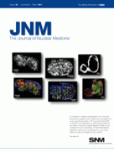
ON THE COVER

A comparison of digital autoradiography and composite fluorescence images of tumor hypoxia and vasculature. In a study evaluating the ability of 18F-fluoromisonidazole PET to monitor tumor response to an antivascular compound, the pattern of the tracer uptake reflected the effect of the compound on tumor physiology. 18F-fluoromisonidazole distribution was found to be governed by tumor tracer delivery as well as locoregional tumor hypoxia.

See page 441 and supplemental materials (available online at http://jnm.snmjournals.org).

Figure
  • © 2011 by Society of Nuclear Medicine
PreviousNext
Back to top

In this issue

Journal of Nuclear Medicine: 52 (3)
Journal of Nuclear Medicine
Vol. 52, Issue 3
March 1, 2011
  • Table of Contents
  • Table of Contents (PDF)
  • About the Cover
  • Index by author
Print
Download PDF
Article Alerts
Sign In to Email Alerts with your Email Address
Email Article

Thank you for your interest in spreading the word on Journal of Nuclear Medicine.

NOTE: We only request your email address so that the person you are recommending the page to knows that you wanted them to see it, and that it is not junk mail. We do not capture any email address.

Enter multiple addresses on separate lines or separate them with commas.
This Month in JNM
(Your Name) has sent you a message from Journal of Nuclear Medicine
(Your Name) thought you would like to see the Journal of Nuclear Medicine web site.
Citation Tools
This Month in JNM
Journal of Nuclear Medicine Mar 2011, 52 (3) 11A-12A;

Citation Manager Formats

  • BibTeX
  • Bookends
  • EasyBib
  • EndNote (tagged)
  • EndNote 8 (xml)
  • Medlars
  • Mendeley
  • Papers
  • RefWorks Tagged
  • Ref Manager
  • RIS
  • Zotero
Share
This Month in JNM
Journal of Nuclear Medicine Mar 2011, 52 (3) 11A-12A;
Twitter logo Facebook logo LinkedIn logo Mendeley logo
  • Tweet Widget
  • Facebook Like
  • Google Plus One
Bookmark this article

Jump to section

  • Article
    • ON THE COVER
  • Info & Metrics
  • PDF

Related Articles

  • Transport of Selected PET Radiotracers by Human P-Glycoprotein (ABCB1) and Breast Cancer Resistance Protein (ABCG2): An In Vitro Screening
  • Recording Intracellular Molecular Events from the Outside: Glycosylphosphatidylinositol-Anchored Avidin as a Reporter Protein for In Vivo Imaging
  • Shortened Acquisition Protocols for the Quantitative Assessment of the 2-Tissue-Compartment Model Using Dynamic PET/CT 18F-FDG Studies
  • Molecular Design and Optimization of 99mTc-Labeled Recombinant Affibody Molecules Improves Their Biodistribution and Imaging Properties
  • Observation of Glucose Metabolism in the Thalamic Nuclei by Fusion PET/MRI
  • Micro-SPECT/CT–Based Pharmacokinetic Analysis of 99mTc-Diethylenetriaminepentaacetic Acid in Rats with Blood–Brain Barrier Disruption Induced by Focused Ultrasound
  • The Sentinel Node Procedure in Breast Cancer: Nuclear Medicine as the Starting Point
  • Preliminary Study of 11C-Choline PET/CT for T Staging of Locally Advanced Nasopharyngeal Carcinoma: Comparison with 18F-FDG PET/CT
  • In Vivo Imaging of Mineral Deposition in Carotid Plaque Using 18F-Sodium Fluoride PET/CT: Correlation with Atherogenic Risk Factors
  • Radiation Dosimetry of 82Rb in Humans Under Pharmacologic Stress
  • Detection and Characterization of Tumor Changes in 18F-FDG PET Patient Monitoring Using Parametric Imaging
  • 18F-FDG PET/CT for Early Response Assessment in Diffuse Large B-Cell Lymphoma: Poor Predictive Value of International Harmonization Project Interpretation
  • Total Abdominal 18F-FDG Uptake Reflects Intestinal Adenoma Burden in Apc Mutant Mice
  • In Vitro and In Vivo Evaluation of 64Cu-Labeled SarAr-Bombesin Analogs in Gastrin-Releasing Peptide Receptor–Expressing Prostate Cancer
  • Avoiding Barriers to PET Radioligand Development: Cellular Assays of Brain Efflux Transporters
  • Molecular Imaging in Radiotherapy Planning for Head and Neck Tumors
  • Intratumor Heterogeneity Characterized by Textural Features on Baseline 18F-FDG PET Images Predicts Response to Concomitant Radiochemotherapy in Esophageal Cancer
  • Accuracy and Reproducibility of Absolute Quantification of Myocardial Focal Tracer Uptake from Molecularly Targeted SPECT/CT: A Canine Validation
  • Monitoring Tumor Response to Antiangiogenic Sunitinib Therapy with 18F-Fluciclatide, an 18F-Labeled αVβ3-Integrin and αVβ5-Integrin Imaging Agent
  • PET of αvβ3-Integrin and αvβ5-Integrin Expression with 18F-Fluciclatide for Assessment of Response to Targeted Therapy: Ready for Prime Time?
  • Improvement in Lesion Detection with Whole-Body Oncologic Time-of-Flight PET
  • Dual-Biomarker Imaging of Regional Cerebral Amyloid Load and Neuronal Activity in Dementia with PET and 11C-Labeled Pittsburgh Compound B
  • 18F-Fluromisonidazole PET Imaging as a Biomarker for the Response to 5,6-Dimethylxanthenone-4-Acetic Acid in Colorectal Xenograft Tumors
  • Google Scholar

Cited By...

  • No citing articles found.
  • Google Scholar

More in this TOC Section

  • This Month in JNM
  • This Month in JNM
  • This Month in JNM
Show more This Month in JNM

Similar Articles

SNMMI

© 2025 SNMMI

Powered by HighWire