


Abstract
Small-animal PET scanning with 18F-FDG is increasingly used in murine models of human diseases. However, the impact of dietary conditions, mode of anesthesia, and ambient temperature on the biodistribution of 18F-FDG in mice has not been systematically studied so far. The aim of this study was to determine how these factors affect assessment of tumor glucose use by 18F-FDG PET and to develop an imaging protocol that optimizes visualization of tumor xenografts. Methods: Groups of severe combined immunodeficient (SCID) mice were first imaged by microPET with free access to food, at room temperature (20°C), and no anesthesia during the uptake period (reference condition). Subsequently, the impact of (a) fasting for 8–12 h, (b) warming the animals with a heating pad (30°C), and (c) general anesthesia using isoflurane or ketamine/xylazine on the 18F-FDG biodistribution was evaluated. Subcutaneously implanted human A431 epidermoid carcinoma and U251 glioblastoma cells served as tumor models. Results: Depending on the study conditions, 18F-FDG uptake by normal tissues varied 3-fold for skeletal muscle, 13-fold for brown adipose tissue, and 15-fold for myocardium. Warming and fasting significantly reduced the intense 18F-FDG uptake by brown adipose tissue observed under the reference condition and markedly improved visualization of tumor xenografts. Although tumor 18F-FDG uptake was not above background activity under the reference condition, tumors demonstrated marked focal 18F-FDG uptake in warmed and fasted animals. Quantitatively, tumor 18F-FDG uptake increased 4-fold and tumor-to-organ ratios were increased up to 17-fold. Ketamine/xylazine anesthesia caused marked hyperglycemia and was not further evaluated. Isoflurane anesthesia only mildly increased blood glucose levels and had no significant effect on tumor 18F-FDG uptake. Isoflurane markedly reduced 18F-FDG uptake by brown adipose tissue and skeletal muscle but increased the activity concentration in liver, myocardium, and kidney. Conclusion: Animal handling has a dramatic effect on 18F-FDG biodistribution and significantly influences the results of microPET studies in tumor-bearing mice. To improve tumor visualization mice should be fasted and warmed before 18F-FDG injection and during the uptake period. Isoflurane appears well suited for anesthesia of tumor-bearing mice, whereas ketamine/xylazine should be used with caution, as it may induce marked hyperglycemia.
PET with the glucose analog 18F-FDG (18F-FDG PET) is increasingly used in murine models of human diseases. Specifically, 18F-FDG PET is rapidly gaining importance for monitoring progression and transformation of tumors in mice (1), biologic characterization of tumor tissue (2), and studying the effectiveness of anticancer drugs (3,4). Furthermore, 18F-FDG is frequently used as a reference standard when evaluating other imaging agents in mice (3–9). For human 18F-FDG PET studies, standard protocols have been established that optimize contrast between tumor and normal tissues (10,11). Protocols for animal imaging, on the other hand, vary widely (1–9,12).
Despite their rapid growth, malignant tumor xenografts frequently exhibit only modestly higher 18F-FDG uptake than most normal tissues. For example, we found in a recent study that A431 tumor xenografts (volume doubling time <1 wk) were only barely visible in 18F-FDG PET studies (4). Upon more careful review we realized that glucose metabolic activity of various background tissues was high, thereby possibly masking glucose metabolic activity of the tumors. We hypothesized that the dietary state, ambient temperature, or muscle activity might influence tumor detectability in small animals by changing 18F-FDG uptake of normal tissues. These factors are known to significantly affect the biodistribution of 18F-FDG in humans. Because mice have approximately 7-fold higher basal metabolic rates per body weight than humans (13), the effect of dietary state and ambient temperature on 18F-FDG biodistribution may be even more pronounced than in humans. Preliminary studies by Akhurst et al. (14) have suggested that isoflurane anesthesia and fasting may improve biodistribution of 18F-FDG for tumor imaging. However, to our knowledge, no systematic studies on this issue have been published so far. The aim of this study was therefore to investigate how 18F-FDG biodistribution and tumor detectability could be manipulated in mice by altering dietary state, ambient temperature, and mode of anesthesia.
MATERIALS AND METHODS
Animal Preparation
All animal handling was performed in accordance with and approved by the University of California Animal Research Committee guidelines. Eight- to 10-wk-old male severe combined immunodeficient (C.B.-17 Scid/Scid) mice were obtained from Taconic Farms.
Tumor Model
The human epidermoid carcinoma cell line A431 (15) was acquired from the American Type Culture Collection. The human glioma cell line U251 (16) was obtained from Dr. Charles Sawyer's laboratory, Department of Medicine, UCLA, Los Angeles, CA. Both were cultivated in Dulbecco's modification of Eagle's medium supplemented with 10% fetal bovine serum. All animal manipulations were performed under sterile conditions. Cells growing exponentially in vitro were trypsinized, resuspended in phosphate-buffered saline and Matrigel (Collaborative Research), and injected subcutaneously into the right shoulder area of SCID mice (∼106 cells per mouse). Mice were imaged when tumor diameter was at least 5 mm.
Measurement of Physiologic Parameters
Rectal temperature was measured with a thermistor probe. One group of animals was kept under isoflurane anesthesia and on a heating pad for 60 min. The heating pad we used is a plastic pad (41 × 31 cm), with water-filled chambers (Baxter Healthcare Corp.). Warm water of a defined temperature is continuously being pumped through the chambers. Another group of animals was kept under isoflurane anesthesia at room temperature. To avoid severe hypothermia, this experiment was stopped in this group after 30 min. Serum glucose levels were assayed in fasted and nonfasted conscious mice before and after isoflurane and ketamine anesthesia for 60 min. Blood samples (∼10 μL per mouse) were collected from the tail vein and glucose concentration (2 samples per condition) was measured using the Freestyle glucose meter by TheraSense.
Influence of Animal Preparation and Handling on Biodistribution of 18F-FDG
To determine the impact of dietary state, ambient temperature, and anesthesia on the biodistribution of 18F-FDG, groups of 3–6 mice each were studied under the experimental conditions summarized in Table 1. At the time of PET mice were 10–12 wk old with an average body weight ± SD of 24.2 ± 2.4 g. 18F-FDG (7.4 MBq [200 μCi] in 0.2 mL) was injected intraperitoneally after a short (∼5 min) isoflurane (2% in 100% oxygen) anesthesia period unless otherwise indicated in Table 1. PET was started 60 min after 18F-FDG injection. As a reference condition, we imaged the mice with no special preparation—that is, mice not fasted and kept conscious at room temperature—during the uptake period. The biodistribution of 18F-FDG during all other conditions (Table 1) was compared with this reference condition. For the fasting condition, mice were deprived of food for 8–12 h before 18F-FDG injection. Mice had access to drinking water at all times. Warming was achieved by placing the entire cage, including 5 or 6 animals, on the heating pad kept at 30°C. Warming was started at least 30 min before 18F-FDG injection and continued throughout the uptake and imaging period. To evaluate the influence of anesthesia on 18F-FDG biodistribution, mice were either conscious during the uptake period or anesthetized by either isoflurane inhalation anesthesia (2% in 100% oxygen, IsoFlo; Abbott Laboratories) or intraperitoneal injection of a ketamine/xylazine solution (200 mg/kg ketamine and 10 mg/kg xylazine; Fort Dodge Animals Health, Division of Wyeth).
Summary of Study Conditions
microPET was performed with the P4 microPET scanner (Concorde Microsystems Inc.). The characteristics of this device have been reported previously (17). In brief, the device has a ring diameter of 26 cm and a 7.8-cm axial field of view. The intrinsic spatial resolution ranges from 1.56 to 2.01 mm, with a mean of 1.75 mm. The reconstructed resolution is 1.8-mm full width at half maximum in the center of the field of view and 3 mm at 4-cm radial offset. For the PET scans the mice were kept under isoflurane anesthesia and placed in an imaging chamber with an ambient temperature of 30°C (18). 18F-FDG was synthesized by a previously described method (19) that is routinely used in our facility. Quality control procedures were similar to the ones given by Hung (20).
Image Reconstruction
Images were reconstructed using filtered backprojection without scatter or attenuation correction. We chose a ramp filter with a cutoff frequency of 0.5 and a zoom of 5 to give a voxel size of 0.379 mm3. For cross-calibration of the dose calibrator and the microPET scanner, a 3.5-cm cylinder phantom filled with a known concentration of 18F-FDG was imaged. From this scan a system calibration factor was derived by dividing the known activity concentration in the phantom by the measured mean counts per voxel in the reconstructed PET images.
Quantitative Image Analysis
Regions of interest were manually drawn over the following organs: brain, brown adipose tissue, heart, liver, paraspinal muscle, kidney, Harderian glands, and subcutaneous tumors. Tracer uptake by various organs was quantified as standardized uptake values (SUVs) using the formula: SUV = tissue activity concentration (Bq/mL)/injected dose (Bq) × body weight (g).
Intravenous Versus Intraperitoneal Injection of 18F-FDG
Because of the very small caliber of the murine tail veins, partial paravenous injection is common if 18F-FDG is administered by tail vein injection (intravenous). This could have significantly biased our comparison of the biodistribution of 18F-FDG under various conditions. Therefore, we used intraperitoneal injection of 18F-FDG for our experiments evaluating the influence of animal handling on 18F-FDG biodistribution. To compare the biodistribution of 18F-FDG after intravenous or intraperitoneal injection, a dynamic PET scan of 60-min duration (12 × 5 s, 4 × 1 min, 1 × 5 min, 5 × 10 min) was acquired in 12 fasted and warmed mice bearing U251 xenografts; half had 18F-FDG injected intravenously and half had intraperitoneal injections. In a second experiment we compared intravenous and intraperitoneal injection of 18F-FDG in not-fasted and not-warmed mice that were not kept under anesthesia during the uptake period. In these animals, we acquired 10-min static images 60 min after injection of 18F-FDG. Thus, comparison of intravenous and intraperitoneal injection of 18F-FDG was performed for the 2 most diverse experimental conditions studied (fasted, warmed, and anesthesia vs. not fasted, not warmed, and no anesthesia).
Statistical Analysis
Results are presented as mean ± 1 SD. Differences among the experimental groups in the SUVs of the various tissues and tumor-to-organ ratios were statistically evaluated by ANOVA and Bonferroni post hoc tests. Statistical significance was established at the 95% level.
RESULTS
Changes in Physiologic Parameters During Anesthesia and PET
When animals were kept under anesthesia for 30 min at room temperature, the mean rectal temperature dropped from 32.1°C ± 0.8°C to 24.1°C ± 0.3°C. This dramatic decline of body temperature was avoided when mice were kept on a heating pad (body temperature after 30 and 60 min of anesthesia, 35°C ± 0.7°C and 35°C ± 0.7°C, respectively).
Serum glucose levels averaged 122 ± 21 mg/dL in the nonfasted state and 73 ± 34 mg/dL in the fasted state. One hour of isoflurane anesthesia caused a modest increase of blood glucose levels to 147 ± 33 mg/dL. A similar effect was observed in fasted animals (blood glucose, 104 ± 49 mg/dL after 1 h of anesthesia). Anesthetizing the mice with ketamine/xylazine markedly increased the serum glucose level in the fasted as well as in the nonfasted animals (335 ± 73 mg/dL and 363 ± 59 mg/dL, respectively).
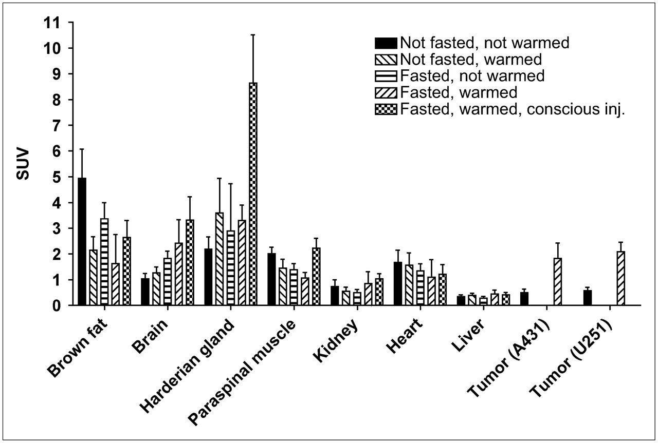
Influence of Warming and Fasting on Biodistribution of 18F-FDG in Mice Without Anesthesia During Uptake Period
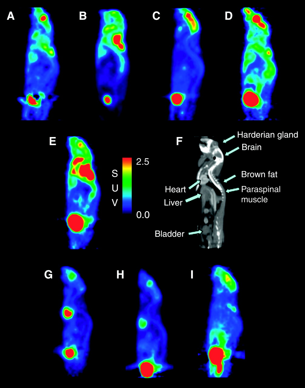
Figure 1 shows typical examples for PET scans acquired under the various conditions in nonanesthetized animals. The results of the quantitative data analysis are summarized in Figure 2. Under the reference condition (no warming and no fasting, Fig. 1E), the highest 18F-FDG uptake was seen in brown fat (SUV, 4.9 ± 1.1), Harderian glands (SUV, 2.2 ± 0.5), skeletal muscle (SUV, 2.0 ± 0.26), and myocardium (SUV, 1.7 ± 0.5).

Typical examples of biodistribution of 18F-FDG under various conditions. Images show sagittal sections through mice. A contrast-enhanced microCT scan is shown for anatomic reference. (A) Not fasted, warmed, no anesthesia. (B) Fasted, not warmed, no anesthesia. (C) Fasted, warmed, no anesthesia. (D) Fasted, warmed, no anesthesia, conscious injection. (E) Reference conditions: not fasted, not warmed, no anesthesia. (F) microCT, sagittal view for anatomic reference. (G) Not fasted, warmed, isoflurane. (H) Fasted, warmed, isoflurane. (I) Fasted, warmed, ketamine.

Biodistribution of 18F-FDG in mice that were not anesthetized during uptake period for various studied conditions. Error bars show SD.
Warming of the animals (Fig. 1A) reduced 18F-FDG uptake of brown fat by 56% (P = 0.0001) and in skeletal muscle by 28% (P = 0.0001, Fig. 2). Fasting (Fig. 1B) reduced 18F-FDG uptake of skeletal muscle by 31% (P = 0.001) and of brown fat by 32% (P = 0.006). Combining warming and fasting (Fig. 1C) reduced 18F-FDG uptake of brown fat by 67% (P = 0.0001) and of skeletal muscle by 47% (P = 0.0001) as compared with the reference condition. 18F-FDG uptake by brown fat was significantly lower after combined warming and fasting than after fasting alone (P = 0.001). Conversely, 18F-FDG uptake by skeletal muscle was significantly lower after combined warming and fasting than after warming alone (P = 0.04). The effects of warming and fasting on 18F-FDG uptake by skeletal muscle were offset when no anesthesia was used during 18F-FDG injection. Under this condition (Fig. 1D), 18F-FDG uptake by muscle was not significantly different from the reference condition (SUV, 2.2 ± 0.4, P = 0.23). As expected, there was also a significant increase in cerebral 18F-FDG uptake when no anesthesia was used for 18F-FDG injection (SUV, 3.3 ± 0.9, P = 0.04).
18F-FDG uptake by Harderian glands was not significantly influenced by warming or fasting but was markedly increased when no anesthesia was used for 18F-FDG injection (SUV, 8.6 ± 1.9, P < 0.001). None of the other analyzed organs (kidney, myocardium, liver) showed significant differences in 18F-FDG uptake in conscious mice. For myocardium and kidney, this was caused mainly by a large interindividual variability of tracer uptake within all study conditions. In contrast, mean liver 18F-FDG uptake showed little variability within and across the different study conditions (Fig. 2).
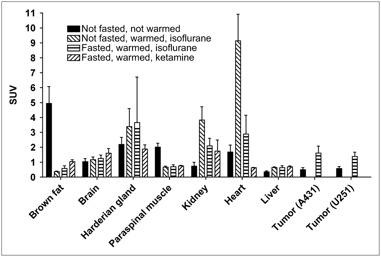
18F-FDG Biodistribution in Anesthetized Mice
Isoflurane Anesthesia.
Isoflurane anesthesia in nonfasted (Fig. 1G) mice caused a 5.5-fold increase of myocardial 18F-FDG uptake, when compared with the reference condition (P = 0.0001, Fig. 3). Isoflurane anesthesia also caused a significant increase in 18F-FDG uptake by liver and kidneys (P < 0.002, Fig. 3). In contrast, 18F-FDG uptake in brown fat and skeletal muscle was markedly reduced (−92% and −67%, respectively, P = 0.0001).

Biodistribution of 18F-FDG in mice that were anesthetized during uptake period. Reference condition (not fasted, not warmed, no anesthesia during uptake period) is shown as a comparison. Error bars show SD.
When mice were fasted before isoflurane anesthesia (Fig. 1H), 18F-FDG uptake by the myocardium was reduced by a factor of 3 compared with nonfasted animals, but still remained higher (1.7-fold, P = 0.03) than in the reference condition (no anesthesia, no fasting). Under isoflurane anesthesia, fasting had no significant effect on 18F-FDG uptake by brown fat and muscle (Fig. 3).
Ketamine/Xylazine Anesthesia.
Ketamine/xylazine anesthesia (Fig. 1I) had a similar effect as isoflurane on the 18F-FDG uptake in brown fat, skeletal muscle, kidneys, and liver (Fig. 3). Interestingly, ketamine/xylazine had the opposite effect on myocardial 18F-FDG uptake than isoflurane. Isoflurane increased myocardial 18F-FDG uptake up to 5.5-fold, whereas ketamine/xylazine decreased myocardial 18F-FDG uptake by 62%.
Comparison of Intravenous Versus Intraperitoneal Injection of 18F-FDG
Figure 4 shows the time course of 18F-FDG accumulation by various tissues after intraperitoneal and intravenous injection in fasted and warmed mice anesthetized by isoflurane (n = 6 per group). Though tracer uptake is slower after intraperitoneal injection, all organs and the U251 tumors reach comparable activity concentrations within 60 min after injection. Similarly, no significant differences were found for tissue 18F-FDG uptake of not-fasted and not-warmed mice that were not kept under anesthesia during the uptake period (Table 2, n = 4 per group). Thus, these data indicate that at 60 min after injection 18F-FDG biodistribution is comparable for intravenous and intraperitoneal injection.

(A–D) 18F-FDG uptake of various normal tissues and U251 xenografts after intravenous (iv) and intraperitoneal (ip) injection of 18F-FDG (n = 6 per group). Error bars shown as SEs of the mean.
18F-FDG Uptake of Various Tissues 60 Minutes After Intravenous and Intraperitoneal Injection of 18F-FDG (n = 4 per group)
Impact of Warming, Fasting, and Anesthesia on Tumor Uptake of 18F-FDG
In summary, our data in nontumor–bearing mice indicated that warming, fasting, and isoflurane anesthesia were likely to improve the biodistribution of 18F-FDG for tumor imaging. We therefore evaluated whether fasting, warming, and isoflurane anesthesia significantly improve visualization of A431 and U251 tumor xenografts by reducing background activity. Groups of 3 (A431) and 6 (U251) tumor-bearing mice each were imaged under the reference condition as well as after combined warming and fasting with and without isoflurane anesthesia. Because of the marked increase in blood glucose levels caused by ketamine/xylazine, no further experiments were performed with this form of anesthesia. At the time of imaging tumor size was 5–11 mm and did not show a significant difference among the 3 groups of animals. Figure 5 illustrates the biodistribution of 18F-FDG in mice bearing A431 xenografts. Though tumors did not show focal 18F-FDG above background activity under the reference condition, they demonstrated marked focal 18F-FDG uptake, when animals were fasted and warmed. The average SUV of the tumors under reference conditions was 0.5 ± 0.1. In fasted, warmed, and not-anesthetized animals the tumor SUV was 3.7 times higher (1.8 ± 0.6, P = 0.008). Keeping fasted and warmed mice under isoflurane during the uptake period increased tumor SUV to the same level (1.60 ± 0.47, P = 0.04 for comparison with the reference condition).

Tumor 18F-FDG uptake under various conditions. Coronal and axial sections are shown. White lines in the coronal sections indicate position of axial sections. (A) Not fasted, not warmed, no anesthesia. (B) Fasted, warmed, no anesthesia. (C) Fasted, warmed, isoflurane anesthesia. Red arrow indicates tumor; brown arrow indicates brown fat; white arrow indicates paraspinal muscle; yellow arrow indicates myocardium.
To assess image contrast, we calculated various tumor-to-organ ratios as shown in Table 3. Tumor-to-organ ratios were lowest under the reference condition. Warming and fasting (without isoflurane anesthesia) significantly improved the tumor-to-muscle ratio (7.9-fold, P = 0.008) and the tumor-to-brown fat ratio (17.4-fold, P = 0.01). A similar increase in 18F-FDG uptake was seen in warmed, fasted, and anesthetized animals. Under this condition, the tumor-to-muscle ratio increased 8.3-fold (P = 0.01) and the tumor-to-brown fat ratio increased 15.5-fold (P = 0.04).
Tumor-to-Organ Ratios for Groups of Mice (n = 3–6) Imaged Under 3 Different Conditions
Experiments with mice bearing U251 xenografts provided similar results (Table 3). There was a highly significant increase in the tumor-to-muscle and tumor-to-brown-fat ratios in fasted and warmed mice (P < 0.001 in anesthetized and in not-anesthetized mice).
DISCUSSION
This study demonstrates that animal handling has a profound impact on the biodistribution of 18F-FDG in mice and significantly influences the visualization of xenotransplanted tumors. Varying fasting state, body temperature, and mode of anesthesia caused >10-fold changes in the 18F-FDG uptake of normal organs. Tumor 18F-FDG uptake varied by a factor of 3.7. Depending on the conditions used for animal handling, tumors were either barely visible in the microPET studies or demonstrated marked focal 18F-FDG uptake.
The influence of blood glucose and insulin levels on 18F-FDG biodistribution is well known from human PET studies and previous studies in rats using tissue sampling to assess regional 18F-FDG uptake (21–23). Because 18F-FDG competes with glucose for intracellular transport and phosphorylation, tumor 18F-FDG uptake decreases with increasing blood glucose levels. Furthermore, insulin markedly increases 18F-FDG uptake by skeletal muscles and myocardium though it has generally no effect on 18F-FDG uptake of cancer cells. Therefore, tumor 18F-FDG uptake and image contrast are lower in the nonfasted state (high insulin and glucose levels) than in the fasted state (low insulin and glucose levels).
More recently the effect of ambient temperature on the 18F-FDG uptake by brown adipose tissue has been described in patients (24). Our data show that the ambient temperature has a much more pronounced effect on 18F-FDG biodistribution in mice. For mice the so-called zone of thermoneutrality lies between 30°C and 34°C (25). At this temperature body temperature is controlled by heat convection and no active processes are needed to maintain body temperature. At room temperature (21°C) mice need to generate heat by activation of brown adipose tissue and muscle activity to maintain a stable body temperature. Accordingly, metabolic rates have been shown to be 67% higher at room temperature (15 W/kg) than at the zone of thermoneutrality (9 W/kg) (25). Consistent with these previous observations, mice that were kept at 30°C showed markedly lower 18F-FDG uptake by brown fat and muscle in our study (Fig. 2). Because the zone of thermoneutrality varies between different mouse strains (25) and we were unable to find specific data for SCID mice, we arbitrarily selected an ambient temperature of 30°C for our experiments. At higher temperatures a further reduction in 18F-FDG uptake by brown adipose tissue might have been achieved. However, keeping mice above the zone of thermoneutrality represents a considerable heat stress. For example, C57BL/6J mice exposed to 39.5°C for 4 h demonstrate dehydration, hypoglycemia, and renal tubular necrosis (26). Therefore, we selected the lower end of the zone of thermoneutrality for our experiments.
18F-FDG uptake by brown fat was also reduced by fasting the animals overnight (Figs. 1 and 2). It is known from previous studies that feeding increases the metabolic activity of brown fat in rodents. This is considered to represent a mechanism for stabilization of body weight: excess caloric intake is converted to heat by the brown adipose tissue. Conversely, overnight fasting has been shown to decrease perfusion of brown adipose tissue as well as heat production (27).
In addition to its effect on 18F-FDG uptake by normal tissues, warming and fasting lead to a >3-fold increase in tumor 18F-FDG uptake. This finding is likely explained by a combination of lower plasma glucose levels and decreased 18F-FDG uptake by normal organs.
The effect of anesthesia on 18F-FDG biodistribution has very recently been studied by Lee et al. for ketamine/xylazine and pentobarbital (28). In the present study we extended these observations to isoflurane. Both xylazine and isoflurane are known to suppress insulin secretion (28–30). However, the effects of xylazine appear to be much more pronounced, as xylazine induced marked hyperglycemia (>300 mg/dL) even when mice were fasted for at least 8 h. Fasting mice for 20 h before 18F-FDG injection has been shown to attenuate xylazine-induced hyperglycemia (28). However, prolonged fasting leads to weight loss and may therefore be impractical, when animals need to be repeatedly imaged within a short period of time—for example, for treatment monitoring. In contrast to xylazine, isoflurane anesthesia caused only a modest increase in blood glucose levels in fasted and nonfasted animals, suggesting that its effect on insulin secretion is relatively mild.
In addition to affecting insulin secretion, anesthetic drugs also have specific effects on the glucose use of various tissues. Brown adipose tissue has a dense sympathetic innervation and its metabolic activity is regulated by β3 and α2 receptors. Activation of β3 receptors increases and activation of α2 receptors decreases perfusion and metabolic activity. Norepinephrine binds to both types of receptors, but its effect on β3 receptors is predominant and norepinephrine markedly stimulates metabolic activity (31). Accordingly, ketamine, which increases norepinephrine plasma levels, has been shown to stimulate metabolic activity and 18F-FDG uptake of brown adipose tissue (32). However, our data indicate that during anesthesia with a combination of ketamine and xylazine the effects of α2 receptor stimulation by xylazine are predominant and lead to a marked decrease in 18F-FDG uptake.
Isoflurane anesthesia also decreased 18F-FDG uptake by brown adipose tissue, which is consistent with its inhibiting effect on thermogenesis by brown adipose tissue (33). As observed in a previous study for BALB/c mice, isoflurane markedly (up to 5.4-fold) increased 18F-FDG uptake by the myocardium (12). The mechanisms underlying the high myocardial 18F-FDG uptake during isoflurane anesthesia are currently unknown, but 18F-FDG uptake could be significantly decreased by fasting of the animals and using isoflurane only during 18F-FDG injection.
CONCLUSION
On the basis of our data we propose the following protocol for imaging tumor xenografts with 18F-FDG. Mice should be fasted the night before the 18F-FDG PET scan and warmed on a heating pad before and after 18F-FDG injection. This approach markedly reduces 18F-FDG uptake by brown adipose tissue and skeletal muscle, which otherwise significantly interferes with the visualization of tumor tissue. 18F-FDG should be administered under anesthesia to further decrease skeletal muscle uptake. Keeping the mice under isoflurane anesthesia for the whole uptake period not only minimizes 18F-FDG uptake by skeletal muscle and brown fat but also decreases blood clearance resulting in higher activity concentrations in liver and kidneys. Whereas isoflurane anesthesia demonstrates a favorable effect on 18F-FDG biodistribution, ketamine/xylazine anesthesia should be used with caution as it induces marked hyperglycemia. Standardization of animal handling and anesthesia will be essential to ensure that reproducible and comparable data are obtained from 18F-FDG PET scans of mice performed at different institutions.
Acknowledgments
We thank Waldemar Ladno and Judy Edwards for their support with animal handling and imaging. This research was supported by the UCLA Center for In-Vivo Imaging in Cancer Biology (National Institutes of Health [NIH] grant P50 CA86306), the UCLA Institute of Molecular Medicine (Department of Energy grant DE-FC03-87E60615), UCLA Lung Specialized Programs of Research Excellence (NIH grant P50 CA90388), and the Austrian Science Fund through an Erwin Schroedinger Scholarship.
References
- Received for publication November 2, 2005.
- Accepted for publication March 10, 2006.


Jump to section
Related Articles
Cited By...
- Addressing Biological Questions with Preclinical Cancer Imaging
- In Vivo Evaluation of Brain [18F]F-FDG Uptake Pattern Under Different Anaesthesia Protocols
- Integrin {alpha}5 Is Regulated by miR-218-5p in Endothelial Progenitor Cells
- Slow TCA flux implies low ATP production in tumors
- Preclinical PERCIST and 25% of SUVmax Threshold: Precision Imaging of Response to Therapy in Co-clinical 18F-FDG PET Imaging of Triple-Negative Breast Cancer Patient-Derived Tumor Xenografts
- Assessment of Brain Glucose Metabolism Following Cardiac Arrest by [18F]FDG Positron Emission Tomography
- High-Throughput PET/CT Imaging Using a Multiple-Mouse Imaging System
- Hepatic stearoyl CoA desaturase 1 deficiency increases glucose uptake in adipose tissue partially through the PGC-1{alpha}-FGF21 axis in mice
- Reproducibility and Comparability of Preclinical PET Imaging Data: A Multicenter Small-Animal PET Study
- Reporting animal research: Explanation and Elaboration for the ARRIVE guidelines 2019
- Awake 18F-FDG PET Imaging of Memantine-Induced Brain Activation and Test-Retest in Freely Running Mice
- High Throughput PET/CT Imaging Using a Multiple Mouse Imaging System
- Brown adipocyte glucose metabolism: a heated subject
- 68Ga-NOTA-Functionalized Ubiquicidin: Cytotoxicity, Biodistribution, Radiation Dosimetry, and First-in-Human PET/CT Imaging of Infections
- Influence of Animal Heating on PET Imaging Quantification and Kinetics: Biodistribution of 18F-Tetrafluoroborate and 18F-FDG in Mice
- Dissociation Between Brown Adipose Tissue 18F-FDG Uptake and Thermogenesis in Uncoupling Protein 1-Deficient Mice
- A novel thermoregulatory role for PDE10A in mouse and human adipocytes
- Impact of Image-Derived Input Function and Fit Time Intervals on Patlak Quantification of Myocardial Glucose Uptake in Mice
- 18F-FDG-PET/CT Imaging of Drug-Induced Metabolic Changes in Genetically Engineered Mouse Lung Cancer Models
- PET/CT with 18F-FDG- and 18F-FBEM-Labeled Leukocytes for Metabolic Activity and Leukocyte Recruitment Monitoring in a Mouse Model of Pulmonary Fibrosis
- Mapping Changes in Mouse Brain Metabolism with PET/CT
- Optical Metabolic Imaging Identifies Glycolytic Levels, Subtypes, and Early-Treatment Response in Breast Cancer
- A PET-Compatible Tissue Bioreactor for Research, Discovery, and Validation of Imaging Biomarkers and Radiopharmaceuticals: System Design and Proof-of-Concept Studies
- In Vivo PET/CT in a Human Glioblastoma Chicken Chorioallantoic Membrane Model: A New Tool for Oncology and Radiotracer Development
- Functional Lysine Modification by an Intrinsically Reactive Primary Glycolytic Metabolite
- Evaluation of the Genisys4, a Bench-Top Preclinical PET Scanner
- Mapping Brain Metabolic Connectivity in Awake Rats with {mu}PET and Optogenetic Stimulation
- In Vivo Imaging of Cell Proliferation Enables the Detection of the Extent of Experimental Rheumatoid Arthritis by 3'-Deoxy-3'-18F-Fluorothymidine and Small-Animal PET
- Brain imaging reveals neuronal circuitry underlying the crow's perception of human faces
- PET of HER2-Positive Pulmonary Metastases with 18F-ZHER2:342 Affibody in a Murine Model of Breast Cancer: Comparison with 18F-FDG
- Oxygen Breathing Affects 3'-Deoxy-3'-18F-Fluorothymidine Uptake in Mouse Models of Arthritis and Cancer
- Evaluation of Mouse Tail-Vein Injections Both Qualitatively and Quantitatively on Small-Animal PET Tail Scans
- Specific PET Imaging of xC- Transporter Activity Using a 18F-Labeled Glutamate Derivative Reveals a Dominant Pathway in Tumor Metabolism
- 18FDG-PET Predicts Pharmacodynamic Response to OSI-906, a Dual IGF-1R/IR Inhibitor, in Preclinical Mouse Models of Lung Cancer
- Effects of Administration Route, Dietary Condition, and Blood Glucose Level on Kinetics and Uptake of 18F-FDG in Mice
- Total Abdominal 18F-FDG Uptake Reflects Intestinal Adenoma Burden in Apc Mutant Mice
- PET Imaging of Tumor Neovascularization in a Transgenic Mouse Model with a Novel 64Cu-DOTA-Knottin Peptide
- Establishment of In Vivo Brain Imaging Method in Conscious Mice
- Specific estrogen sulfotransferase (SULT1E1) substrates and molecular imaging probe candidates
- 18F-FDG Small-Animal PET/CT Differentiates Trastuzumab-Responsive from Unresponsive Human Breast Cancer Xenografts in Athymic Mice
- Imaging Biomarkers Predict Response to Anti-HER2 (ErbB2) Therapy in Preclinical Models of Breast Cancer
- Quantification of Left Ventricular Volumes and Ejection Fraction in Mice Using PET, Compared with MRI
- Gene Expression Patterns and Tumor Uptake of 18F-FDG, 18F-FLT, and 18F-FEC in PET/MRI of an Orthotopic Mouse Xenotransplantation Model of Pancreatic Cancer
- Changes in Tumor Metabolism as Readout for Mammalian Target of Rapamycin Kinase Inhibition by Rapamycin in Glioblastoma
- Preclinical Efficacy of the c-Met Inhibitor CE-355621 in a U87 MG Mouse Xenograft Model Evaluated by 18F-FDG Small-Animal PET
- Microglial Activation in Perinatal Rabbit Brain Induced by Intrauterine Inflammation: Detection with 11C-(R)-PK11195 and Small-Animal PET
- Reproducibility of 18F-FDG microPET Studies in Mouse Tumor Xenografts