
Main menu

  • Home
  • Content
    • Current
    • Ahead of print
    • Past Issues
    • JNM Supplement
    • SNMMI Annual Meeting Abstracts
    • Continuing Education
    • JNM Podcasts
  • Subscriptions
    • Subscribers
    • Institutional and Non-member
    • Rates
    • Journal Claims
    • Corporate & Special Sales
  • Authors
    • Submit to JNM
    • Information for Authors
    • Assignment of Copyright
    • AQARA requirements
  • Info
    • Reviewers
    • Permissions
    • Advertisers
  • About
    • About Us
    • Editorial Board
    • Contact Information
  • More
    • Alerts
    • Feedback
    • Help
    • SNMMI Journals
  • SNMMI
    • JNM
    • JNMT
    • SNMMI Journals
    • SNMMI

User menu

  • Subscribe
  • My alerts
  • Log in
  • My Cart

Search

  • Advanced search
Journal of Nuclear Medicine
  • SNMMI
    • JNM
    • JNMT
    • SNMMI Journals
    • SNMMI
  • Subscribe
  • My alerts
  • Log in
  • My Cart
Journal of Nuclear Medicine

Advanced Search

  • Home
  • Content
    • Current
    • Ahead of print
    • Past Issues
    • JNM Supplement
    • SNMMI Annual Meeting Abstracts
    • Continuing Education
    • JNM Podcasts
  • Subscriptions
    • Subscribers
    • Institutional and Non-member
    • Rates
    • Journal Claims
    • Corporate & Special Sales
  • Authors
    • Submit to JNM
    • Information for Authors
    • Assignment of Copyright
    • AQARA requirements
  • Info
    • Reviewers
    • Permissions
    • Advertisers
  • About
    • About Us
    • Editorial Board
    • Contact Information
  • More
    • Alerts
    • Feedback
    • Help
    • SNMMI Journals
  • View or Listen to JNM Podcast
  • Visit JNM on Facebook
  • Join JNM on LinkedIn
  • Follow JNM on Twitter
  • Subscribe to our RSS feeds
Research ArticleEndocrinology

Dissociation Between Brown Adipose Tissue 18F-FDG Uptake and Thermogenesis in Uncoupling Protein 1–Deficient Mice

Mohammed K. Hankir, Mathias Kranz, Susanne Keipert, Juliane Weiner, Sille G. Andreasen, Matthias Kern, Marianne Patt, Nora Klöting, John T. Heiker, Peter Brust, Swen Hesse, Martin Jastroch and Wiebke K. Fenske
Journal of Nuclear Medicine July 2017, 58 (7) 1100-1103; DOI: https://doi.org/10.2967/jnumed.116.186460
Mohammed K. Hankir
1Integrated Research and Treatment Centre for Adiposity Diseases, Department of Medicine, University of Leipzig, Leipzig, Germany
  • Find this author on Google Scholar
  • Find this author on PubMed
  • Search for this author on this site
Mathias Kranz
2Institute of Radiopharmaceutical Cancer Research, Department of Neuroradiopharmaceuticals, Helmholtz Centre Dresden-Rossendorf, Leipzig, Germany
  • Find this author on Google Scholar
  • Find this author on PubMed
  • Search for this author on this site
Susanne Keipert
3Helmholtz Diabetes Center, Helmholtz Center Munich, Neuherburg, Germany
  • Find this author on Google Scholar
  • Find this author on PubMed
  • Search for this author on this site
Juliane Weiner
4Collaborative Research Center for Obesity Mechanisms, University of Leipzig, Leipzig, Germany; and
  • Find this author on Google Scholar
  • Find this author on PubMed
  • Search for this author on this site
Sille G. Andreasen
1Integrated Research and Treatment Centre for Adiposity Diseases, Department of Medicine, University of Leipzig, Leipzig, Germany
  • Find this author on Google Scholar
  • Find this author on PubMed
  • Search for this author on this site
Matthias Kern
1Integrated Research and Treatment Centre for Adiposity Diseases, Department of Medicine, University of Leipzig, Leipzig, Germany
  • Find this author on Google Scholar
  • Find this author on PubMed
  • Search for this author on this site
Marianne Patt
5Department of Nuclear Medicine, Leipzig University Hospital, Leipzig, Germany
  • Find this author on Google Scholar
  • Find this author on PubMed
  • Search for this author on this site
Nora Klöting
1Integrated Research and Treatment Centre for Adiposity Diseases, Department of Medicine, University of Leipzig, Leipzig, Germany
  • Find this author on Google Scholar
  • Find this author on PubMed
  • Search for this author on this site
John T. Heiker
4Collaborative Research Center for Obesity Mechanisms, University of Leipzig, Leipzig, Germany; and
  • Find this author on Google Scholar
  • Find this author on PubMed
  • Search for this author on this site
Peter Brust
2Institute of Radiopharmaceutical Cancer Research, Department of Neuroradiopharmaceuticals, Helmholtz Centre Dresden-Rossendorf, Leipzig, Germany
  • Find this author on Google Scholar
  • Find this author on PubMed
  • Search for this author on this site
Swen Hesse
1Integrated Research and Treatment Centre for Adiposity Diseases, Department of Medicine, University of Leipzig, Leipzig, Germany
5Department of Nuclear Medicine, Leipzig University Hospital, Leipzig, Germany
  • Find this author on Google Scholar
  • Find this author on PubMed
  • Search for this author on this site
Martin Jastroch
3Helmholtz Diabetes Center, Helmholtz Center Munich, Neuherburg, Germany
  • Find this author on Google Scholar
  • Find this author on PubMed
  • Search for this author on this site
Wiebke K. Fenske
1Integrated Research and Treatment Centre for Adiposity Diseases, Department of Medicine, University of Leipzig, Leipzig, Germany
  • Find this author on Google Scholar
  • Find this author on PubMed
  • Search for this author on this site
  • Article
  • Figures & Data
  • Info & Metrics
  • PDF
Loading

Abstract

18F-FDG PET imaging is routinely used to investigate brown adipose tissue (BAT) thermogenesis, which requires mitochondrial uncoupling protein 1 (UCP1). It remains uncertain, however, whether BAT 18F-FDG uptake is a reliable surrogate measure of UCP1-mediated heat production. Methods: UCP1 knockout (KO) and wild-type (WT) mice housed at thermoneutrality were treated with the selective β3 adrenergic receptor agonist CL 316, 243 and underwent metabolic cage, infrared thermal imaging and 18F-FDG PET/MRI experiments. Primary brown adipocytes were additionally examined for their bioenergetics by extracellular flux analysis as well as their uptake of 2-deoxy-3H-glucose. Results: In response to CL 316, 243 treatments, oxygen consumption, and BAT thermogenesis were diminished in UCP1 KO mice, but BAT 18F-FDG uptake was fully retained. Isolated UCP1 KO brown adipocytes exhibited defective induction of uncoupled respiration whereas their glycolytic flux and 2-deoxy-3H-glucose uptake rates were largely unaffected. Conclusion: Adrenergic stimulation can increase BAT 18F-FDG uptake independently of UCP1 thermogenic function.

  • animal imaging
  • neuroendocrine
  • PET/MRI
  • brown adipose tissue
  • glucose metabolism
  • thermogenesis
  • uncoupling protein 1

In human PET imaging studies, brown adipose tissue (BAT) 18F-FDG uptake has been shown to be markedly increased by controlled cold exposure (1,2) and agonism of β3 adrenergic receptors (3), correlating positively with changes in energy expenditure (1,3). Similar findings have been made in small-animal PET imaging studies performed on mice (4,5).

Mitochondrial uncoupling protein 1 (UCP1) endows both human and mouse brown adipocytes with thermogenic capacity (6). It is widely assumed that BAT 18F-FDG uptake provides an indirect quantitative measure of thermogenesis. We therefore sought to formally test this basic assumption by performing 18F-FDG PET/MRI experiments on UCP1-deficient mice that exhibit defective BAT thermogenesis and uncoupled respiration in response to the selective β3 adrenergic receptor agonist CL 316, 243 (7,8).

MATERIALS AND METHODS

Animals

Experiments were performed on female wild-type (WT) C57BL/6J mice and UCP1 knockout (KO) littermates (Jackson Laboratory) aged 3–9 mo and were all approved by the Institutional Animal Care and Use Committee at the University of Leipzig (TVV 63/13 and TVV 04/12). Animals were housed in an incubator (Memmert GmbH & Co. KG) set at thermoneutrality (30°C) and 60% humidity on a 12-h light–dark cycle and had free access to a high-fat high-sugar diet (Sniff GmbH) to potentiate thermogenic responses to adrenergic stimulation (8) unless otherwise indicated. In line with the findings of Feldmann et al. (8), UCP1 KO mice housed under these conditions consistently showed higher body weights than WT littermates (Table 1).

View this table:
  • View inline
  • View popup
TABLE 1

Weight of UCP1 KO Mice Compared with Their WT Littermates at Time of Experiments

Indirect Calorimetry

Animals were transferred to metabolic cages (TSE Systems) immediately after intraperitoneal injection of the selective β3 adrenergic receptor agonist CL 316, 243 (1 mg/kg) (Tocris) (7), and oxygen consumption was measured for 3 h by indirect calorimetry.

Core Body Temperature Measurements

Core body temperature was measured using a rectal probe (Thermalert) at baseline and at 15-min intervals for 1 h after intraperitoneal injection of CL 316, 243 (1 mg/kg) (7).

Infrared Thermal Imaging

Infrared thermal imaging (9) was performed using a VarioCAM thermal camera (InfraTec GmbH). Each mouse was placed on a cage top at a fixed distance away from the camera lens. Serial 1-s images (10 Hz) were taken in triplicate at baseline and at 15-min intervals for 1 h after intraperitoneal injection of CL 316, 243 (1 mg/kg). For analysis, a constantly sized circular region of interest was drawn over BAT, and the average temperature was recorded.

PET/MRI

Small-animal PET/MRI (Mediso Medical Imaging Systems) was performed as previously described (10). Overnight fasted mice received intraperitoneal injections of either CL 316, 243 (1 mg/kg) (4) or 0.9% saline along with an intraperitoneal injection of 18.8 ± 0.4 MBq of 18F-FDG (Department of Nuclear Medicine, University Hospital Leipzig). Animals were then returned to their home cages for 45 min and subsequently anesthetized with isoflurane (1.8%, 0.35 L/min) delivered in a 60% oxygen–40% air mixture (MCQ Instruments) and transferred to the PET/MRI scanner. A 15-min static PET scan was initiated, during which animals were maintained at 37°C under isoflurane anesthesia. For analysis, mean SUVs of 18F-FDG by BAT were calculated.

Cellular Bioenergetics and 2-Deoxy-3H-Glucose Assay

Stromal vascular cells were isolated from interscapular BAT and differentiated as previously described (11). On day 7 of differentiation, mature brown adipocytes were washed twice with assay medium (XF Dulbecco modified Eagle medium supplemented with 25 mM glucose, 2 mM pyruvate, and 4% bovine serum albumin [w/v]), followed by incubation at 37°C without CO2 for 20 min. Cells were transferred to an XF96 Extracellular Flux Analyzer (Seahorse Bioscience), and after 5 basal values oligomycin (5 μg/mL) was injected to induce uncoupled respiration followed by an injection of 100 μM 2-,4-dinitrophenol. To correct for nonmitochondrial respiration and nonglycolytic sources of changes in extracurricular acidification rate, a cocktail of 2.5 μM rotenone, 2.5 μM antimycin A, and 2.5 μM 2-deoxyglucose was injected. For analysis, basal values were averaged as were the 3 values immediately after oligomycin treatment and the 3 values at 1 h after oligomycin treatment.

For the 2-deoxy-3H-glucose uptake assay, mature brown adipocytes were cultivated overnight in serum-free medium and were stimulated with 1 μM CL 316, 243 (Sigma-Aldrich GmbH) (12) or distilled water followed by incubation at 37°C in 5% CO2 for 110 min. Cells were washed once with prewarmed phosphate-buffered saline, then 2-deoxy-3H-glucose (0.5 kBq/mL) and 2-deoxy-glucose (100 μM) were added, followed by incubation at 37°C in 5% CO2 for 10 min. After being washed with ice-cold phosphate-buffered saline, cells were lysed with radioimmunoprecipitation assay buffer and transferred to a liquid scintillation analyzer (PerkinElmer) for 3 h. Uptake rates of 2-deoxy-3H-glucose were normalized to protein concentration.

Statistics

Two-way ANOVA with the Sidak post hoc test was used to assess differences between groups (GraphPad Software Inc.). P values of less than 0.05 were considered significant.

RESULTS

The increase in whole-body oxygen consumption caused by CL 316, 243 treatment was blunted in UCP1 KO compared with WT mice (F1, 90 = 554.8, P < 0.0001) (Fig. 1A) as was the rise in core body temperature (F1, 35 = 10.77, P = 0.0023) (Fig. 1B).

FIGURE 1.
  • Download figure
  • Open in new tab
  • Download powerpoint
FIGURE 1.

(A) Diminished oxygen consumption (VO2) and (B) core body temperature (Tb) changes in response to adrenergic stimulation in UCP1 KO mice. Data points represent mean ± SEM of 3–5 animals per group. *P < 0.05. **P < 0.01. ****P < 0.0001.

Representative thermal images are presented in Figure 2A. Unlike UCP1 KO mice, WT mice showed an increase in interscapular BAT temperature in response to CL 316, 243 treatment (F1, 30 = 18.53, P = 0.0002) (Fig. 2B).

FIGURE 2.
  • Download figure
  • Open in new tab
  • Download powerpoint
FIGURE 2.

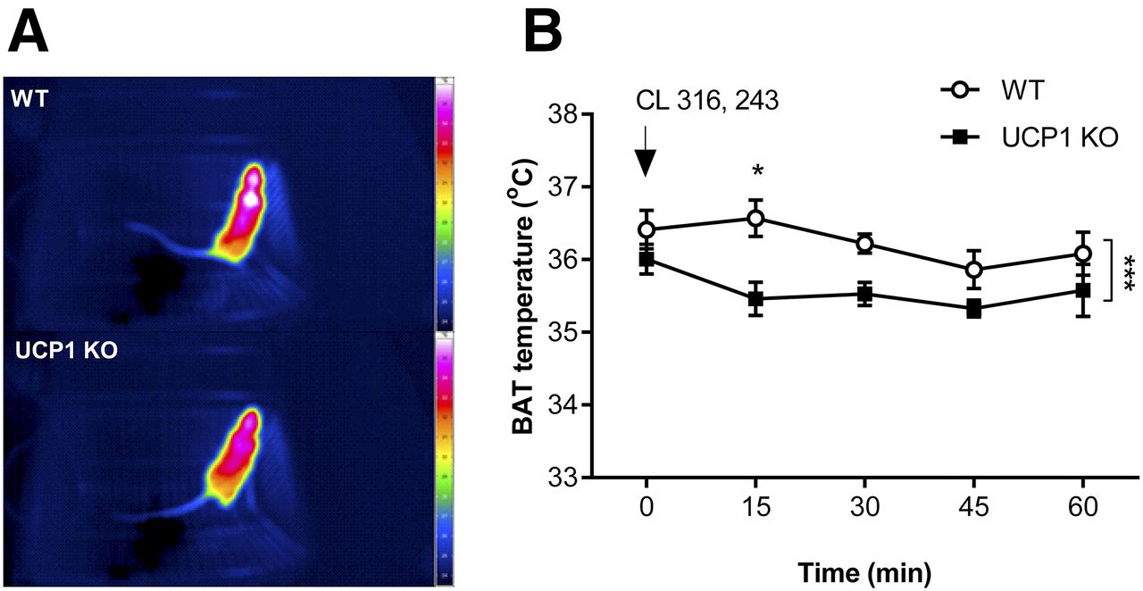
(A) Representative thermal images of WT (top) and UCP1 KO (bottom) mice taken 15 min after CL 316, 243 injections. (B) Defective BAT thermogenesis in response to adrenergic stimulation in UCP1 KO mice. Data points represent mean ± SEM of 4 animals per group. *P < 0.05. ***P < 0.001.

Representative 18F-FDG PET/MRI data are presented in Figure 3A. Both WT and UCP1 KO mice showed an increased mean SUV for18F-FDG in BAT in response to CL 316, 243 treatment relative to vehicle treatment (P < 0.0001) (Fig. 3B), with no significant effect of genotype found (F1, 14 = 0.17, P = 0.68).

FIGURE 3.
  • Download figure
  • Open in new tab
  • Download powerpoint
FIGURE 3.

(A) Representative PET/MRI data of WT and UCP1 KO mice taken 45–60 min after CL 316, 243/saline and 18F-FDG injections. White arrows in each image point to interscapular BAT. (B) Retained BAT 18F-FDG uptake in response to adrenergic stimulation in UCP1 KO mice. Bars represent mean ± SEM of 4–5 animals per group. ****P < 0.0001.

Extracellular flux analysis of cultured primary brown adipocytes revealed that 1 h after oligomycin treatment, reporting induction of uncoupled respiration (13), oxygen consumption rates were significantly lower for UCP1 KO than WT cells (P < 0.001) (Fig. 4A). In contrast, extracellular acidification rates, reporting glycolytic activity (13), tended to be similar between UCP1 KO and WT cells at this time point after oligomycin treatment (P = 0.07) (Fig. 4B). Both WT and UCP1 KO brown adipocytes showed an increase in 2-deoxy-3H-glucose uptake in response to CL 316, 243 treatment compared with vehicle (P = 0.01 and P < 0.0001, respectively) (Fig. 4C), with no significant effect of genotype found (F1, 12 = 0.00045, P = 0.97).

FIGURE 4.
  • Download figure
  • Open in new tab
  • Download powerpoint
FIGURE 4.

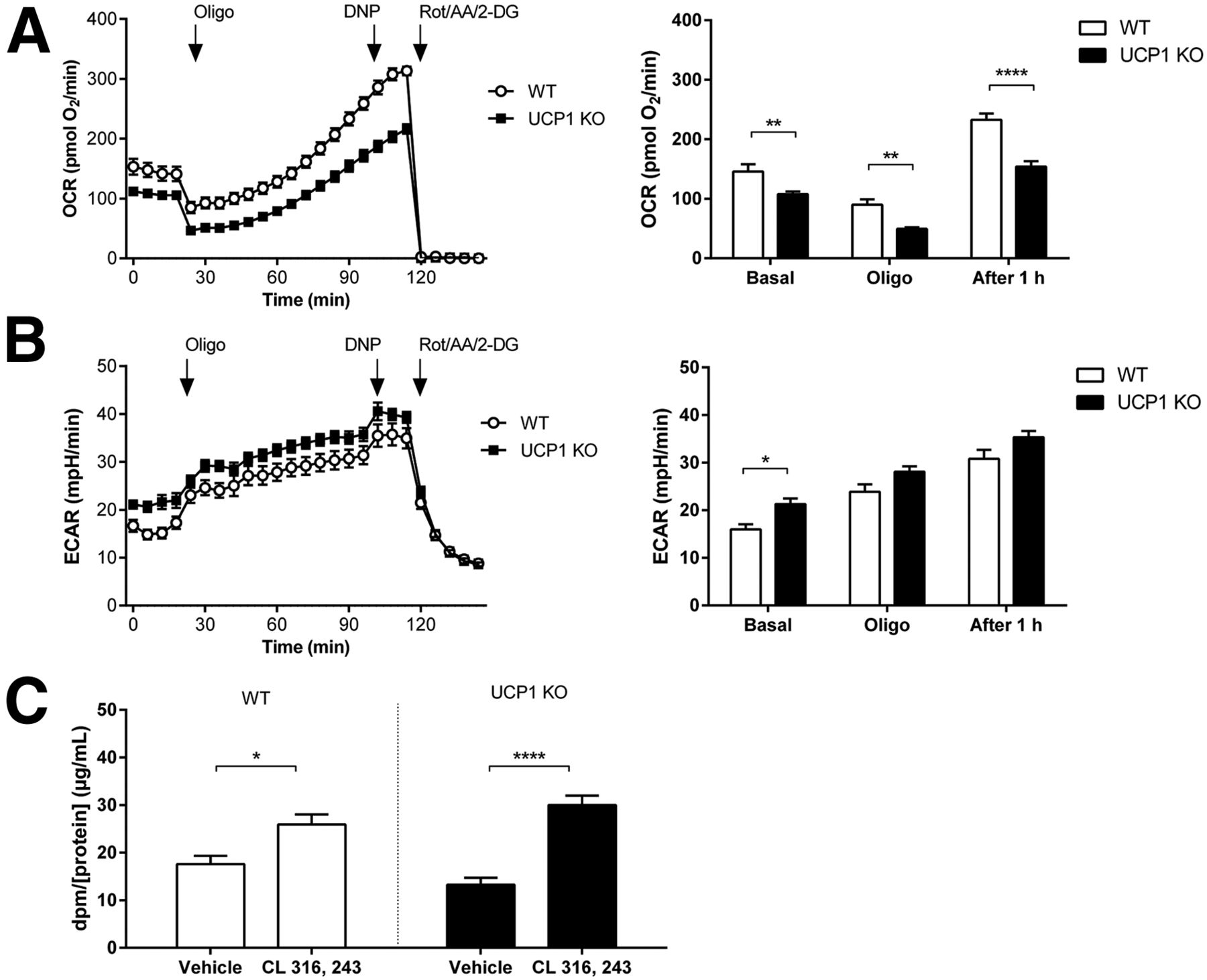
(A) Impaired induction of uncoupled respiration but normal (B) glycolysis and (C) 2-deoxy-3H-glucose uptake in UCP1 KO mouse primary brown adipocytes. Data points represent mean ± SEM of 4–6 cultures per group. 2-DG = 2-deoxyglucose; AA = antimycin A; DNP = dinitrophenol; dpm = dose per minute; ECAR = extracellular acidification rate; Rot = rotenone. *P < 0.05. **P < 0.01. ****P < 0.0001.

DISCUSSION

The findings from the present study demonstrate that glucose metabolism can increase in brown adipocytes in the absence of UCP1-mediated uncoupled respiration and heat production.

In mouse primary brown adipocytes treated with CL 316,243, glucose is transported into the cell and through the action of diacylglycerol acyltransferase 2 feeds into specialized lipid droplet pools, which are simultaneously hydrolyzed (14). The released free fatty acids are then either rapidly oxidized or activate UCP1 in mitochondria (14). Our findings of similar β3 adrenergic receptor–stimulated 18F-FDG/2-deoxy-3H-glucose uptake between WT and UCP1 KO brown adipocytes suggest that UCP1 (thermogenic) function does not provide feedback, on the initial step at least, of this concerted process.

Noradrenaline treatment was previously found not to stimulate BAT 2-deoxy-3H-glucose uptake in male UCP1 KO mice (15). The discordance with the findings from the present study could be explained by our use of female UCP1 KO mice. Indeed, cold-stimulated BAT 18F-FDG uptake is diminished in male UCP1 KO mice but not in female counterparts (16).

It is unlikely that the low BAT 18F-FDG uptake in the present study is diet related (17). This is because we found in preliminary experiments performed on high-fat high-sugar fed mice a much higher amount of BAT 18F-FDG uptake when animals were housed and scanned at room temperature (22°C). This suggests that experimental conditions even only mildly below thermoneutrality (30°C for mice) (8) can significantly stimulate BAT. Therefore, studies under thermoneutral conditions are optimal because the low BAT activity constitutes a true basal state allowing the effects of novel stimulators of BAT to be examined in isolation.

CONCLUSION

BAT 18F-FDG uptake and UCP1-mediated heat production can be dissociated. Additional techniques that measure BAT thermogenesis apart from 18F-FDG PET imaging and blood flow measurements (18) are required in model systems in which UCP1 is expected not to be functional. Because UCP1 deficiency is not the standard physiologic scenario in humans, caution is, however, warranted when translating our findings.

DISCLOSURE

The work in this manuscript was funded by the German Federal Ministry of Education and Research, the German Research Foundation, and the German Centre for Diabetes Research. No other potential conflict of interest relevant to this article was reported.

Acknowledgments

We thank Dr. Kerstin Krause for assistance with the experiments.

Footnotes

  • ↵* Contributed equally to this work.

  • Published online Jan. 12, 2017.

  • © 2017 by the Society of Nuclear Medicine and Molecular Imaging.

REFERENCES

  1. 1.↵
    1. van der Lans AA,
    2. Hoeks J,
    3. Brans B,
    4. et al
    . Cold acclimation recruits human brown fat and increases nonshivering thermogenesis. J Clin Invest. 2013;123:3395–3403.
    OpenUrlCrossRefPubMed
  2. 2.↵
    1. Virtanen KA,
    2. Lidell ME,
    3. Orava J,
    4. et al
    . Functional brown adipose tissue in healthy adults. N Engl J Med. 2009;360:1518–1525.
    OpenUrlCrossRefPubMed
  3. 3.↵
    1. Cypess AM,
    2. Weiner LS,
    3. Roberts-Toler C,
    4. et al
    . Activation of human brown adipose tissue by a β3-adrenergic receptor agonist. Cell Metab. 2015;21:33–38.
    OpenUrlCrossRefPubMed
  4. 4.↵
    1. Cypess AM,
    2. Doyle AN,
    3. Sass CA,
    4. et al
    . Quantification of human and rodent brown adipose tissue function using 99mTc-methoxyisobutylisonitrile SPECT/CT and 18F-FDG PET/CT. J Nucl Med. 2013;54:1896–1901.
    OpenUrlAbstract/FREE Full Text
  5. 5.↵
    1. Fueger BJ,
    2. Czernin J,
    3. Hildebrandt I,
    4. et al
    . Impact of animal handling on the results of 18F-FDG PET studies in mice. J Nucl Med. 2006;47:999–1006.
    OpenUrlAbstract/FREE Full Text
  6. 6.↵
    1. Porter C,
    2. Herndon DN,
    3. Chondronikola M,
    4. et al
    . Human and mouse brown adipose tissue mitochondria have comparable UCP1 function. Cell Metab. 2016;24:246–255.
    OpenUrlCrossRefPubMed
  7. 7.↵
    1. Inokuma K,
    2. Okamatsu-Ogura Y,
    3. Omachi A,
    4. et al
    . Indispensable role of mitochondrial UCP1 for antiobesity effect of beta3-adrenergic stimulation. Am J Physiol Endocrinol Metab. 2006;290:E1014–E1021.
    OpenUrlAbstract/FREE Full Text
  8. 8.↵
    1. Feldmann HM,
    2. Golozoubova V,
    3. Cannon B,
    4. et al
    . UCP1 ablation induces obesity and abolishes diet-induced thermogenesis in mice exempt from thermal stress by living at thermoneutrality. Cell Metab. 2009;9:203–209.
    OpenUrlCrossRefPubMed
  9. 9.↵
    1. Crane JD,
    2. Mottillo EP,
    3. Farncombe TH,
    4. et al
    . A standardized infrared imaging technique that specifically detects UCP1-mediated thermogenesis in vivo. Mol Metab. 2014;3:490–494.
    OpenUrl
  10. 10.↵
    1. Hankir MK,
    2. Kranz M,
    3. Gnad T,
    4. et al
    . A novel thermoregulatory role for PDE10A in mouse and human adipocytes. EMBO Mol Med. 2016;8:796–812.
    OpenUrlAbstract/FREE Full Text
  11. 11.↵
    1. Fisher FM,
    2. Kleiner S,
    3. Douris N,
    4. et al
    . FGF21 regulates PGC-1α and browning of white adipose tissues in adaptive thermogenesis. Genes Dev. 2012;26:271–281.
    OpenUrlAbstract/FREE Full Text
  12. 12.↵
    1. Chernogubova E,
    2. Cannon B,
    3. Bengtsson T
    . Norepinephrine increases glucose transport in brown adipocytes via beta3-adrenoceptors through a cAMP, PKA, and PI3-kinase-dependent pathway stimulating conventional and novel PKCs. Endocrinology. 2004;145:269–280.
    OpenUrlCrossRefPubMed
  13. 13.↵
    1. Divakaruni AS,
    2. Paradyse A,
    3. Ferrick DA,
    4. et al
    . Analysis and interpretation of microplate-based oxygen consumption and pH data. Methods Enzymol. 2014;547:309–354.
    OpenUrlCrossRefPubMed
  14. 14.↵
    1. Irshad Z,
    2. Dimitri F,
    3. Christian M,
    4. et al
    . Diacylglycerol acyltransferase 2 (DGAT2) links glucose utilization to fatty acid oxidation in the brown adipocytes. J Lip Res. 2017;58:15–30.
    OpenUrlAbstract/FREE Full Text
  15. 15.↵
    1. Inokuma K,
    2. Ogura-Okamatsu Y,
    3. Toda C,
    4. et al
    . Uncoupling protein 1 is necessary for norepinephrine-induced glucose utilization in brown adipose tissue. Diabetes. 2005;54:1385–1391.
    OpenUrlAbstract/FREE Full Text
  16. 16.↵
    1. Jeanguillaume C,
    2. Metrard G,
    3. Ricquier D,
    4. et al
    . Visualization of activated BAT in mice, with FDG-PET and its relation to UCP1. Adv J Mol Imaging. 2013;3:19–22.
    OpenUrl
  17. 17.↵
    1. Roberts-Toler C,
    2. O’Neill BT,
    3. Cypess AM
    . Diet-induced obesity causes insulin resistance in mouse brown adipose tissue. Obesity (Silver Spring). 2015;23:1765–1770.
    OpenUrl
  18. 18.↵
    1. Abreu-Vieira G,
    2. Hagberg CE,
    3. Spalding KL,
    4. et al
    . Adrenergically stimulated blood flow in brown adipose tissue is not dependent on thermogenesis. Am J Physiol Endocrinol Metab. 2015;308:E822–E829.
    OpenUrlAbstract/FREE Full Text
  • Received for publication November 2, 2016.
  • Accepted for publication December 16, 2016.
PreviousNext
Back to top

In this issue

Journal of Nuclear Medicine: 58 (7)
Journal of Nuclear Medicine
Vol. 58, Issue 7
July 1, 2017
  • Table of Contents
  • Table of Contents (PDF)
  • About the Cover
  • Index by author
Print
Download PDF
Article Alerts
Sign In to Email Alerts with your Email Address
Email Article

Thank you for your interest in spreading the word on Journal of Nuclear Medicine.

NOTE: We only request your email address so that the person you are recommending the page to knows that you wanted them to see it, and that it is not junk mail. We do not capture any email address.

Enter multiple addresses on separate lines or separate them with commas.
Dissociation Between Brown Adipose Tissue 18F-FDG Uptake and Thermogenesis in Uncoupling Protein 1–Deficient Mice
(Your Name) has sent you a message from Journal of Nuclear Medicine
(Your Name) thought you would like to see the Journal of Nuclear Medicine web site.
Citation Tools
Dissociation Between Brown Adipose Tissue 18F-FDG Uptake and Thermogenesis in Uncoupling Protein 1–Deficient Mice
Mohammed K. Hankir, Mathias Kranz, Susanne Keipert, Juliane Weiner, Sille G. Andreasen, Matthias Kern, Marianne Patt, Nora Klöting, John T. Heiker, Peter Brust, Swen Hesse, Martin Jastroch, Wiebke K. Fenske
Journal of Nuclear Medicine Jul 2017, 58 (7) 1100-1103; DOI: 10.2967/jnumed.116.186460

Citation Manager Formats

  • BibTeX
  • Bookends
  • EasyBib
  • EndNote (tagged)
  • EndNote 8 (xml)
  • Medlars
  • Mendeley
  • Papers
  • RefWorks Tagged
  • Ref Manager
  • RIS
  • Zotero
Share
Dissociation Between Brown Adipose Tissue 18F-FDG Uptake and Thermogenesis in Uncoupling Protein 1–Deficient Mice
Mohammed K. Hankir, Mathias Kranz, Susanne Keipert, Juliane Weiner, Sille G. Andreasen, Matthias Kern, Marianne Patt, Nora Klöting, John T. Heiker, Peter Brust, Swen Hesse, Martin Jastroch, Wiebke K. Fenske
Journal of Nuclear Medicine Jul 2017, 58 (7) 1100-1103; DOI: 10.2967/jnumed.116.186460
Twitter logo Facebook logo LinkedIn logo Mendeley logo
  • Tweet Widget
  • Facebook Like
  • Google Plus One
Bookmark this article

Jump to section

  • Article
    • Abstract
    • MATERIALS AND METHODS
    • RESULTS
    • DISCUSSION
    • CONCLUSION
    • DISCLOSURE
    • Acknowledgments
    • Footnotes
    • REFERENCES
  • Figures & Data
  • Info & Metrics
  • PDF

Related Articles

  • This Month in JNM
  • PubMed
  • Google Scholar

Cited By...

  • High-fructose feeding suppresses cold-stimulated brown adipose tissue glucose uptake in young men independently of changes in thermogenesis and the gut microbiome
  • Brown adipose tissue lipoprotein and glucose disposal is not determined by thermogenesis in uncoupling protein 1-deficient mice
  • Blocking mitochondrial pyruvate import causes energy wasting via futile lipid cycling in brown fat
  • Substrate fluxes in brown adipocytes upon adrenergic stimulation and uncoupling protein 1 ablation
  • CPEB2-dependent translation of long 3'-UTR Ucp1 mRNA promotes thermogenesis in brown adipose tissue
  • Brown adipocyte glucose metabolism: a heated subject
  • Accurate quantification of brown adipose tissue mass by xenon-enhanced computed tomography
  • Google Scholar

More in this TOC Section

Endocrinology

  • Assessment and Comparison of 18F-Fluorocholine PET and 99mTc-Sestamibi Scans in Identifying Parathyroid Adenomas: A Metaanalysis
  • Targeted Optical Imaging of the Glucagonlike Peptide 1 Receptor Using Exendin-4-IRDye 800CW
  • 18F-Fluorocholine PET/CT in Primary Hyperparathyroidism: Superior Diagnostic Performance to Conventional Scintigraphic Imaging for Localization of Hyperfunctioning Parathyroid Glands
Show more Endocrinology

Basic

  • Dopamine D1 Receptor Agonist PET Tracer Development: Assessment in Nonhuman Primates
  • Optical Navigation of the Drop-In γ-Probe as a Means to Strengthen the Connection Between Robot-Assisted and Radioguided Surgery
  • Synthesis and Preclinical Evaluation of a 68Ga-Labeled Adnectin, 68Ga-BMS-986192, as a PET Agent for Imaging PD-L1 Expression
Show more Basic

Similar Articles

Keywords

  • Animal Imaging
  • neuroendocrine
  • PET/MRI
  • Brown adipose tissue
  • glucose metabolism
  • Thermogenesis
  • Uncoupling protein 1
SNMMI

© 2025 SNMMI

Powered by HighWire