


Abstract
Radiolabeled peptides are of increasing interest in nuclear oncology. Special emphasis has been given to the development of peptides labeled with positron emitters. Among these, 68Ga deserves special attention, because it is available from an inhouse generator rendering 68Ga radiopharmacy independent of an onsite cyclotron. 68Ga has a half-life of 68 min and decays by 89% through positron emission. The parent, 68Ge, is accelerator produced and decays with a half-life of 270.8 d by electron capture. Currently, at least 1 commercial and several in-house generators are available. 68Ge is strongly absorbed on metal oxides or organic material, making a 68Ge-breakthrough highly unlikely. Several groups continue to further develop generators to remove cationic impurities from the eluate. Several bifunctional chelators based on 1,4,7-triazacyclononane-N,N′,N″-triacetic acid and 1,4,7,10-tetraazacyclododecane-N,N′,N″,N‴-tetraacetic acid (DOTA) macrocycles are available for coupling to peptides and other biomolecules. In addition to these hydrophilic chelators, a lipophilic tetradentate S3N ligand was developed. Radiopeptides for 68Ga labeling have been developed and tested preclinically for the targeting of somatostatin receptors, the melanocortin 1 receptor, and the bombesin receptor. Clinical studies were performed with 68Ga-DOTA,Tyr3-octreotide, localizing neuroendocrine tumors with higher sensitivity than 111In-diethylenetriaminepentaacetic acid-octreotide. In addition, 68Ga-DOTA-based bombesin derivatives are being investigated with some success in patients with prostate cancer. Conclusion: Generator-produced 68Ga and the development of small chelator-coupled peptides (and other small biomolecules) may open a new generation of freeze-dried, good manufacturing practice–produced, kit-formulated PET radiopharmaceuticals similar to 99Mo-/99mTc-generator–based, 99mTc-labeled radiopharmaceuticals.
Radiopeptides are of increasing interest in imaging and targeted radiotherapy of tumors (1–5). The major goal in the past was the development of radiopeptides labeled with 99mTc and 111In for SPECT and with 90Y and 177Lu for targeted radiotherapy (6–13).
Recently, peptides labeled with positron emitters based on 68Ga, 66Ga, 18F, 86Y, and 64Cu have been investigated (12–19). Interest in using nonphysiologic metallic positron emitters for clinical PET comes mainly from the availability and additional advantages of 68Ga and from the use of 86Y to quantitate the biodistribution of 90Y-labeled vector-targeted radiotherapy. 68Ga is available from an inhouse generator, rendering it independent of an onsite cyclotron. With a half-life of 68 min, it decays by 89% through positron emission of 1.92 MeV (max. energy) and 11% orbital electron capture. In addition, 68Ga can be labeled to a chelator-conjugated biomolecule, allowing kit production and enabling wide availability. Deutsch (20) proposed in a 1993 editorial in The Journal of Nuclear Medicine that the time had come to use the 68Ge/68Ga generator and cold kit formulations for labeling with 68Ga, similar to the successfully used 99Mo/99mTc generator system in 99mTc radiopharmacy. This, he suggested, would allow PET centers to benefit from the generator for important clinical studies. Deutsch’s editorial was included as a commentary on an article by Tsang et al. (21) on a cationic 68Ga(III) complex with a N4O22− Schiff-base ligand that showed promising properties as a radiopharmaceutical for cardiac PET. Unfortunately, little activity was focused on 68Ga-based PET radiopharmaceuticals over the last 10 y. With the appearance of small radiolabeled peptides as a new class of radiopharmaceuticals, however, a renaissance in 68Ga-based PET radiopharmaceuticals is underway. These peptides show very fast blood clearance and fast target localization, making the short half-life ideal for clinical studies.
THE 68Ge/68Ga GENERATOR
Data on the 68Ge/68Ga generator system have recently been summarized by Mirzadeh and Lambrecht (22). The parent 68Ge is accelerator produced on Ga2O3 targets by a (p,2n) reaction. It decays with a half-life of 270.8 d by electron capture. 68Ge is strongly absorbed to different solid supports, such as metal oxides and organic pyrogallol-formaldehyde resins.
An important aspect for wide use of 68Ga in clinical PET is its chemical form and concentration after elution from the generator. In addition, there is concern about 68Ge-breakthrough and contamination of the generator column material. As indicated, different 68Ge-carrier column materials were proposed and used in the past, among them inorganic oxides such as Al2O3, TiO2, or SnO2. Recently, a TiO2-based generator (Cyclotron Co.) has become commercially available and is being eluted with 0.1 mol/L HCl. Several groups are currently using this generator and modifying it to allow safe handling and remove potential cationic impurities.
Meyer et al. (23) used a microchromatography column filled with a strong basic anion-exchange resin to purify and concentrate the generator eluate. They designed a very sophisticated system to monitor the accumulation of radioactivity in the microcolumn and to survey 68Ge breakthrough. Their final labeling solution contained 10–20 nmol of 1,4,7,10-tetraazacyclododecane-N,N′,N″,N‴-tetraacetic acid (DOTA)-peptides in 400 μL N-(2-hydroxyethyl)piperazine-N′-(2-ethanesulfonic acid) buffer.
Velikyan et al. (24) used microwave heating and, like Meyer et al. (23), purified and concentrated the eluate from potential cationic impurities, such as a breakthrough of 68Ge, using 68GaCl4− to be adsorbed on anion exchange resins from HCl solutions. The adsorption was found to be 100% in 3.8 mol/L HCl. Deionized water was used to elute. The parent 68Ge4+ was not retained on the anion exchange column. The concentration step was also seen as a purification of 68Ga3+ from 68Ge4+ breakthrough.
Under microwave heating, labeling yields of >99% were obtained at 1 min with as low as 0.5 nmol DOTA-Tyr3-octreotide (DOTATOC).
In addition, Breeman et al. (25) reported using the same TiO2 generator to label specific activities of 1 GBq/nmol DOTATOC within 5 min at 80°C without the need for purification of the eluate.
Schuhmacher et al. (26) used a pyrogallol-formaldehyde copolymer, an organic ion-exchange resin, as a matrix for their homemade generator. They eluted the generator with 5.5 mol/L HCl and purified and concentrated the 68Ga by adsorbing it on a small Dowex anion-exchange column as 68GaCl4−, which they eluted with 0.5 mol/L HCl.
AQUEOUS COORDINATION CHEMISTRY OF Ga(III) AND BIFUNCTIONAL CHELATORS
Ga(III) chemistry in radiopharmaceutical applications has been reviewed elsewhere in the literature (27).
In aqueous solution, gallium occurs solely in the oxidation state +3. Ga3+ is classified as a hard acid metal, bonding to highly ionic hard base ligand donors, such as carboxylic acids, amino nitrogens, hydroxamates, and phenolates. Thiols also have been shown to be good coordinating groups. In addition, the aqueous solution chemistry is determined by the easy hydrolysis of the aquo ion when the pH is raised above about 3 (depending on concentration). Ultimately, hydrolysis leads to the precipitation of Ga(OH)3. The other important competitor in physiologic fluid for Ga3+ radiometals is transferrin. This Fe3+-carrying protein binds Ga3+ with high affinity (log K = 23.7). Any application of a 68Ga radiopharmaceutical, then, needs to resist the exchange of Ga3+ from its chelate with transferrin and OH− ions.
Several suitable bifunctional chelators were proposed, developed, and coupled to biomolecules for gallium labeling. For radiopharmaceutical studies, 67Ga is often used as a surrogate for 68Ga.
One suitable chelator is desferal, which has high affinity for Fe3+ and also for Ga3+. It has 3 hydroxamate groups as metal binding sites. Desferal was coupled with 67Ga to octreotide via a succinyl spacer and studied in tumor-bearing rats and in vitro (28–30). Promising preclinical data prompted investigators to perform clinical studies. In humans, Bihl et al. (H. Bihl, unpublished data, 1994) studied the radiopeptide along with 111In-diethylenetriaminepentaacetic acid (DTPA)-octreotide (Octreoscan; Mallinckrodt, Inc.) in 8 patients with somewhat disappointing results because of slow blood clearance (31).
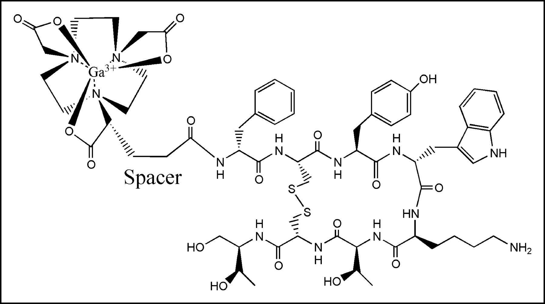
DOTA was also used as a monofunctional version for linking to peptides. The synthesis of a DOTA-tris(tBu) ester allowed convenient linking to the N-terminus of peptides assembled an a solid support (8). If coupled to Tyr3-octreotide to yield DOTA0-Tyr3-octreotide (Fig. 1) and labeled with 67Ga, the resulting radiopeptide showed not only 5 times higher binding affinity to the somatostatin-receptor subtype 2 (SSTR2) but also about 2.5 times higher tumor uptake in a mouse model and lower kidney uptake than the 111In/90Y-DOTATOC. Clinical data with this PET radiopharmaceutical are very promising.
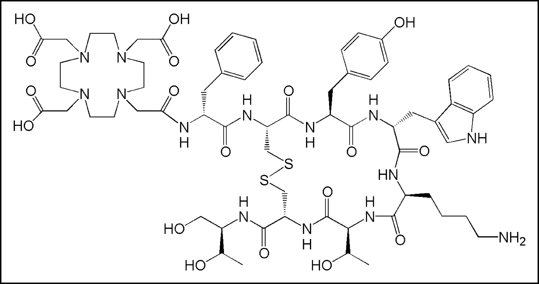
Structural formula of DOTATOC.
Other small molecules coupled to DOTA have also been developed for 68Ga labeling. Roivainen et al. (32) synthesized DOTA-coupled 17-mer-oligonucleotides as imaging agents for tumors containing K-ras point mutations in codon 12. In addition, hybridization properties were not altered by the 68Ga-DOTA coupling, and labeling was possible with high specific activity. Griffiths et al. (33) synthesized and studied a bivalent peptidic hapten, again coupled to DOTA, and labeled it with 68Ga. They used it along with a bispecific antibody against carcinoembryonic antigen and against the hapten in a 2-step pretargeting approach. The preclinical study was summarized by noting that the outstanding target-to-tissue ratios obtained with this approach encourage its development for improved cancer imaging in the future.
In addition to DOTA as a chelator for 68Ga, 1,4,7-tricarboxymethyl-1,4,7-triazacyclononane (NOTA) was coupled to Tyr3-octreotide as the bifunctional version, (1-(1-carboxy-3-carboxy-propyl)-4,7-(carboxy-methyl)-1,4,7-triazacyclononane (NODAGA); Fig. 2). NODAGATOC has not been tested clinically yet but is at least equivalent to 68Ga-DOTATOC in preclinical studies (34).

Structural formula of NODAGATOC.
A tetradentate tripodal S3N ligand was described forming lipophilic tetrahedral Ga(III) complexes (35). The bifunctional version bis(2-(benzylthio)benzyl)(2-(benzylthio)-4-aminobenzyl)amine was coupled to model peptides to show the usefulness of the approach. The bioconjugate complex was found to be stable to ligand exchange, indicating its suitability for in vivo use.
PRECLINICAL DEVELOPMENT OF DOTA-COUPLED PEPTIDES FOR 68Ga-LABELING AND DIAGNOSTIC PET IMAGING
Targeting of the Somatostatin Receptors
As indicated previously, 68Ga-DOTATOC is the gold standard for 68Ga-based PET peptide radiopharmaceuticals, and other chelator-based somatostatin analogs are being developed for clinical studies.
Targeting of Melanoma Using the Melanocortin System
The melanocortin system consists of the melanocortin peptides α-, β-, and γ-melanocyte-stimulating hormone (α-, β-, and γ-MSH) and adrenocorticotropic hormone. The melanocortins are involved in diverse physiologic functions, including pigmentation, steroidogenesis, energy homeostasis, exocrine secretion, sexual function, analgesia, inflammation, immunomodulation, temperature control, cardiovascular regulation, and neuromuscular regeneration. Their action is mediated by a family of 5 7-transmembrane G-protein-coupled melanocortin receptors and the endogenous melanocortin antagonists agouti and agouti-related protein.
MC1R is expressed by cutaneous melanocytes, where it has a key role in determining skin and hair pigmentation, in keratinocytes, fibroblasts, endothelial cells, and antigen-presenting cells. The receptor is also expressed by leukocytes, where it mediates the antiinflammatory and immunomodulatory properties of melanocortins.
Because both melanotic and amelanotic melanomas overexpress MC1R, radiolabeled α-MSH analogs were developed for tumor imaging and staging but also with the intention of using them for peptide-based radionuclide therapy (36).
Studies using α-MSH analogs labeled with 111In after conjugation to the metal chelator DTPA were performed preclinically and in patients (37,38). They revealed targeting of melanomas, however, with high nonspecific accumulation of these compounds in several tissues including the liver.
Another α-MSH analog, [Nle4,Asp5,D-Phe7]-α-MSH (4–11) (NAPamide), was conjugated to DOTA. After labeling with 67Ga and 68Ga, DOTA-NAPamide was characterized in vitro and in vivo in the mouse B16F1 melanoma model. Both the 111In-DOTA-NAPamide and 67Ga-DOTA-NAPamide showed high tumor and relatively low kidney uptake. PET studies using 68Ga-DOTA-NAPamide revealed high contrast images even at 1 h after tracer administration (39) (Fig. 3). However, receptor density in human melanomas is expected to be much lower than in the murine tumor model. Consequently, the first clinical scans in 5 patients with melanoma were negative.
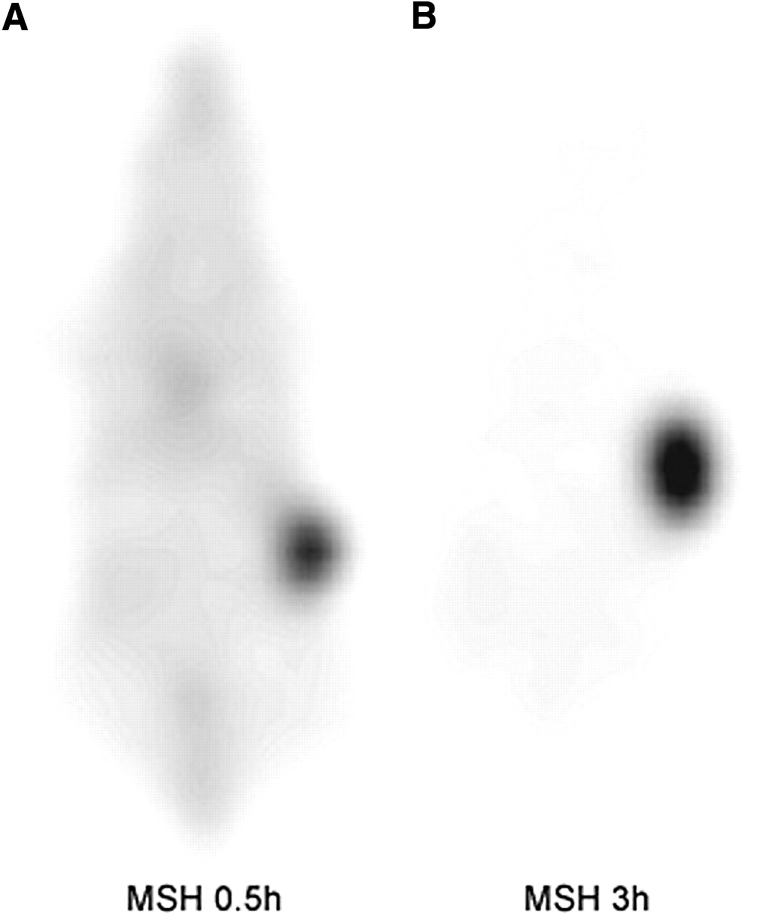
Coronal images at 0.5 h (A) and 3 h (B) after administration of 68Ga-DOTA-NAPamide in a melanoma-bearing mouse. Tracer uptake is seen predominantly in tumor and bladder.
Bombesin Derivatives and Bombesin Receptors
Bombesin receptors belong to the group of G-protein-coupled receptors and are overexpressed on major human tumors such as prostate and breast cancer (40,41). Therefore, radioligands based on bombesin are currently being developed and studied preclinically and in patients (42–45). Several of these new bombesin-based radiopeptides are conjugated to DOTA and can be labeled with 68Ga. As an example, in a pancreatic carcinoma model (AR42J), DOTA-PEG2-[d-Tyr6,βAla11,Thi13,Nle14]bombesin(6–14) was studied after radiolabeling with 67Ga or 68Ga. The compound showed high affinity and rapid internalization in vitro with >85% endocytosed radioactivity after a 1-h incubation period. Tumor uptake in vivo ranged between 5.5 and 11 %ID/g (depending on the injected peptide mass) at 1 h after tracer administration, with a biologic half-life of 15 h. Scintigraphic images at 1 h after injection showed specific accumulation in the tumor, kidneys, bowel, and pancreas (Fig. 4), which was confirmed by biodistribution data including blocking studies (46).

Coronal PET images of AR42J-tumor bearing mice at 90 min after administration of 0.5 MBq 68Ga-BZH3. Tracer uptake in tumors, pancreas, and duodenum.
FUTURE DIRECTIONS: PHAGE DISPLAY FOR THE IDENTIFICATION OF NEW TUMOR TARGETING PEPTIDES
Bioengineering will lead to the design of new biomolecules by methods such as phage display, which may be used for new approaches in isotope-based diagnosis and treatment of disease. The principle behind phage-displayed peptide libraries is the display of these libraries fused with the carboxy terminal domain of the minor coat protein (gene III protein fragment) on the surface of a filamentous phage. The relevant molecule is then directly detected and screened using the target molecules and amplified after infection with E. coli. This allows a rapid selection (within weeks) of specific clones from large pools (>1010 clones) and determination of the amino acid sequence of a peptide displayed on a phage by sequencing the relevant section of the phage genome. This technique has been used for searching antibodies, receptors for new drug discovery and cancer therapy (either as an antagonist or an agonist of a natural ligand–receptor interaction), and custom-made enzymes for gene therapy (47).
The breakthrough with regard to clinical PET studies with 68Ga was the development of 68Ga-DOTATOC (48). Three communications and an “image of the month” using 68Ga-DOTATOC have been published recently (14–16,49). In 3 patients with meningiomas, Henze et al. (15) showed very fast uptake of the tracer with standardized uptake values (SUVs) reaching a plateau at 60–120 min after injection (mean SUV = 10.6). No tracer was found in the healthy surrounding brain tissue. All meningiomas, including the smallest (<7 mm diameter), showed high tracer uptake and were clearly visualized. These studies provided useful information about the extent of meningiomas located beneath osseous structures, especially at the base of the skull. An example of a PET image fused with a CT image is shown in Figure 5.
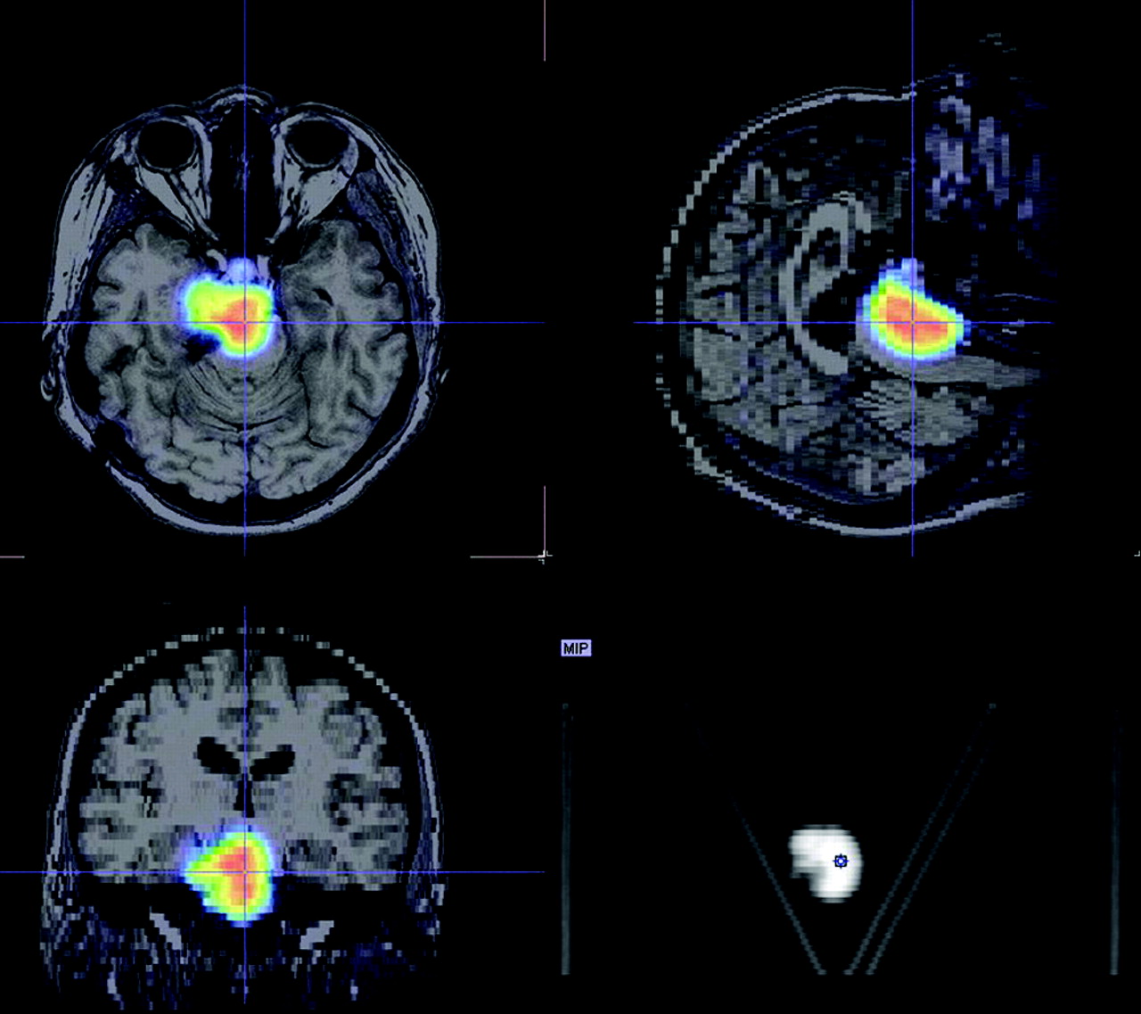
Fusion image of MR scan and 68Ga-DOTATOC PET in patient with meningioma. PET scan was acquired 1 h after tracer administration.
Hofmann et al. (14) studied 8 patients with histologically proven carcinoid tumors. They found a biexponential blood clearance with half-lives of 2 ± 0.3 min and 48 ± 7 min. Tumor accumulation reached a maximum after 70 ± 20 min. In all, 40 lesions were predefined by CT or MR imaging, and all lesions were found on 68Ga-DOTATOC PET images. In addition, >30% more lesions were detected with this tracer. 111In-DTPA-octreotide showed a lower detection rate. The authors concluded that 68Ga-DOTATOC PET showed a high tumor-to-nontumor contrast at early times and a detection rate superior to 111In-DTPA-octreotide. Kowalski et al. (16) also studied 4 patients with metastasizing neuroendocrine tumors. The pharmacokinetic data were found to be very similar to the studies mentioned here and the diagnostic sensitivity higher than that with 111In-DTPA-octreotide.
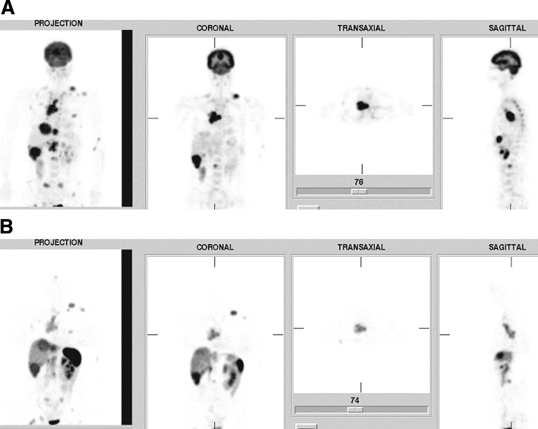
Figures 6A–6C compare 18F-FDG PET and 68Ga-DOTATOC in a patient with abdominal carcinoid. Unlike 18F-FDG, 68Ga-DOTATOC PET shows high uptake in the primary tumor and in several lymph nodes. Figure 6C shows the tracer uptake in the primary tumor and the bowel with high SUVs at early time points.
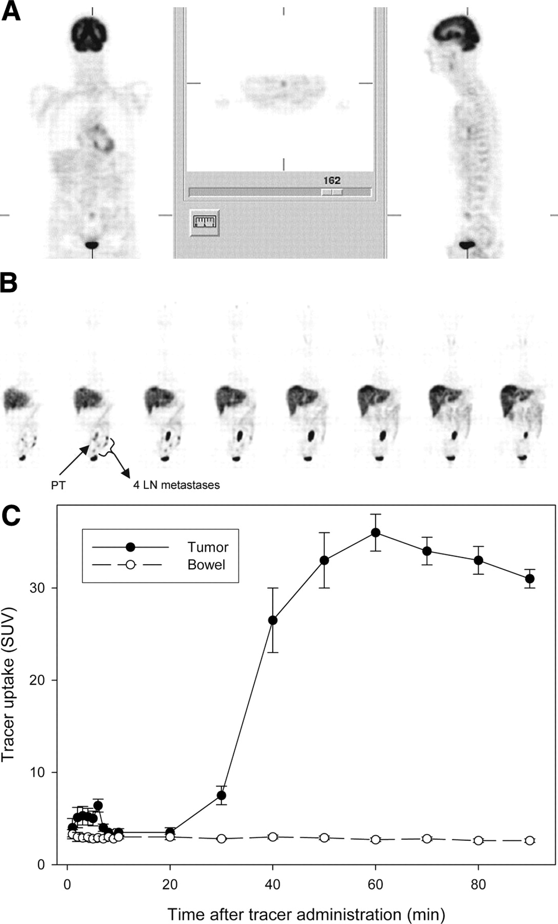
18F-FDG PET (320 MBq injected dose) (A) and 68Ga-DOTATOC PET (169 MBq) (B) images obtained at 60 min after tracer administration in patient with abdominal carcinoid. 68Ga-DOTATOC scan showed higher accumulation of tracer in tumor (SUV in primary tumor [PT] = 38) than did 18F-FDG (SUV = 1.6). Four lymph node (LN) metastases are shown with 68Ga that were missed with 18F-FDG. PT and LN metastases were proven histologically. (C) Tracer kinetics for 68Ga-DOTATOC in primary tumor and bowel.
Because small cell lung cancer also shows expression of SSTR, therapeutic uses for 90Y-DOTATOC may be promising. However, for the identification of patients who are likely to receive therapeutically sufficient doses, pretherapeutic dosimetry with 68Ga-DOTATOC is useful. Figures 7A–7B show an example of a patient with metastasizing small cell lung cancer imaged with 18F-FDG and 68Ga-DOTATOC.
18F-FDG PET (350 MBq injected dose) (A) and 68Ga-DOTATOC PET (250 MBq injected dose) (B) images obtained at 60 min after tracer administration in a patient with a small cell lung carcinoma. 18F-FDG shows higher accumulation (SUV = 10.6) than 68Ga-DOTATOC (SUV = 6.0).
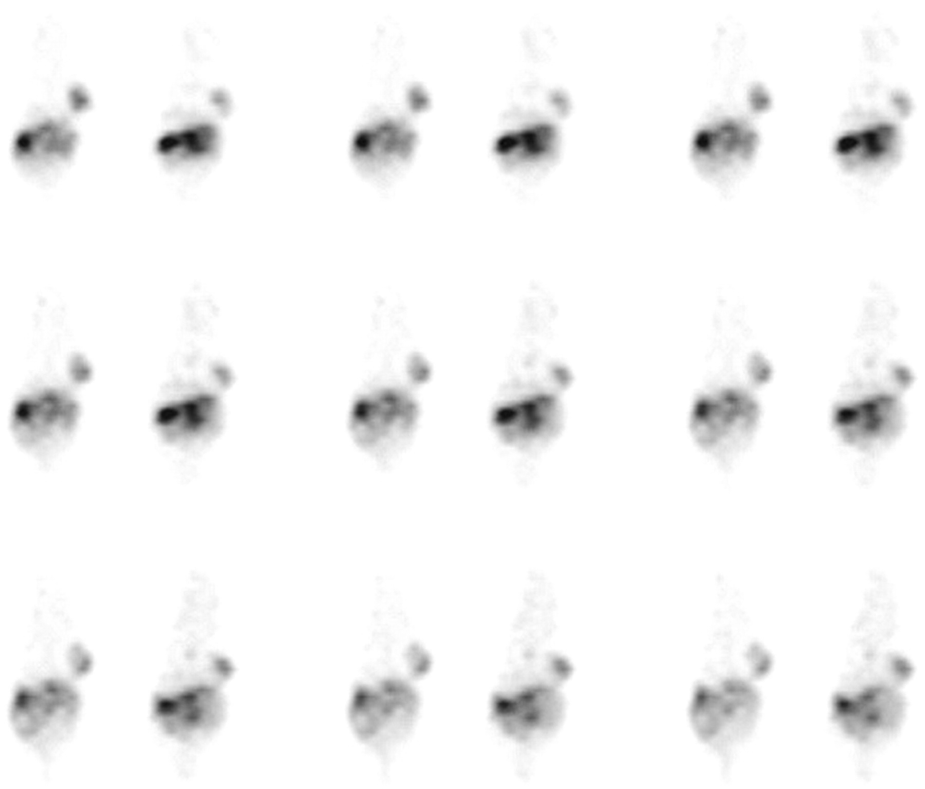
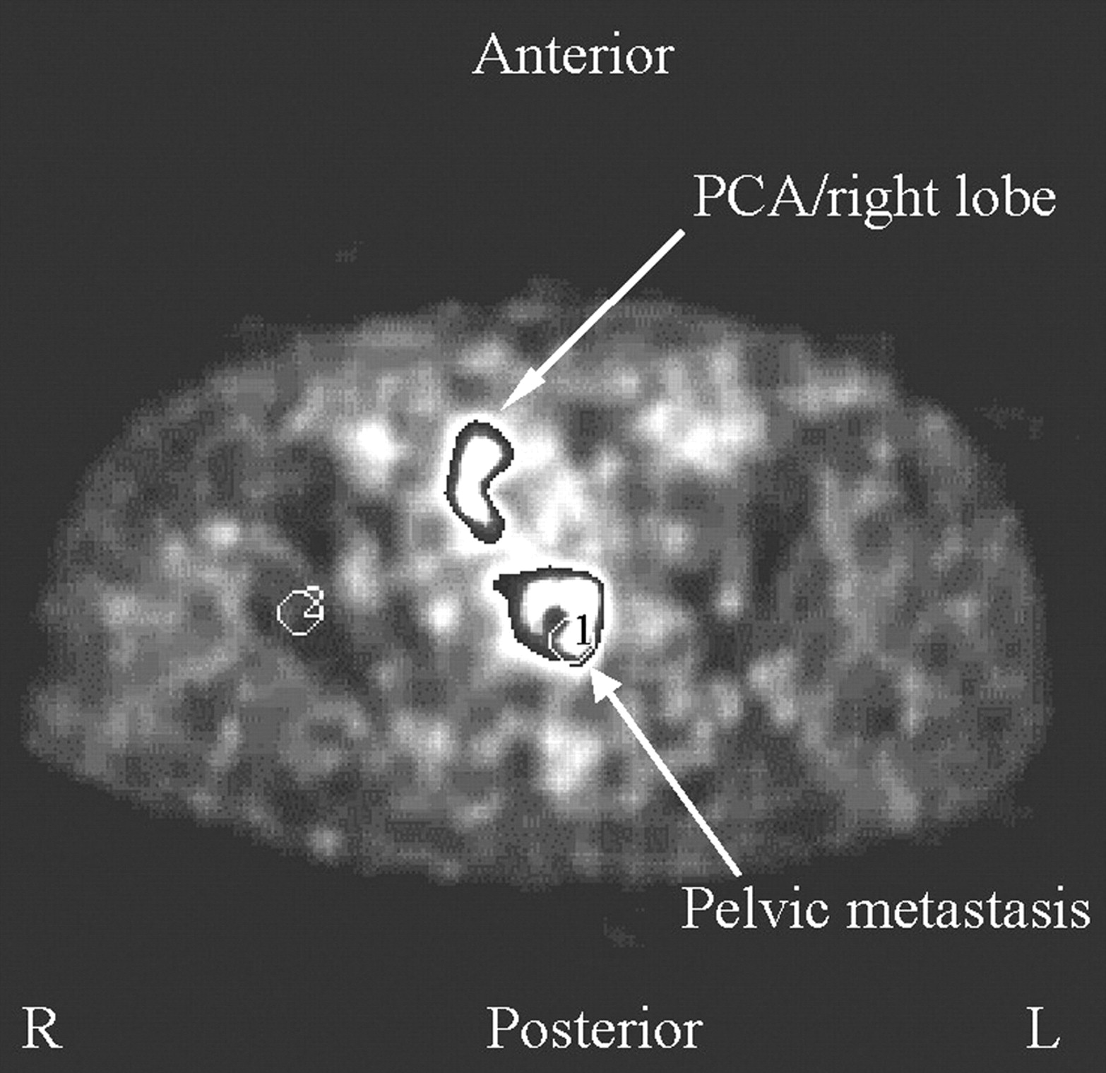
PET studies with a 68Ga-labeled bombesin analog were performed in 11 patients with prostate cancer (50). In 4 of these patients, mild reversible systolic blood pressure reduction was observed in the first 2 min after a slow intravenous administration. Renal secretion was rapid, with >75% of injected dose recovered in the urine at 60 min after injection. In all patients, primary tumors were visible, with the smallest tumor size being 5 mm and a plateau of tumor uptake at 15–25 min after injection. In 3 of these patients lymph node metastases also were found. However, in 4 patients a significant nonspecific enrichment was observed in the upper abdomen, which was interpreted as the pancreas. A typical example of primary tumor and lymph node metastases is shown in Figure 8.

68Ga-labeled bombesin analog image showing primary prostate carcinoma (PCA) and a pelvic metastasis (region of interest [ROI] 2:ROI 1 = 1:202).
CONCLUSION
Generator-produced 68Ga and the development of small chelator-coupled peptides with affinity to receptors overexpressed on a variety of human tumors may open a new generation of kit-formulated PET radiopharmaceuticals. This would allow good manufacturing practice–produced kits and an onsite generator to produce radiopharmaceuticals similar to routinely used 99mTc-based radiopharmaceuticals. Along with the long half-life of the generator, which can be used for more than a year, 68Ga-based radiopharmaceuticals may also become a very cost-effective alternative to cyclotron-based tracers.
The short half-life of 68Ga and the fast localization of small peptides make this an ideal combination to study receptor regulation in patients. The 68-min half-life allows generator elution every 2–3 h and several applications in patients per day. This allows, for example, determination of the ideal time point for a therapeutic application of 90Y-DOTATOC or 177Lu-DOTA-Tyr3-Thr8-octreotide after discontinuation of cold octreotide therapy in patients with neuroendocrine tumors.
Acknowledgments
Helmut Maecke acknowledges the financial support of the Swiss National Science Foundation (grant 33100A0–100390). Uwe Haberkorn is supported by the Deutsche Forschungsgemeinschaft grants HA2901/3–1 and 3–2. We thank Jochen Schuhmacher and Michael Henze for providing 68Ga-DOTATOC scans.
Footnotes
Received Aug. 12, 2004; revision accepted Oct. 26, 2004.
For correspondence or reprints contact: Helmut R. Maecke, PhD, Division of Radiological Chemistry, Department of Radiology, University Hospital Basel, Petersgraben 4, CH-4031 Basel, Switzerland.
E-mail: hmaecke{at}uhbs.ch
REFERENCES
In this issue


Jump to section
- Article
- Abstract
- THE 68Ge/68Ga GENERATOR
- AQUEOUS COORDINATION CHEMISTRY OF Ga(III) AND BIFUNCTIONAL CHELATORS
- PRECLINICAL DEVELOPMENT OF DOTA-COUPLED PEPTIDES FOR 68Ga-LABELING AND DIAGNOSTIC PET IMAGING
- FUTURE DIRECTIONS: PHAGE DISPLAY FOR THE IDENTIFICATION OF NEW TUMOR TARGETING PEPTIDES
- CONCLUSION
- Acknowledgments
- Footnotes
- REFERENCES
- Figures & Data
- Info & Metrics
Related Articles
Cited By...
- Primary Preclinical and Clinical Evaluation of 68Ga-DOTA-TMVP1 as a Novel VEGFR-3 PET Imaging Radiotracer in Gynecological Cancer
- Succinylated Gelatin Improves the Theranostic Potential of Radiolabeled Exendin-4 in Insulinoma Patients
- The Glucose-Dependent Insulinotropic Polypeptide Receptor: A Novel Target for Neuroendocrine Tumor Imaging--First Preclinical Studies
- Quantitative and Qualitative Intrapatient Comparison of 68Ga-DOTATOC and 68Ga-DOTATATE: Net Uptake Rate for Accurate Quantification
- Targeted Radiotherapy of Prostate Cancer with a Gastrin-Releasing Peptide Receptor Antagonist Is Effective as Monotherapy and in Combination with Rapamycin
- Bombesin Antagonist-Based Radioligands for Translational Nuclear Imaging of Gastrin-Releasing Peptide Receptor-Positive Tumors
- Nuclear medicine techniques for the imaging and treatment of neuroendocrine tumours
- Patient-Specific Radiation Dosimetry of 99mTc-HYNIC-Tyr3-Octreotide in Neuroendocrine Tumors
- A Universally Applicable 68Ga-Labeling Technique for Proteins
- Optimization of Hapten-Peptide Labeling for Pretargeted ImmunoPET of Bispecific Antibody Using Generator-Produced 68Ga
- 18F-Labeled Bombesin Analog for Specific and Effective Targeting of Prostate Tumors Expressing Gastrin-Releasing Peptide Receptors
- Radiolabeled Bicyclic Somatostatin-Based Analogs: A Novel Class of Potential Radiotracers for SPECT/PET of Neuroendocrine Tumors
- Exendin-4-Based Radiopharmaceuticals for Glucagonlike Peptide-1 Receptor PET/CT and SPECT/CT
- Bone Metastases in Patients with Neuroendocrine Tumor: 68Ga-DOTA-Tyr3-Octreotide PET in Comparison to CT and Bone Scintigraphy
- Peptide-Based Probes for Cancer Imaging
- Molecular Imaging of Metastatic Potential
- Somatostatin Receptor Imaging in Patients with Neuroendocrine Tumors: Not Only SPECT?
- Dosimetry in Peptide Radionuclide Receptor Therapy: A Review
- 177Lu-AMBA: Synthesis and Characterization of a Selective 177Lu-Labeled GRP-R Agonist for Systemic Radiotherapy of Prostate Cancer
- 111In-Benzyl-DTPA-ZHER2:342, an Affibody-Based Conjugate for In Vivo Imaging of HER2 Expression in Malignant Tumors
- Preparation and Evaluation of 68Ga-DOTA-hEGF for Visualization of EGFR Expression in Malignant Tumors
- Molecular Imaging as In Vivo Molecular Pathology for Gastroenteropancreatic Neuroendocrine Tumors: Implications for Follow-Up After Therapy