


Abstract
Bispecific antibody pretargeting is highly sensitive and specific for cancer detection by PET. In this study, the preparation of a high-specific-activity 68Ga-labeled hapten-peptide, IMP288, was evaluated. Methods: IMP288 (DOTA-D-Tyr-D-Lys(histamine-succinyl-glycine [HSG])-D-glu-D-Lys(HSG)-NH2) was added to buffered 68Ga and then heated in boiling water and purified on a reversed-phase cartridge. Tumor-bearing nude mice were used for biodistribution and tumor localization studies. Results: 68Ga-IMP288 was prepared at a starting specific activity up to 1.78 GBq/nmol, with final yields of 0.74 GBq/nmol (decay-corrected) and less than 1% unbound 68Ga. Purification was essential to remove unbound 68Ga and 68Ge breakthrough. Pretargeted animals showed a high 68Ga-IMP288 uptake (27.5 ± 5.8 percentage injected dose per gram), with ratios of 13.6 ± 4.8, 66.8 ± 14.5, and 325.9 ± 61.9 for the kidneys, liver, and blood, respectively, at 1.5 h after peptide injection. Conclusion: High-specific-activity labeling of DOTA-hapten-peptide was obtained from the 68Ga/68Ge generator for approximately 1 y, yielding products suitable for immunoPET.
The PET radionuclide 68Ga can be obtained from commercially available 68Ge generators with no carrier added, allowing compounds to be prepared at high specific activity. The half-life of 68Ge (270.8 d) allows the generator to be used for an extended period, which reduces the unit-dose cost, and the half-life of 68Ga (67.6 min) is suitable for rapidly clearing molecules, such as peptides. Interest in 68Ga-labeled compounds has been growing over the past 10 y, with promising initial clinical results (1–11).
We are interested in using a small radiolabeled hapten-peptide as part of a bispecific antibody (bsmAb) pretargeting procedure for PET. Previous studies reported high tumor uptake with minimal accretion in normal tissues, producing targeting superior to that of directly radiolabeled antibody fragments and greater sensitivity and specificity than 18F-FDG (12–14). Like directly radiolabeled peptides, the hapten-peptide used in pretargeting would benefit from high-specific-activity labeling. Thus, the main objective of the study was to evaluate the suitability of 68Ga for pretargeting.
MATERIALS AND METHODS
Reagents
Humanized tri-Fab bsmAb TF2 and IMP288 (DOTA-D-Tyr-D-Lys(histamine-succinyl-glycine [HSG]-D-Glu-D-Lys(HSG)-NH2) were described previously (12,15). A 1.3 mM stock solution of IMP288 was diluted to 6.5 × 10⁻⁵ M in 1 M N-(2-hydroxyethyl)piperazine-N′-(2-ethanesulfonic acid) (HEPES), pH 6.9, and stored at −20°C.
68Ga Generator
The IGG-100, 1.85-GBq (50-mCi) 68Ga generator was purchased from Eckert-Ziegler Isotope Products Eurotope GmbH and eluted according to the manufacturer's recommendations, using 0.1 M HCl. Three fractions (1.5, 1.0, and 2.5 mL) were isolated, with fraction 2 containing the highest concentration of 68Ga.
Within 2–17 d of the 68Ga elution, aliquots of fraction 2 and a sample of the radiolabeled product before and after purification were counted in an open window using a Wizard 3″ automatic γ-counter (Perkin Elmer). To approximate the amount of the 68Ge counts in fraction 2, we assumed 100% counting efficiency and expressed the activity as Becquerels of 68Ge/37 MBq of 68Ga. The 68Ge counts in the labeled product were normalized as a percentage of the activity in fraction 2 used to prepare the labeled product.
Radiolabeling
IMP288 was radiolabeled with 111InCl3, according to methods published previously (specific activity, 36.8 MBq/nmol [0.995 mCi/nmol]; <3% unbound by instant thin-layer chromatography) (12).
The 68Ga-labeling procedure was modeled after that reported previously, with some modifications (13). IMP288 was added to all or a portion of fraction 2, along with 1.0 M HEPES, pH 6.9. After being heated in a boiling water bath for 12 min, the vial was cooled in an ice bath to room temperature, and then 0.1 M ethylenediaminetetraacetic acid, pH 5.5, was added to a final concentration of 5 mM. The mixture was transferred to a reversed-phase polymeric-sorbent packed, 1-mL Oasis HLB cartridge for purification (Waters). After it was washed with three 1.0-mL aliquots of water, the product was eluted with two 200-μL aliquots of water:ethanol (1:1) into a vial containing 50 μL of ascorbic acid (300 mg/mL).
The peptide concentration ranged from 400 to 650 nM and from 200 to 250 nM for preparations that started with a specific activity of 0.888 and 1.776 GBq/nmol, respectively (116- and 56-fold molar excess to the 68Ga activity, respectively). Final specific activity assumed full recovery of IMP288 from the HLB cartridge.
Unbound 68Ga was determined by reverse-phase HPLC (RP-HPLC) (Nova-Pak C18, 4 μm, 8 × 100 μm Radial-Pak; Waters).
Additional details can be found in the supplemental data (available online only at http://jnm.snmjournals.org).
Animal Studies
Nude mice bearing subcutaneous implants of human colonic cancer cell lines were given intravenous injections of the radiolabeled product. Specific information regarding the doses administered and methods are provided in the “Results” section and supplemental data.
RESULTS
Generator Elution
Over a period of 350 d from generator calibration, 79.9% ± 3.1% of the expected 68Ga activity was accounted for in the total elution volume (Fig. 1). Fraction 2 (1 mL) contained 63%–83% of the total eluted activity. An approximation of 68Ge activity, based on an estimated counting efficiency of 100%, revealed that 0.05–0.22 Bq of 68Ge/MBq of 68Ga was present in fraction 2, except for 2 instances in which it was 0.64 and 0.49 Bq of 68Ge/MBq of 68Ga between days 160 and 350. Assays performed on samples taken before and after purification indicated the HLB purification assisted in the removal of unbound 68Ga but also of 68Ge. For example, most often 68Ge was not detected in aliquots of purified 68Ga-IMP288, but when present, more than 95% of the 68Ge in the fraction 2 used for the labeling had been removed.
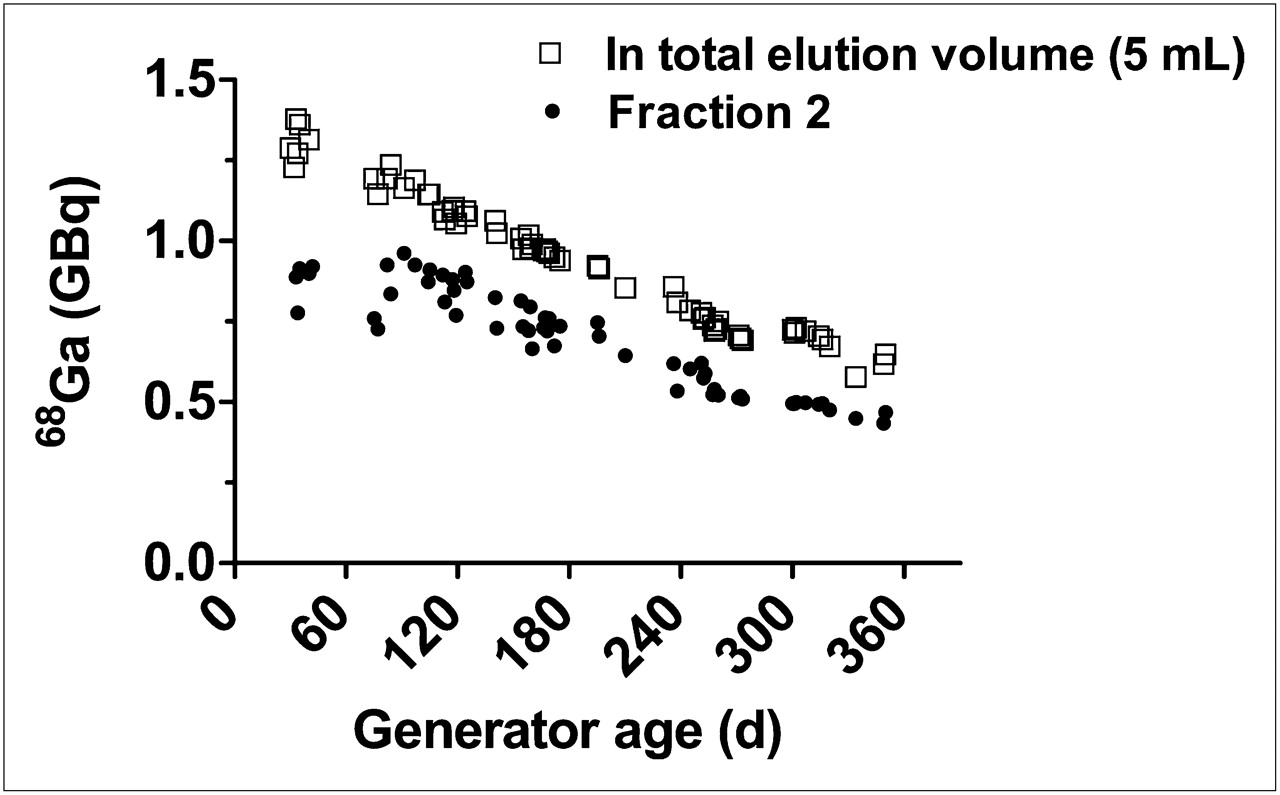
68Ga generator was eluted over 350 d with 0.1 M HCl in 3 fractions, starting 28 d after its calibration date. Activity in 1-mL fraction 2 and total in all fractions are shown.
68Ga-IMP288 Radiolabeling
68Ga-IMP288 preparation was completed within 30–40 min. Variable product recovery was encountered when following the procedure of Schoffelen et al. (13), suggesting that colloidal forms of gallium were formed. Three labeling procedures were performed using IMP288 (0.444 GBq/nmol) with HEPES added at one half, one quarter, or one eighth of the 68Ga volume. Mock mixtures found that the pH was 4.6 at one half, 3.7 at one quarter, and 3.1 at one eighth of the volume. Analysis without HLB purification showed that each label had approximately 0.9% unbound 68Ga by RP-HPLC, but the RP-HPLC recoveries were 60%, 74%, and 87%, respectively, suggesting colloid formation at a higher pH. Therefore, all subsequent labeling procedures were performed with HEPES buffer at one eighth of the 68Ga volume.
The starting specific activity was increased to as high as 1.776 GBq/nmol using peptide concentrations ranging from approximately 200 to approximately 650 nM. High specific activity could be achieved when the generator was 350 d old using a starting ratio of 0.888 GBq/nmol (unbound 68Ga, ∼1%).
Initially, ascorbic acid was added only to the HLB-purified product, but later several products were prepared by adding ascorbic acid to the reaction mixture before heating at a final concentration of 6.7 mg/mL. This step improved recovery without compromising product quality (e.g., at initial specific activity of 0.88 GBq/nmol, 68Ga-IMP288 recovery improved from 59.1% ± 8.8% [n = 11] to 70.4% ± 16.8% [n = 6]). A mock-labeling mixture showed that the addition of ascorbic acid had little effect on the pH. Cenolate (Hospira Worldwide, Inc.), pH 6.18, ascorbic acid for human use, could not be added during radiolabeling, presumably because of the presence of excessive amounts of aluminum or the higher pH.
Seven products, all including ascorbic acid in the reaction mixture, were prepared at the initial specific activity of 1.7 GBq/nmol. The overall recovery of the purified product without decay correction was 38.3% ± 8.9% (54.1% ± 10.5% decay-corrected; starting activity was 407–444 MBq). These 7 labeled products were stable for 2 h at room temperature (i.e., RP-HPLC indicated no change in unbound or the molecular character of IMP288, and size-exclusion HPLC showed that HSG-binding was retained) (data not shown).
Biodistribution
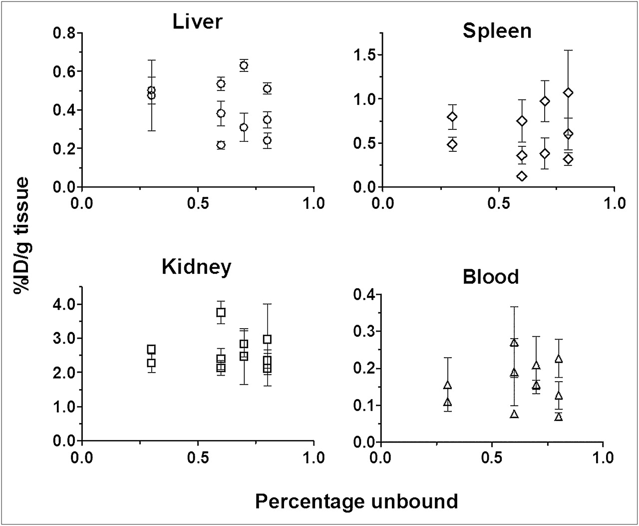
One animal injected with 68Ga-IMP288 before HLB purification (3.5% unbound by RP-HPLC and 14% colloidal 68Ga, as indicated by the activity retained on the RP-HPLC) had 14.1 percentage injected dose per gram (%ID/g), 4.5 %ID/g, and 2.1 %ID/g, respectively, in the liver, spleen, and bone at 1 h after injection. Most of the purified products had less than 1% unbound 68Ga by RP-HPLC, and in 10 separate studies with purified product prepared without ascorbic acid, uptake in the liver, spleen, kidneys, and blood at 1 h after injection averaged 0.42 ± 0.05, 0.59 ± 0.13, 2.60 ± 0.29, and 0.16 ± 0.04 %ID/g, respectively (Fig. 2). Thus, purification was considered essential to minimize normal-tissue uptake.

Evaluation of 68Ga-IMP288 tissue uptake in 10 separate studies. Each study consisted of groups of 2–5 mice that were given 25 pmole (7.77–11.1 MBq [210–300 μCi]) of 68Ga-IMP288 and necropsied 1 h later.
TF2 anti-CEACAM5 pretargeting was assessed in mice bearing either subcutaneous LS 174T or HT-29 xenografts. A TF2 dose-finding study was performed with a fixed amount of 111In-IMP288 given at 16 h after the TF2 injections, using 10:1, 20 or 25:1, and 40:1 TF2:111In IMP288 mol ratios. In each model, 25:1 to 40:1 was preferred (Fig. 3).

Tumor-to-nontumor ratios of 111In-IMP288 in mice bearing subcutaneous LS 174T (A) and HT-29 (B) human colon cancer xenografts. For pretargeting, TF2 anti-CEACAM5 bsmAb was given intravenously at 3 doses and then, 16 h later, 111In-IMP288 (2.53 × 10⁻¹¹ mol, 0.925 MBq [25 μCi]) was given to all groups. For LS 174T, TF2 dose was 40 μg (2.53 × 10⁻¹⁰ mol) for 10:1, 100 μg (6.34 × 10⁻¹⁰ mol) for 25:1, and 160 μg (1.01 × 10⁻⁹ mol) for 40:1 groups. For HT-29, TF2 doses were 40, 80, and 160 μg for 10:1, 20:1, and 40:1 groups, respectively. Mice were necropsied 1.5 h after peptide injection. Inserts show tumor-to-kidney ratios. Tumor-to-kidney ratio for 111In-IMP288 (without TF2) was 0.08 ± 0.03. T/NT = tumor to nontumor.
Pretargeted 68Ga-IMP288 tumor uptake averaged 27.5 ± 5.8 %ID/g in LS 174T, with low tissue uptake providing high tumor-to-nontumor ratios at 1.5 h after injection (Table 1). 68Ga-IMP288 alone was 0.33 ± 0.07 %ID/g. Tumor and tissue uptake of the 68Ga-IMP288 compared favorably to LS 174T–bearing animals that had been necropsied at 1.0 h after being given TF2-pretargeted 111In-IMP288; however, there was a suggestion that liver and spleen uptake was higher for the 68Ga group (P < 0.05) and a suggestion of somewhat higher renal uptake for the 111In-IMP288 (P = 0.04).
Tissue Uptake of 68Ga-IMP288 in Mice Bearing Subcutaneous LS 174T Human Colon Cancer Xenografts
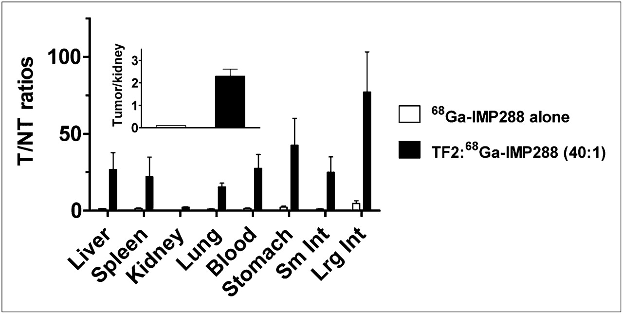
HT-29 accretion in the TF2–68Ga-IMP288 pretargeted group was 8.6 ± 1.3 %ID/g (0.20 ± 0.03 %ID/g with 68Ga-IMP-288 alone). Tumor-to-nontumor ratios were again favorable (e.g., tumor-to-liver, tumor-to-spleen, tumor-to-kidney, and tumor-to-blood ratios were 27 ± 11, 22 ± 13, 2.3 ± 0.3, and 28 ± 9, respectively) (Fig. 4). Tumor-to-liver, tumor-to-blood, and tumor-to-kidney ratios were 1.3 ± 0.1, 1.6 ± 0.3 and 0.065 ± 0.003, respectively, for 68Ga-IMP-288 alone.

Tumor-to-nontumor ratios in nude mice bearing subcutaneous HT-29 human colon cancer xenografts (%ID/g). Mice (n = 5) were given TF2 (160 μg, 1.01 × 10⁻⁹ mol) intravenously, and after 16 h 68Ga-IMP288 (2.53 × 10⁻¹¹ mol, 11.1 MBq [300 μCi]) was given intravenously. Another group received the same 68Ga-IMP288 alone (no bsmAb). Specific activity of purified 68Ga-IMP288 was 0.516 GBq/nmol, and it was prepared with ascorbic acid. Mice were necropsied 1 h after peptide injection. Lrg Int = large intestine; Sm Int = small intestine; T/NT = tumor-to-nontumor.
DISCUSSION
The short half-life of 68Ga is matched well for rapidly clearing molecules, cost, procedural simplicity, and the ability to prepare products at high specific activities, contributing to its appeal for PET.
Our 68Ga-IMP288 labeling experience was favorable. The generator performed according to the manufacturer's specifications, with high elution efficiency remaining at approximately 1 y after calibration (98 elutions). Isolating the peak amount of radioactivity in a 1.0-mL fraction minimized the total elution volume. A more concentrated form was not required for successful radiolabeling. Ascorbic acid added during the radiolabeling procedure appeared to enhance recovery without altering the pH, but we also routinely added it to the final product, which was stable for 2 h.
111In-, 90Y-, and 177Lu-IMP288 have been prepared without purification, but 68Ga-IMP288 required HLB purification. Purification removed unwanted forms of 68Ga3+, but it also removed 68Ge. Because gallium exists in various forms based on the pH and concentration (16), our goal was to retain the pH near 3.0, which is lower than most other 68Ga-DOTA-peptides (6,10,17–19). For example, Bauwens et al. found labeling efficiencies or more than 90% for 68Ga-DOTATOC using 1 M HEPES, pH 3.75, whereas with 0.5 M sodium succinate, pH approximately 4.0, labeling efficiencies suffered when 10 μg of the peptide was used (19). IMP288 labeling was always performed with less than 10 μg, and at pH 3.7 and 4.6, recovery from analytic RP-HPLC was low, but better recoveries were possible at the lower pH.
Because unbound forms of 68Ga3+ can be difficult to quantify, and to ensure 68Ga was retained sufficiently by DOTA-IMP288, we repeatedly administered randomly selected products into mice. Hepatic and splenic uptake was reduced substantially by HLB purification, but even then it was somewhat higher than with 111In-IMP288, suggesting there may be a small amount of transchelation. A similar observation was reported, in a comparison study in rats, in which 68Ga-DOTATOC showed higher uptake in the liver than did 111In-DOTATOC (6).
CONCLUSION
68Ga-IMP288 specific activities as high as 0.84 GBq/nmol (i.e., starting specific activity, 1.776 GBq/nmol) were achieved without any tedious handling of 68Ga.68Ga-IMP288 had excellent tumor localization properties in a bsmAb pretargeting setting and is easily and conveniently prepared, but a final purification procedure using an HLB reversed-phase cartridge is necessary to remove unwanted unbound 68Ga3+ forms. Acceptable products could be prepared over a 1-y period from the generator's calibration date.
Acknowledgments
This article is dedicated to the memory of Prof. Thomas M. Behr, who spent an important time in his short but productive career working at our center, before returning to Germany to become the youngest director of an academic nuclear medicine department. We thank Ali Mostafa, Tom Jackson, Dion Yeldell, and Jayson Jebsen for their technical assistance. William J. McBride, Edmund A. Rossi, Chien-Hsing Chang, and David M. Goldenberg are employed or have a financial interest in Immunomedics, Inc., or IBC Pharmaceuticals, Inc. This study was supported in part by NCI grant 2R44CA123985 and NJ CCR grant 10-8-CCR-EO.
- © 2011 by Society of Nuclear Medicine
REFERENCES
- Received for publication September 22, 2010.
- Accepted for publication December 17, 2010.


Jump to section
Related Articles
Cited By...
- Quantitative Immuno-SPECT Monitoring of Pretargeted Radioimmunotherapy with a Bispecific Antibody in an Intraperitoneal Nude Mouse Model of Human Colon Cancer
- Phase II Trial of Anticarcinoembryonic Antigen Pretargeted Radioimmunotherapy in Progressive Metastatic Medullary Thyroid Carcinoma: Biomarker Response and Survival Improvement