


Abstract
Gastrin-releasing peptide receptors (GRP-R) are upregulated in many cancers, including prostate, breast, and lung. We describe a new radiolabeled bombesin (BBN) analog for imaging and systemic radiotherapy that has improved pharmacokinetics (PK) and better retention of radioactivity in the tumor. Methods: DO3A-CH2CO-G-4-aminobenzoyl-Q-W-A-V-G-H-L-M-NH2 (AMBA) was synthesized and radiolabeled. The human prostate cancer cell line PC-3 was used to determine the binding (Kd), retention, and efflux of 177Lu-AMBA. Receptor specificity was determined by in vitro autoradiography in human tissues. PK and radiotherapy studies were performed in PC-3 tumor-bearing male nude mice. Results: 177Lu-AMBA has a high affinity for the GRP-R (Kd, 1.02 nmol/L), with a maximum binding capacity (Bmax) of 414 fmol/106 cells (2.5 × 10⁵ GRP-R/cell). Internalization was similar for 177Lu-AMBA (76.8%), 177Lu-BBN8 (72.9%), and 125I-[Tyr4]-BBN (74.9%). Efflux was markedly lower for 177Lu-AMBA (2.9%) compared with 177Lu-BBN8 (15.9%) and 125I-[Tyr4]-BBN (46.1%). By receptor autoradiography, Lu-AMBA binds specifically to GRP-R (0.8 nmol/L) and to the neuromedin B receptor (NMB-R) (0.9 nmol/L), with no affinity for the bb3 receptor (>1,000 nmol/L). 177Lu-AMBA was renally excreted (55 %ID 1 h [percentage injected dose at 1 h]); tumor uptake at 1 and 24 h was 6.35 %ID/g and 3.39 %ID/g, respectively. One or 2 doses of 177Lu-AMBA (27.75 MBq/dose) significantly prolonged the life span of PC-3 tumor-bearing mice (P < 0.001 and P < 0.0001, respectively) and decreased PC-3 tumor growth rate over controls. When compared using World Health Organization criteria, mice receiving 2 doses versus 1 dose of 177Lu-AMBA demonstrated a shift away from stable/progressive disease toward complete/partial response; by RECIST (Response Evaluation Criteria in Solid Tumors), median survival increased by 36% and time to progression/progression-free survival increased by 65%. Conclusion: 177Lu-AMBA binds with nanomolar affinity to GRP-R and NMB-R, has low retention of radioactivity in kidney, demonstrates a very favorable risk–benefit profile, and is in phase I clinical trials.
Prostate cancer is the most frequently diagnosed, and one of the most deadly forms of cancer in men in the United States, second only to cancer of the lung and bronchus. The National Cancer Institute estimates that 1 in 6 men will be diagnosed with prostate cancer in their lifetime. If the diagnosis is made while the cancer is still localized, radical prostatectomy or localized radiation therapy increases the likelihood of cure. However, once it has metastasized, treatment with chemical or medical castration becomes less effective, and the cancer likely progresses to a hormone refractory state characterized by bony metastases with high morbidity and mortality.
The expression and distribution of mammalian bombesin (BBN)-like peptides and their receptors have been extensively reviewed (1,2). These gut–brain peptides elicit a broad spectrum of biologic responses, including secretion of adrenal, pituitary, and gastrointestinal hormones; gastric acid secretion; modulation of neuronal firing rate; and regulation of smooth muscle contraction. BBN-like peptides exert their effects on cells by binding to members of a superfamily of G-protein–coupled receptors (GPCR), characterized by 7 transmembrane domains that cluster to form the ligand-binding pocket. There are 4 known subtypes of BBN-related peptide receptors, including GRP-R ([gastrin-releasing peptide receptor] BB2, BRS-2), NMB-R ([neuromedin B receptor] BB1, BRS-1), the orphan receptor bb3-R (BRS-3), and the amphibian receptor bb4-R, although cognate ligands for the last 2 have yet to be described for mammals (1).
GRP-R expression is normally restricted to nonneuroendocrine tissues of the pancreas and breast, and neuroendocrine cells of the brain, gastrointestinal tract, lung, and prostate, but it is not normally expressed by epithelial cells present in the colon, lung, or prostate (1–4). GRP-R demonstrates frequent ectopic expression in a variety of human malignancies, including tumors from breast, colon, and prostate (5–8). When aberrantly expressed as a functional protein in cancer cells, GRP-R activation regulates tumor cell morphology, differentiation, and proliferation (7) as well as upregulating proangiogenic gene expression (9).
GRP-R is a promising target for molecular imaging and targeted cancer therapy, as on binding of an agonist ligand, the receptor–ligand complex is internalized. GRP-R have been shown to be present in primary and metastatic prostate cancer, and peptides binding this receptor have attracted considerable interest for imaging and treatment (10–18). Clinical studies with radiolabeled derivatives targeting GRP-R have been reported (10,11). Studies with antagonists and agonists that target the 3 relevant receptors for humans (GRP-R, NMB-R, and bb3-R) have been reported (5,10) and include pan-BBN peptides and the “universal ligand” that bind to all BBN-like receptors (15). Despite the variety of compounds synthesized, one with the optimal characteristics for systemic radiotherapy—including maximal tumor uptake and retention and minimal nontumor tissue uptake and retention—has yet to be reported. The optimal pharmacokinetics (PK) are influenced by the choice of radionuclide, and there will be differences depending on whether the intended use is for diagnosis (imaging), therapy, or a combination of the two. 177Lu (half-life [t1/2] = 6.7 d) has both γ-emissions suitable for imaging and medium-energy β-emissions for radiotherapy. Clinical studies with 177Lu-labeled peptides have demonstrated reduced normal tissue damage by virtue of the short β-particle pathlength of 177Lu and the ability to use a single radiolabeled agent for both therapy and imaging (19).
The present study describes the synthesis, radiolabeling, and in vitro and in vivo characteristics of a novel BBN-related peptide agonist, 177Lu-labeled DO3A-CH2CO-G-4-aminobenzoyl-Q-W-A-V-G-H-L-M-NH2 (177Lu-AMBA) (Fig. 1).
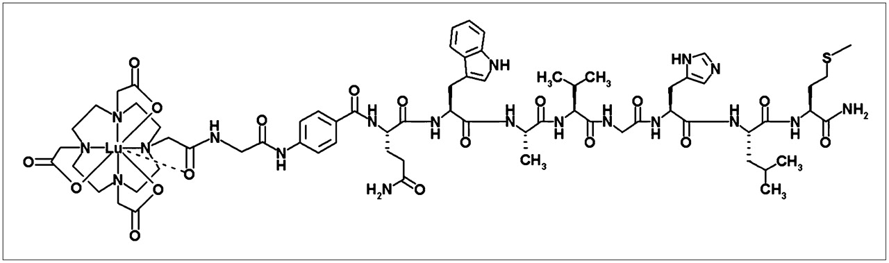
Chemical structure of 177Lu-AMBA shows proposed coordination in lutetium chelate.
MATERIALS AND METHODS
The Fmoc-protected amino acids were purchased from Nova-Biochem, Advanced ChemTech, Chem-Impex International, and Multiple Peptide Systems. The resin was procured from Nova-Biochem. Other chemicals, reagents, and chemicals required for the syntheses were procured from Aldrich Chemical Co. and VWR Scientific Products. Solvents for peptide synthesis were obtained from Pharmco Co. DO3A(tri-t-butyl)-CH2COOH was purchased from Macrocycles Inc.
Synthesis of AMBA
AMBA was synthesized using solid-phase peptide synthesis. Cleavage of the peptide with reagent B (trifluoroacetic acid [TFA]/water/phenol/triisopropylsilane, 88:5:5:2; 4 h) and purification by reversed-phase (RP) high-performance liquid chromatography (HPLC) completed the synthesis, with a 14.75% yield.
Analytic HPLC conditions are as follows. Column: XTerra MS-C18 (Waters Corp.); 4.6-mm inner diameter × 50 mm; 5-μm particle; eluent A, water (HPLC grade with 0.1% TFA [w/w]); eluent B, acetonitrile (0.1% TFA [w/w]). Retention time, 6.15 min; elution, initial conditions: 10% eluent B, linear gradient 10%–40% eluent B over 10 min; elution rate, 3 mL/min; detection, ultraviolet (UV) at 220 nm. Mass Spectrum (MS): m/z 1,502.6 [M+H]+, 752.0 [M+2H]/22+. Anal. Calcd for C68H99N19O18S·3TFA·5H2O: C, 45.96; H, 5.79; N, 13.76. Found: C, 45.97; H, 5.61; N, 13.78.
Synthesis of DO3A-CH2CO-8-Aminooctanoyl- Q-W-A-V-G-H-L-M-NH2 (BBN8)
BBN8 was synthesized according to the method of Smith et al. (14), with a 21% yield. MS: m/z 1,467.6 [M+H]+, 734.5 [M+2H]/22+. Anal. Calcd for C67H106N18O17S·3TFA· 5.39 H2O: C, 45.98; H, 6.33; N, 13.22. Found: C, 45.65; H, 6.33; N, 13.13.
Preparation of Radiotracer
AMBA or BBN8 (60 μg), 0.2 mol/L sodium acetate buffer (0.5 mL, pH 4.8), and ∼2.22 GBq 177LuCl3 (in 0.05N HCl; specific activity, 103.6–151.3 GBq/μmol; Missouri University Research Reactor) were heated at 100°C for 10 min. The radiocomplexes were separated on a Zorbax Bonus-RP HPLC column (250 × 4.6 mm, 5 μm, 80-Å pore) eluted with a gradient mixture of water, water with 30 mmol/L ammonium sulfate, and 0.1% TFA (v/v), methanol, and acetonitrile. The retention times for AMBA and Lu-AMBA were ∼24 and ∼30 min, respectively; BBN8 and Lu-BBN8 had retention times of ∼20 and ∼23 min. The HPLC-purified radiocomplexes were collected into a radiolysis-protecting buffer comprised of either 0.5 mol/L citrate buffer, pH 5.3, containing 0.2% human serum albumin, 5% ascorbic acid, and 0.9% (w/w) benzyl alcohol or a 9:1 (v/v) mixture of bacteriostatic 0.9% sodium chloride injection (USP) and ASCOR L500 (Ascorbic Acid Injection, USP) for efficacy studies. Organics were removed under reduced pressure and the complexes were diluted to the required radioconcentration using the appropriate radiolysis-protecting buffer. The radiochemical purity of all samples was ≥95% with no detectable free ligand seen by UV absorbance.
Cell Culture
In vitro assays used the human prostate cancer cell line PC-3 (androgen-resistant prostate adenocarcinoma; American Type Culture Collection), which has been shown to overexpress human GRP-R (7,20). PC-3 cells were maintained in RPMI 1640 medium (10% heat-inactivated fetal bovine serum, 10 mmol/L N-(2-hydroxyethyl)piperazine-N′-(2-ethanesulfonic acid)) at 37°C in 5% CO2/95% air and subcultured using 0.05% trypsin/ethylenediaminetetraacetic acid once per week. In vitro assays/general conditions: 96-well plate assays seeded at 6.0 × 10⁴ cells/cm2 and used 2–3 d after plating at 9.0 × 10⁴ cells/cm2. All receptor assays were conducted in binding buffer containing protease inhibitors (0.5 mmol/L phenylmethylsulfonyl fluoride, 100 μg/mL bacitracin, pH 7.2). The following experiments were conducted as described (15) with minor modifications as noted.
Receptor Competition Binding and Saturation Binding
Competition and saturation binding studies were performed at 4°C to ensure an accurate measurement for receptor binding without interference by the kinetics of internalization and degradation (21). The inhibitory concentration of 50% (IC50) of GRP analogs to adherent PC-3 cells was determined using ligand alone (BBN, BBN8, AMBA), or cold metalated ligand (175Lu-BBN8, 175Lu-AMBA), at 6 concentrations (1.25 × 10⁻⁹ mol/L to 5.0 × 10⁻⁸ mol/L), to inhibit the binding of 125I-[Tyr4]-BBN (125I-BBN; specific activity, 81.4 GBq/μmol; Perkin-Elmer Life Sciences). Direct saturation binding studies to determine the binding affinity (Kd) of 177Lu-AMBA, and the maximum binding capacity (Bmax, fmol/106 cells), used 10 concentrations of 177Lu-AMBA (∼133.2 GBq/μmol; 0.0–0.37 MBq/mL final concentration), with or without the presence of 175Lu-AMBA or AMBA ligand alone (1 μmol/L) to determine nonspecific binding. 125I-BBN was used as the positive control.
Internalization and Efflux Studies
As internalization of GPCRs are temperature sensitive, studies were performed at 37°C on adherent PC-3 cells, using 177Lu-AMBA (∼118.4 GBq/μmol), 177Lu-BBN8 (∼103.6 GBq/μmol), or the standard 125I-BBN (∼81.4 GBq/μmol). Some assays were also conducted in the presence of chloroquine (100 μmol/L), an inhibitor of lysosomal degradation (22).
Efflux studies were conducted on PC-3 cells in 24-well plates (9.0 × 10⁴ cells/cm2). After the initial 40-min incubation with 177Lu-AMBA, receptor-bound material was removed using a mild acid prewash (pH 2.5), followed by incubation in fresh binding buffer for 2 h at 37°C. The culture supernatants were collected and analyzed by HPLC using 177Lu-AMBA as the standard.
Stability in Plasma
To determine the in vitro stability of 177Lu-AMBA, plasma samples (human and mouse) were spiked with 177Lu-AMBA (0.37 MBq/0.1 mL) and incubated up to 48 h at 37°C, followed by HPLC analysis.
In Vitro Downregulation and Regeneration of GRP-R
Downregulation and regeneration of GRP-R was evaluated in PC-3 cells with unlabeled ligand (BBN or AMBA; 6 nmol/L) in cells pretreated for up to 24 h, followed by direct binding with 177Lu-AMBA (∼118.4 GBq/μmol; 2.3 kBq/mL) using previously published methods (23).
Receptor Subtype Specificity
The receptor subtype specificity was determined by in vitro receptor autoradiography as described in detail (24). To determine the affinity of Lu-AMBA for each of the 3 BBN receptor subtypes, serial sections of human ileal carcinoid tissue (NMB-R), human prostate carcinoma (GRP-R), and human bronchial carcinoid (bb3-R) were incubated with universal ligand (125I-[d-Tyr6,β-Ala11,Phe13, Nle14]BBN[6–14], 20 pmol/L, 74 TBq/mmol; Anawa) in the presence of GRP or NMB (50 nmol/L; Bachem), or increasing concentrations of cold 175Lu-AMBA, and used unlabeled universal ligand as the control.
PK
Animal studies were conducted in accordance with the U.S. Public Health Service Policy on Humane Care and Use of Laboratory Animals as well as institutional guidelines. The subjects were 4- to 6-wk old Tac:Cr:(NCr)-Foxn1nu homozygous male nude mice (Taconic Farms Inc.) xenografted with human PC-3 cells (2 × 10⁶ /mouse; “PC-3 tumor mice”) in 0.1 mL phosphate-buffered saline/Matrigel (v/v) (BD Biosciences) using standard methods. Biodistributions (1 and 24 h) were performed in PC-3 tumor mice with tumors averaging 0.5 g, at trace doses of radioactivity (0.185 MBq/0.1 mL; ∼118.4 GBq/μmol; peptide mass = 0.32 μg/m2) by intravenous tail vein, n = 4 or 5 per group. Some PC-3 tumor mice were coadministered blocking doses of AMBA ligand (4 or 10 mg/kg) or l-lysine (400–800 mg/kg). At the end of each residence interval, the mice were sacrificed, and the relevant excised organs, tissues, and blood aliquots were assayed for residual radioactivity in a γ-counter (Wizard 1480; Perkin-Elmer). HPLC analysis was performed on select urine samples (1 h).
In Vivo Downregulation and Saturation
For downregulation studies, PC-3 tumor mice were pretreated with 0.1 mL of AMBA (83.2 μg/m2), intravenously for 5, 15, or 60 min, followed by 177Lu-AMBA (0.185 MBq/0.1 mL; ∼118.4 GBq/μmol; 0.32 μg/m2) intravenously. For saturation studies, 4 ligand doses were prepared based on the proposed range of clinical doses (imaging and radiotherapy for a 70-kg man) from 0.0003 to 0.0025 mg/kg, which when scaled to the mouse (25 g) are equivalent to 0.08–0.64 μg/dose (www.fda.gov/cder/cancer/animalframe.htm). PC-3 tumor mice were administered 0.1 mL intravenous injection of 177Lu-AMBA (0.37 MBq/0.1 mL; ∼118.4 GBq/μmol, n = 4/group), which included ligand over the stated range. Control PC-3 tumor mice received 1 intravenous dose of 177Lu-AMBA, 0.37 MBq/0.1 mL. At the end of the residence interval of 177Lu-AMBA (1 h), the relevant excised organs, tissues, and blood aliquots were assayed for residual radioactivity as before.
Absorbed Radiation Dose to Tumor
The absorbed dose to PC-3 tumors was calculated from the area under the curve (AUC) generated from a high-radioactive-dose biodistribution study (27.75 MBq/0.1 mL; ∼118.4 GBq/μmol; peptide mass = 51 μg/m2 mouse) at 1, 3, 24, and 168 h, n = 4 per group. The absorbed dose to tumor was calculated using the OLINDA nodule module assuming spheric geometry (OLINDA/EXM 1.0, Vanderbilt University). Tumor AUC was calculated using SAAM II software (University of Washington); %ID (percentage injected dose)/organ = Ae−(at) + Be−(bt). The equation for AUC includes a correction for radioactive decay: AUC = A/(a + 0.00431) + B/(b + 0.00431). The residence time (h) is defined as A∼/Ao, where A∼ is the cumulative radioactivity and Ao is the amount of injected radioactivity; AUC (%ID·h) is the cumulative radioactivity and the injected radioactivity is 100%. The S value for each tumor was interpolated from the 177Lu nodule module. Absorbed dose (mGy/MBq) is the product of h and the S value (mGy/MBq·s)·3,600 s/h. The dose (Gy) is the product of absorbed dose (mGy/MBq) and the amount of radioactivity injected (e.g., 27.75 MBq).
Radiotherapeutic Efficacy: Long-Term Single-Dose and 2-Dose Studies
To ensure reliable administration of radiotherapy doses to all test animals (i.e., nude mice with fragile tail veins), all doses for long-term studies were administered subcutaneously. An equivalent biodistribution of 177Lu-AMBA at 1 and 24 h was demonstrated regardless of route of administration (intravenous or subcutaneous [data not shown]). Long-term single- and 2-dose efficacy studies (120 d; n = 32 and n = 36, respectively) were conducted in PC-3 tumor mice with a mean tumor weight of 0.1 g. PC-3 tumor mice were dosed subcutaneously with either a single injection of 0.1 mL of 177Lu-AMBA (1.11 GBq/kg; 27.75 MBq/0.1 mL; peptide mass = 41.3 μg/m2) or 2 equal 0.1-mL doses on days 0 and 14 (cumulative dose, 55.5 MBq; peptide mass = 96.1 μg/m2). Single- dose control PC-3 tumor mice (n = 15) received 0.1 mL radioprotective saline buffer subcutaneously on day 0, and 2-dose control PC-3 tumor mice (n = 13) received an identical subcutaneous injection on days 0 and 14. All subjects were observed 3 times per week and data recorded included body weight and tumor measurements. Following standard Animal Use Protocols, termination was mandated on reaching one or both of the following criteria: a tumor weight of ≥2 g or total body weight loss of ≥20%. Upon termination, necropsy was performed and tumors and kidneys were frozen for histology.
Histopathology and Immunohistochemistry (IHC)
Tissues were cryosectioned (10 μm) and evaluated by standard hematoxylin and eosin (H&E) staining. IHC was performed using an anti-vimentin mouse monoclonal antibody (marker for human mesenchymal origin—i.e., PC-3 cells), Clone V9 (no cross-reactivity with mouse vimentin), 1:50 (M0725; DakoCytomation); a bridging kit to reduce nonspecific (mouse) binding (K3954, DAKO ARK; DakoCytomation); visualized with streptavidin-horseradish peroxidase/Romulin AEC (Biocare Medical), and counterstained. Negative controls were performed on serial sections with mouse IgG for the primary antibody. The H&E- and IHC-stained tissue sections were examined with a Nikon Eclipse E800 photomicroscope. Images were captured by digital camera (DXM 1200; Sylvax Scientific) and imported into Adobe Photoshop CS.
Statistical Analysis
Nonlinear regression analysis was performed on in vitro data (GraphPad Prism 3.0 software). All mean values are given as ± SD. Statistical analysis of in vivo studies used the unpaired t test when 2 groups were analyzed and 1-way ANOVA if ≥2 groups were analyzed (GraphPad Prism 3.0 software). The level of significance was set at P < 0.05.
RESULTS
In Vitro Studies
Competition binding and saturation studies demonstrated that Lu-AMBA binds specifically and with nanomolar affinity to GRP-R (Table 1). The Bmax was ∼414 fmol per 106 PC-3 cells; ∼2.5 × 10⁵ GRP-R per PC-3 cell.
Competition with 125I-BBN: IC50 of BBN-Binding Peptides to GRP-R in PC-3 Cells In Vitro
The degree of internalization of 177Lu-AMBA was similar to 125I-BBN and 177Lu-BBN8 (Table 2). Efflux of 177Lu-AMBA was statistically lower than that for cells treated with either 177Lu-BBN8 (P < 0.005) or 125I-BBN (P < 0.001) . Only efflux of 177Lu-BBN8 and 125I-BBN was significantly lower in the presence of chloroquine (Table 2). For 177Lu-AMBA studies, HPLC analysis of radiolabeled material effluxed into the medium (2.9%) demonstrated that the majority was parent 177Lu-AMBA (78%), with no free 177Lu. 177Lu-AMBA was more stable in vitro in human plasma (t1/2 = 38.8 h) than in mouse plasma (t1/2 = 3.1 h).
In Vitro Internalization and Efflux of Radiolabeled GRP-R Binding Peptides 177Lu-AMBA, 177Lu-BBN8, and 125I-BBN, ± Chloroquine
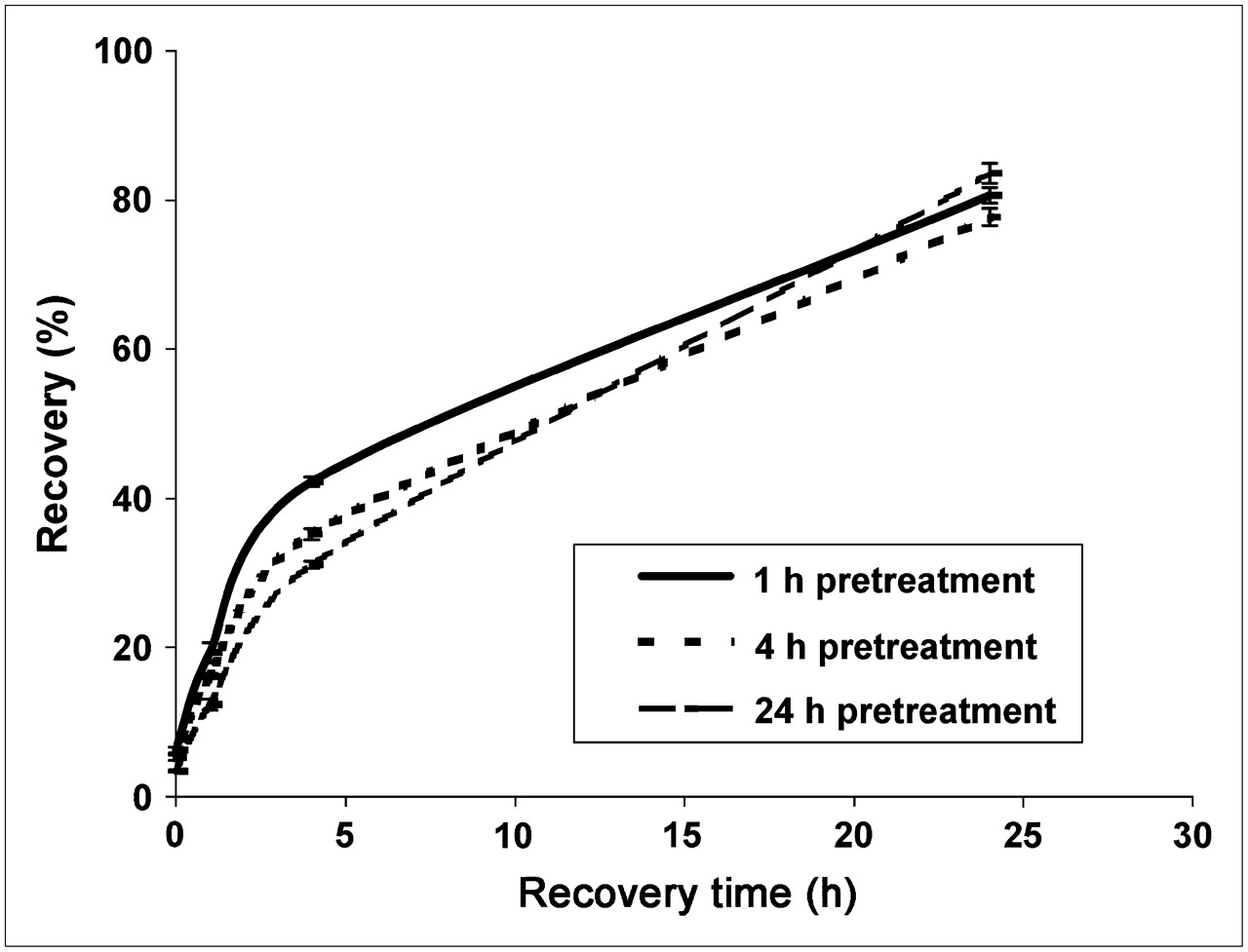
GRP-R in PC-3 cells was rapidly downregulated at similar rates by incubation with 6 nmol/L of AMBA (∼5 times Kd), 175Lu-AMBA, or BBN. GRP-R in PC-3 cells recovered in 24 h (Fig. 2), similar to reported values in Swiss 3T3 cells (23) and recovery of NMB-R reported in C6 cells (25).

Recovery of GRP-R after downregulation in human PC-3 cells by pretreatment with 175Lu-AMBA (6 nmol/L) for 1, 4, and 24 h. Half-maximal downregulation occurred in 2–4 min; ∼90% of GRP-R was lost from the cell surface by 1 h.
Lu-AMBA binds specifically to human GRP-R and NMB-R by in vitro autoradiography (Table 3) but has no affinity or low affinity for the bb3 receptor subtype found predominately in the normal human pancreas (26) and neuroendocrine lung tumors (24). The universal ligand, which binds to all 3 subtypes of mammalian BBN receptors, was used as a positive control.
BBN Receptor Subtype Specificity of 175Lu-AMBA by Receptor Autoradiography in Human Tissues, as Measured by Displacement Assay with Universal 125I-BBN Ligand on Human Tissue Samples
In Vivo Studies
The route of excretion of 177Lu-AMBA and 177Lu-BBN8 was primarily renal (Table 4). The uptake and retention of radioactivity in tumor was notably higher at 1 h for 177Lu-AMBA and was retained at a higher level at 24 h as compared with 177Lu-BBN8. Pancreas was the major nontarget organ for both radiolabeled peptides. Specificity for the receptor was demonstrated when coadministration of excess AMBA ligand (4 mg/kg or 10 mg/kg) effectively blocked tumor uptake by 83% and 87%, respectively (Table 4). The blocking studies also demonstrated 54% and 57% blocking of distribution to kidney, respectively, >96% blocking to gastrointestinal tract, and >99% blocking of receptor-mediated binding in pancreas. Coadministration of l-lysine (400–800 mg/kg) did not alter the already low kidney retention of 177Lu-AMBA at 1 h. HPLC analysis of urine collected after 1 h residence found no parent 177Lu-AMBA.
Biodistribution (0.185 MBq) of 177Lu-AMBA (n = 9) and 177Lu-BBN8 (n = 5) in PC-3 Tumor Mice
Uptake and retention in the tumor was not downregulated by pretreatment with AMBA, whereas a decrease in radioactivity associated with the gastrointestinal tract was seen in all AMBA pretreatment groups (Table 5). Renal effects were transient and only significantly different from control in the 5-min predose group. A significant decrease in pancreatic uptake of radioactivity was seen in the 5- and 15-min predose groups.
Downregulation of 177Lu-AMBA (0.185 MBq) After Administration of AMBA (83.2 μg/m2) in PC-3 Tumor Mice (n = 4)
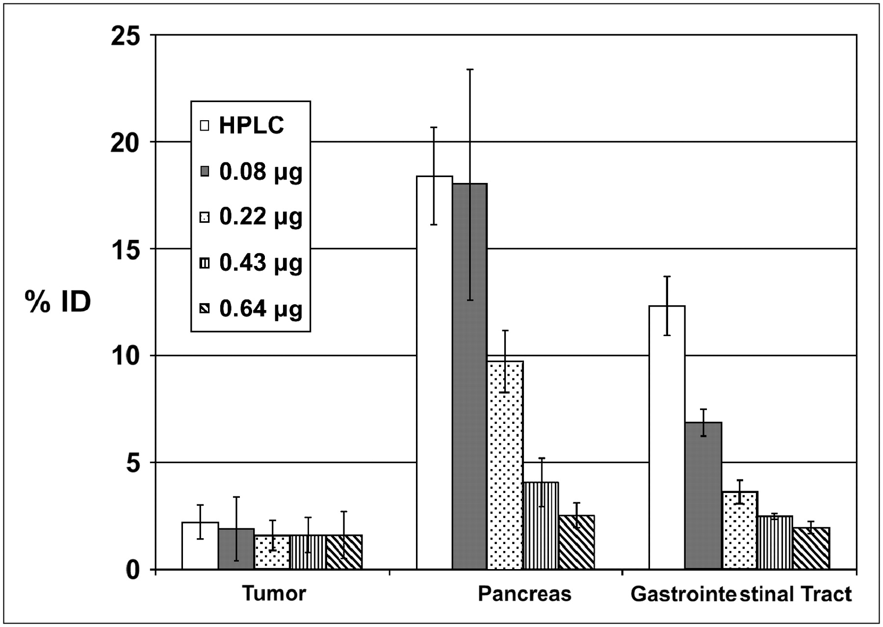
Receptor-specific uptake of Lu-AMBA in the tumor was resistant to saturation, retaining ∼70% of uptake at the highest clinically relevant dose (Fig. 3), whereas pancreas and gastrointestinal tract were reduced by 86% and 84%, respectively.

In vivo saturation studies in PC-3 tumor mice at 1 h after administration. Graph depicts percentage uptake in human PC-3 tumor, mouse pancreas, and mouse gastrointestinal tract with increasing peptide mass (HPLC-purified [0.0025 μg], 0.08, 0.22, 0.43, and 0.64 μg).
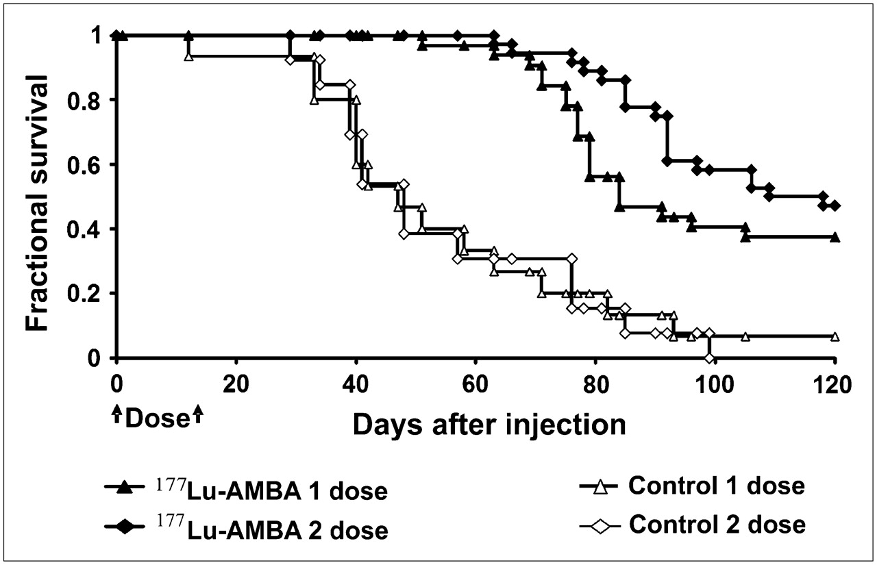
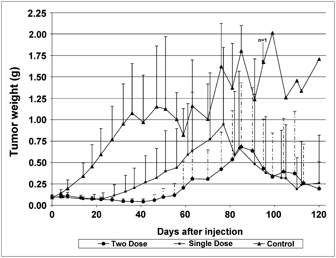
Treatment with 1 or 2 doses of 177Lu-AMBA significantly prolonged the life span of tumor-bearing mice (Fig. 4) and decreased PC-3 tumor growth rate over that of controls (Fig. 5). The histopathology of xenografted tumors was variable. However, in tumors that responded to treatment, the overwhelming histologic finding was necrosis and scarring, with deposition of fibrotic tissue and thickening of the tumor capsule and few residual vimentin-positive PC-3 cells (Fig. 6). In some cases, the actual tumor burden detected by histology was much lower than the gross caliper measurement due to the infiltration of phagocytic cells and fibrosis. In contrast, the histologic pattern in control tumors was that of a solid mass of vimentin-positive PC-3 cells, or centralized necrosis, due to the sheer mass of tumor tissue. However, fibrosis and capsule thickening were not evident in control tumors. Histology of mouse kidney for all radiotherapy studies was within normal parameters.

Kaplan–Meier plot of 177Lu-AMBA radiotherapy shows that single-dose (27.75 MBq; n = 32) or 2-dose (55.5 MBq; n = 36) treatment significantly increased life span and reduced tumor growth rate over that of control (single dose, n = 15; 2 dose, n = 16) (120 d: single dose, P < 0.001; 2 dose, P < 0.0001). Overall survival at 30, 60, 90, and 120 d from single dose was 100%, 97%, 47%, and 38%; overall survival from 2 doses spaced 14 d apart was 100%, 100%, 78%, and 47%.

Average tumor growth over time with 177Lu-AMBA radiotherapy; single-dose (27.75 MBq) or 2-dose (55.5 MBq). Tumor volumes from day 27 to day 80 were significantly different (P < 0.05), with less variability in 2-dose group (day 27 to day 60), most likely due to additional therapeutic effect of second dose.

177Lu-AMBA-treated vs. control tumors. (A) Two-dose treated tumor shows necrosis (n) with few residual vimentin-positive (vim+) PC-3 cells (×20). (B) H&E shows deposition of fibrotic tissue and thickening of tumor capsule, with infiltration of phagocytic cells (×100). (C) Vimentin staining of control tumor demonstrates a solid mass of vimentin-positive (vim+) PC-3 cells (×20). (D) H&E shows a thin capsule without fibrosis (×100).
A comparison of tumors in the single- versus 2-dose groups, using modified World Health Organization (WHO) response criteria confirmed by histology, demonstrated a shift away from stable disease and progressive disease toward complete response and partial response, with no evidence of stable or progressive disease in animals receiving 2 doses of 177Lu-AMBA (Table 6). The rate of tumor growth (Fig. 5) showed that the variability was much smaller in mice treated with 2 doses versus a single dose of 177Lu-AMBA, whereas both treated groups demonstrated a significant reduction in tumor growth over that of controls. The WHO criteria reflect this, showing a 26% increase in complete recoveries and a 39% increase in partial recoveries over the single-dose treated group. The same data compared using RECIST (27) showed that median survival increased by 36% and the time to progression/progression-free survival increased by 65%.
Radiotherapeutic Efficacy of 177Lu-AMBA: Summary of WHO and RECIST Criteria
The average residence time for the tumor was calculated using SAAM II software (0.265 h) and applied to individual mouse tumor weights to calculate absorbed dose (in Gy). When this was applied to tumors in the single- and 2-dose efficacy studies, the following average absorbed doses to tumor were generated: 13.6 ± 5.3 Gy for a single dose of 177Lu-AMBA; and 19.3 ± 12.3 Gy (day 0), plus 17.4 ± 10.1 Gy (day 14), for a cumulative dose of 36.73 Gy for 2 doses of 177Lu-AMBA.
DISCUSSION
Smith et al. (14) reported previously that radiolabeled derivatives of BBN showed receptor-mediated uptake in GRP-R–expressing PC-3 cells in vitro and in vivo and that an optimized compound from this series (BBN8) showed radiotherapeutic efficacy in studies in female PC-3 tumor-bearing SCID (severe combined immunodeficiency) mice. We sought to improve tumor uptake and retention while maintaining the high-affinity binding and good PK properties of BBN8. We maintained the DO3A-CH2CO- chelator (often referred to in generic terms as “DOTA”), as it has been shown to tightly bind to radiometals, and the targeting functionality (Q-W-A-V-G-H-L-M-NH2). By changing the linker we arrived at 177Lu-AMBA (Fig. 1), which contains a glycyl-4-aminobenzoic acid linker in place of the 8-aminooctanoic acid linker used in BBN8. 177Lu-AMBA has nanometer affinity for the GRP-R and is more polar and the linker is more rigid than that of BBN8.
Good internalization of a radiolabeled peptide is advantageous for a successful radiotherapeutic. Internalization for 177Lu-AMBA was much higher than that reported for pan-BBN peptides BZH1 and BZH2 (15). Perhaps more importantly, the amount of radioactivity that remains internalized and therefore available to irradiate the target was significantly higher for 177Lu-AMBA (Table 2). Only 2.9% of the internalized radiopeptide was lost from the cell (Table 2) and HPLC analysis showed that this was predominantly parental 177Lu-AMBA, with no free 177Lu. We obtained comparable results with or without acid prewash but also demonstrated an increase in the membrane-bound fraction in the samples that had undergone acid prewash (data not shown). This is consistent with recycling of intact radiolabeled peptides by tumor cells, presumably for reactivation of membrane-bound receptors by the externalized peptides (28,29). We assume that the fate of the majority of the ligand complex would be similar to that observed by Grady et al., for GRP and GRP-R, in that subsequent to receptor-mediated endocytosis the ligand is hydrolyzed from the receptor and undergoes degradation within lysosomes, whereas GRP-R is recycled to the plasma membrane (30).
Receptor autoradiography studies showed that Lu-AMBA binds specifically to GRP-R– and NMB-R–bearing human tumor tissues, with no binding affinity or low binding affinity for the bb3 receptor. This is better affinity (IC50) than either of the pan-BBN peptides, BZH1 and BZH2, respectively, for GRP-R (3.5 nmol/L and 1.4 nmol/L), and NMB-R (10.5 nmol/L and 4.9 nmol/L), evaluated using the same methodology by one of us (Reubi) (15).
177Lu-AMBA excretion was primarily renal (Table 4). Though the washout of radioactivity from the tumors for complexes of AMBA, BBN8, and the published values for BZH2 (15) were in the 40%–50% range at 24 h, the overall amount of radioactivity retained after uptake of 177Lu-AMBA—and thus capable of delivering therapeutic radiation in the 177Lu-AMBA–targeted tumors at 24 h—was greater. The lack of in vivo downregulation (Table 5) or saturation (Fig. 3) in the tumor target will be beneficial for radiotherapy with 177Lu-AMBA clinically, if the tumor target is also unaffected by predosing or increased mass dose. On the basis of the increased level of targeting and retention of 177Lu-AMBA, we would predict the efficacy of 177Lu-AMBA to be superior to either of the pan-BBN compounds (15) or BBN8.
Greater cellular retention is also an explanation for the higher levels of radioactivity retained in the mouse pancreas (GRP-R+) after 177Lu-AMBA administration. The pancreatic uptake of 177Lu-AMBA in mice is not unexpected, as the predominant receptor subtype in the pancreas of rodents is a GRP-preferring receptor. Interestingly, mice in the radiotherapy studies were not visibly affected by the high pancreatic uptake of 177Lu-AMBA up to 120 d. This phenomenon has been noted before (14), presumably a reflection of the relatively low radiation sensitivity of pancreatic tissues (31).
Peptide radiotherapies have been plagued by problems with kidney retention (32,33). Radiolabeled somatostatin receptor therapy uses coadministration of l-lysine/arginine to increase the therapeutic index (34). When compared with published kidney levels of radiolabeled octreotide with and without coadministration of l-lysine (35,36), biodistribution of 177Lu-AMBA is already ∼50% lower than that of the somatostatin receptor–targeted peptides and is not reduced further with coadministration of l-lysine.
The combination of the physical half-life of the 177Lu (t1/2 = 6.7 d) and the long retention of 177Lu-AMBA in the tumor allows killing radiation to be delivered essentially by hyperfractionation at the radiosensitive tumor target site. Consistent with the energy range of the 177Lu β-emission (37), we believe that the radiation dose delivered to the PC-3 tumors reflects only self-irradiation of the tumor. A single dose of 177Lu-AMBA (13.6 Gy) is more efficacious than an equivalent single administration of external beam radiation delivered as a single administration (∼8 Gy), which achieved a 75% reduction in tumor weight of orthotopic PC-3 tumors (0.4 g) (38). Two doses of 177Lu-AMBA (36.7 Gy) demonstrated an increase in tumor growth delay and improved overall survival as compared with a single dose (Figs. 4 and 5). Moreover, mice administered 2 doses of 177Lu-AMBA demonstrated a marked shift toward complete response and partial response, with no evidence of stable disease or progressive disease.
CONCLUSION
We have described a clinically relevant novel GRP-R/NMB-R–binding peptide agonist, 177Lu-AMBA, which is conducive to both targeted radiotherapy and imaging. The use of 177Lu will allow for individual patient dosing via imaging of the γ-emissions of 177Lu and takes advantage of the short pathlength of the β-emissions of 177Lu for the treatment of metastases. 177Lu-AMBA demonstrates nanomolar affinity in both human prostate tumor tissue and in the human PC-3 cells. Optimized targeting and retention were reflected in the radiotherapeutic efficacy as well as increased efficacy of an appropriately timed second dose of 177Lu-AMBA for systemic radiotherapy. 177Lu-AMBA demonstrates very low uptake and retention in kidney, unlike other reported peptide-based radiotherapies. On the basis of the favorable PK and tumor efficacy in a relevant preclinical model, a phase I single-dose clinical trial with 177Lu-AMBA is in progress.
Acknowledgments
This research was funded by Bracco Research USA.
Footnotes
-
COPYRIGHT © 2006 by the Society of Nuclear Medicine, Inc.
References
- Received for publication November 22, 2005.
- Accepted for publication March 27, 2006.


Jump to section
Related Articles
Cited By...
- Exploring the signaling space of a GPCR using bivalent ligands with a rigid oligoproline backbone
- 68Ga/177Lu-NeoBOMB1, a Novel Radiolabeled GRPR Antagonist for Theranostic Use in Oncology
- Bombesin-Targeted PET of Prostate Cancer
- In Vitro and In Vivo Application of Radiolabeled Gastrin-Releasing Peptide Receptor Ligands in Breast Cancer
- A Comparative Study of Radiolabeled Bombesin Analogs for the PET Imaging of Prostate Cancer
- Targeted Radiotherapy of Prostate Cancer with a Gastrin-Releasing Peptide Receptor Antagonist Is Effective as Monotherapy and in Combination with Rapamycin
- PET of Tumors Expressing Gastrin-Releasing Peptide Receptor with an 18F-Labeled Bombesin Analog
- Bombesin Antagonist-Based Radioligands for Translational Nuclear Imaging of Gastrin-Releasing Peptide Receptor-Positive Tumors
- Alpha- versus Beta-Particle Radiopeptide Therapy in a Human Prostate Cancer Model (213Bi-DOTA-PESIN and 213Bi-AMBA versus177Lu-DOTA-PESIN)
- Multimodality Imaging and Preclinical Evaluation of 177Lu-AMBA for Human Prostate Tumours in a Murine Model
- 177Lu-AMBA Biodistribution, Radiotherapeutic Efficacy, Imaging, and Autoradiography in Prostate Cancer Models with Low GRP-R Expression
- Evaluation of a 1,4,7,10-Tetraazacyclododecane-1,4,7,10-Tetraacetic Acid-Conjugated Bombesin-Based Radioantagonist for the Labeling with Single-Photon Emission Computed Tomography, Positron Emission Tomography, and Therapeutic Radionuclides
- Small-Animal PET of Tumors with 64Cu-Labeled RGD-Bombesin Heterodimer
- High expression of gastrin-releasing peptide receptors in the vascular bed of urinary tract cancers: promising candidates for vascular targeting applications
- Peptide-Based Probes for Cancer Imaging
- International Union of Pharmacology. LXVIII. Mammalian Bombesin Receptors: Nomenclature, Distribution, Pharmacology, Signaling, and Functions in Normal and Disease States
- Bombesin Receptor Antagonists May Be Preferable to Agonists for Tumor Targeting
- In Vivo Evaluation and Small-Animal PET/CT of a Prostate Cancer Mouse Model Using 64Cu Bombesin Analogs: Side-by-Side Comparison of the CB-TE2A and DOTA Chelation Systems