


Abstract
The BB2 receptor subtype, of the bombesin family of receptors, has been shown to be highly overexpressed in a variety of human tumors, including prostate cancer. Bombesin (BBN), a 14-amino acid peptide, has been shown to target the BB2 receptor with high affinity. 64Cu (half-life = 12.7 h, β+: 18%, Eβ+max = 653 keV; β−: 37%, Eβ−max = 578 keV) is a radioisotope that has clinical potential for application in both diagnostic imaging and radionuclide therapy. Recently, new chelation systems such as 1,4,8,11-tetraazabicyclo[6.6.2]hexadecane-4,11-diacetic acid (CB-TE2A) have been reported to significantly stabilize the 64Cu radiometal in vivo. The increased stability of the 64Cu-CB-TE2A chelate complex has been shown to significantly reduce nontarget retention compared with tetraazamacrocycles such as 1,4,7,10-tetraazacyclodoadecane-N,N′,N″,N‴-tetraacetic acid (DOTA). The aim of this study was to determine whether the CB-TE2A chelation system could significantly improve the in vivo stability of 64Cu bombesin analogs. The study directly compares 64Cu bombesin analogs using the CB-TE2A and DOTA chelation systems in a prostate cancer xenograft SCID (severely compromised immunodeficient) mouse model. Methods: The CB-TE2A-8-AOC-BBN(7–14)NH2 and DOTA-8-AOC-BBN(7–14)NH2 conjugates were synthesized and radiolabeled with 64Cu. The receptor-binding affinity and internalization profile of each metallated conjugate was evaluated using PC-3 cells. Pharmacokinetic and small-animal PET/CT studies were performed using female SCID mice bearing PC-3 xenografts. Results: In vivo BB2 receptor targeting was confirmed by tumor uptake values of 6.95 ± 2.27 and 4.95 ± 0.91 %ID/g (percentage injected dose per gram) at the 15-min time point for the 64Cu-CB-TE2A and 64Cu-DOTA radioconjugates, respectively. At the 24-h time point, liver uptake was substantially reduced for the 64Cu-CB-TE2A radioconjugate (0.21 ± 0.06 %ID/g) compared with the 64Cu-DOTA radioconjugate (7.80 ± 1.51 %ID/g). The 64Cu-CB-TE2A-8-AOC-BBN(7–14)NH2 radioconjugate demonstrated significant clearance, 98.60 ± 0.28 %ID, from the mouse at 24 h after injection. In contrast, only 67.84 ± 5.43 %ID of the 64Cu activity was excreted using the 64Cu-DOTA-8-AOC-BBN(7–14)NH2 radioconjugate because of nontarget retention. Conclusion: The pharmacokinetic and small-animal PET/CT studies demonstrate significantly improved nontarget tissue clearance for the 64Cu-CB-TE2A8-AOC-BBN(7–14)NH2. This is attributed to the improved in vivo stability of the 64Cu-CB-TE2A chelate complex as compared with the 64Cu-DOTA chelate complex.
The bombesin family of receptors—in particular, the BB2 receptor—has received a great deal of attention as a potential target for target-directed radiopharmaceuticals due to the high densities of these receptors on a variety of human tumors (1–3). The BB2 receptor subtype has been the most thoroughly studied receptor of the bombesin family and has been shown to be overexpressed in prostate, breast, small cell lung, and pancreatic cancers. Bombesin (BBN) is a 14-amino acid amphibian peptide that has been shown to bind to the BB2 receptor with high affinity (4). Our group and others have been involved in the synthesis of bombesin analogs that incorporate a radionuclide into the structure of the bombesin peptide (5–13). These radiolabeled analogs have been shown to target the BB2 receptor in vivo with high affinity. The potential of bombesin analogs for targeting the BB2 receptors overexpressed on prostate and breast cancer has been demonstrated in human clinical trials by Van de Wiele et al. and others (14–18). Research to find analogs that have optimal retention and clearance properties for diagnostic imaging and radionuclide therapeutic applications continues.
64Cu (half-life [t½] = 12.7 h) is a radionuclide that has received a great deal of interest for both diagnostic imaging and radionuclide therapy due to the radionuclide's dual decay characteristics. The positron (β+: 18%, Eβ+max = 653 keV) and beta (β−: 37%, Eβ−max = 578 keV) emissions of 64Cu could allow clinicians the potential to diagnostically image a patient using PET and, subsequently, give therapeutic treatment using the same 64Cu-targeted radiopharmaceutical. However, the widespread application of 64Cu-based radiopharmaceuticals has been hindered by the lack of in vivo stable copper radiotracers. The uptake and retention of copper-containing compounds in the blood and liver has been known for over half a century (19–24).
Acyclic and macrocyclic polyamines have been widely used as chelators due to the rapid formation of thermodynamically and kinetically inert complexes with a variety of medicinally useful metals. These chelation systems are used today with a great deal of success for stabilizing a variety of medicinal metals for a multitude of clinical applications. Acyclic and macrocyclic polyamines have had limited success stabilizing copper in vivo due to poor kinetic stability (25–27). Macrocyclic polyamine complexes of copper, such as 1,4,8,11-tetraazacyclotetradecane-N, N′,N″,N‴-tetraacetic acid (TETA) and 1,4,7,10-tetraazacyclodoadecane-N,N′,N″,N‴-tetraacetic acid (DOTA) have been shown to exhibit greater kinetic inertness than acyclic analogs, but significant transchelation still occurs (26,27). The design and synthesis of kinetically inert chelation moieties for copper nuclides and other medicinally relevant metals is currently an active area of research (28).
One of several new chelate designs for copper radionuclides focuses on cross-bridged tetraazamacrocycles. 1,4,8,11-Tetraazabicyclo[6.6.2]hexadecane-4,11-diacetic acid (CB-TE2A), reported by Wong et al. in 2000, is a cross-bridge derivative of the TETA macrocycle (29). The cross-bridged tetraamine bicycles were found to be an improvement over their monocyclic counterparts. The rigidity of the cross-bridge systems is accredited for the greater kinetic stability of the copper(II) cross-bridged complexes over traditional DOTA and TETA analogs. Recently, the collaborative work of the Anderson, Weisman, and Wong laboratories has yielded several reports with regard to the in vitro and in vivo properties of copper(II)-CB-TE2A (30–33). This collaboration also yielded the first 64Cu receptor-targeted radiopharmaceutical that incorporated a cross-bridged chelation system, 64Cu-CB-TE2A-Y3-TATE. Anderson et al. reported that the 64Cu-CB-TE2A conjugate poses significantly greater in vivo stability when compared directly with the 64Cu-TETA conjugate in an AR42J tumor-bearing rat model (32).
Our group has reported the synthesis and evaluation of a series of bombesin analogs of the type 111In-DOTA-X-BBN(7–14)NH2, where X = 0-, 3-, 5-, 8-, or 11-atom-length hydrocarbon spacers to determine the effect of spacer length on pharmacokinetics (5). From this work, the conjugates with the tethering lengths between 5 and 8 carbons, 111In-DOTA-5-AVA-BBN(7–14)NH2 and 111In-DOTA-8-AOC-BBN(7–14)NH2, respectively, provided the most favorable pharmacokinetic properties. Using this paradigm, Rogers and coworkers recently reported the synthesis and evaluation of 64Cu-DOTA-8-AOC-BBN(7–14)NH2 (9,34). The 64Cu-DOTA-8-AOC-BBN(7–14)NH2 radioconjugate demonstrated good uptake and impressive retention in the PC-3 tumor mouse model, but the pharmacokinetic properties are indicative of the in vivo instability of 64Cu tetraazamacrocycle chelates (i.e., transchelation to advantageous proteins).
The purpose of the current study was to directly compare bombesin analogs synthesized with the CB-TE2A chelator with traditional tetraazamacrocycle conjugates for in vivo stability. The 64Cu-CB-TE2A-8-AOC-BBN(7–14)NH2 radioconjugate and the previously reported 64Cu-DOTA-8-AOC-BBN(7–14)NH2 radioconjugate were synthesized for the purpose of a direct side-by-side comparison. Here, we present the synthesis of the 64Cu-CB-TE2A-8-AOC-BBN(7–14)NH2 and 64Cu-DOTA-8-AOC-BBN(7–14)NH2 radioconjugates and the in vitro and in vivo evaluation and characterization using small-animal PET technology.
MATERIALS AND METHODS
Unless otherwise specified, all chemicals and reagents were used as received. 64Cu was purchased from MDS Nordion. Fmoc-protected amino acids and resins were purchased from EMD Biosciences Inc. Tris(1,1-dimethylethyl)-1,4,7,10-tetraazacyclododecane-1,4,7,10-tetraacetic acid was purchased from Macrocyclics. 1,4,8,11-Tetraazabicyclo[6.6.2]hexadecane-4,11-diacetic acid was prepared according to literature procedures (29,35,36). Fmoc-8-Aminooctanoic acid was purchased from Advanced ChemTech. Naturally abundant copper(II) sulfate pentahydrate, N,N-dimethylformamide (DMF), N-methyl-2-pyrrolidone (NMP), and diisopropylethylamine (DIEA) were purchased from Fischer Scientific. HPLC (high-performance liquid chromatography)-grade acetonitrile and trifluoroacetic acid were purchased from Fischer Scientific for HPLC analyses. Deionized water used for HPLC analyses and all other chemical applications in this work was obtained from an in-house Millipore Milli-Q Biocel water purification system. RPMI 1640 medium was purchased from Invitrogen/GIBCO. 4-(2-Hydroxyethyl)-1-piperazineethanesulfonic acid (HEPES) and bovine serum albumin (BSA) were purchased from Fisher Scientific (Fisher Bioreagents, Fisher Chemical). [125I-Tyr4]BBN (370 kBq [10 μCi]) was purchased from Perkin Elmer.
Solid-phase peptide synthesis (SPPS) was performed using an Applied Biosystems model 432 automated peptide synthesizer using standard Fmoc chemistry. Reversed-phase HPLC (RP-HPLC) purification of the conjugates, natCu-conjugates and 64Cu-conjugates, was accomplished using a Waters 600E controller equipped with an Eppendorf TC-50 column heater. A Phenomenex Jupiter 5-μm C18 300-Å, 250 × 10 mm semipreparative column was used for the purification of bulk amounts of CB-TE2A-8-AOC- BBN(7–14)NH2. A Phenomenex Jupiter 5-μm C18 300-Å, 250 × 4.60 mm analytic column was used for the purification of all conjugates for in vitro and in vivo studies. Detection of the conjugates and natCu-conjugates during HPLC purification was accomplished using a JASCO UV 975 absorbance detector, whereas the radioconjugates were detected during HPLC purification using a JASCO UV 1575 absorbance detector and an ORTEC (Oak Ridge National Laboratory) NaI(Tl) scintillation detector. During the preparation of the radioconjugates, 3M Empore High Performance Extraction Disk Cartridges 4215(HD) were used. Mass spectrometric determination of the conjugates and natCu conjugates was achieved by nanospray time-of-flight mass spectrometry (QqTOF MS) using an Applied Biosystems/MDS Sciex QStar/Pulsar/i instrument. Radiation measurements for the in vitro binding studies were accomplished using the Packard Riastar γ-counter. Pharmacokinetic radiation measurements were made using a well counter equipped with a NaI(Tl) scintillation detector.
Four- to 5-wk-old ICR SCID (Institute of Cancer Research severely compromised immunodeficient) female mice were obtained from Taconic Farms. The mice were supplied with sterilized water and irradiated rodent chow (Ralston Purina Co.) ad libitum. Five mice were housed per microisolator cage (Alternative Design) in a humidity- and temperature-controlled room with a 12-h light/dark cycle. The human prostate cancer PC-3 cell line was purchased from the American Type Culture Collection. The PC-3 cell line was grown and maintained by the University of Missouri Cell and Immunobiology Core Facility. PC-3 cells were grown in custom RPMI medium 1640 (Invitrogen Corp.) supplemented with 10% fetal bovine serum (U.S. Bio-Technologies Inc.) and gentamicin (American Pharmaceutical Partners, Inc.). To prepare the cells for inoculation, the PC-3 cells with media were centrifuged, the media was decanted, and the cell pellet was combined with Dulbecco's phosphate-buffered saline (Invitrogen Corp.) to reach a concentration of 5 million cells/100 μL. Under gas anesthesia, each SCID mouse was subcutaneously inoculated (bilateral flank) with 5 million PC-3 tumor cells in each flank. Using a nonrebreathing apparatus (Summit Medical Equipment Co.), gas anesthesia was administered at a vaporizer setting of ∼3.5% isoflurane (Baxter Healthcare Corp.) with ∼1 L/min oxygen. The flank tumors were allowed to grow for 3–4 wk before studies were performed. All studies involving animals were conducted in accordance with protocols approved by the institutional animal care and use committee in accordance with U.S. Public Health Service guidelines.
SPPS
The conjugates were assembled using automated peptide synthesis. The crude conjugate was dried by vacuum, weighed, and analyzed by RP-HPLC. Purity of the crude peptides, as determined by RP-HPLC, varied depending on the conjugate. For DOTA-8-AOC-BBN(7–14)NH2, the unpurified peptide was determined to be 70% pure by HPLC. The synthesis of CB-TE2A-8-AOC-BBN(7–14)NH2 was found to be more problematic, with purity determinations of 10% by HPLC. Due to the low yields for CB-TE2A-8-AOC-BBN(7–14)NH2, the conjugate was purified using semipreparative RP-HPLC before studies were performed. All conjugates were peak purified to ≥93% purity and quantified by analytic RP-HPLC before mass spectrometric determination and in vitro studies. Confirmation of the desired conjugates' constitution was determined by mass spectrometry.
Syntheses of natCu-Conjugates
For DOTA-8-AOC-BBN(7–14)NH2 and CB-TE2A-8-AOC- BBN(7–14)NH2, a 1-mg sample of the conjugate was dissolved in 500 μL of a 0.1 M ammonium acetate/0.80 mM copper(II) sulfate pentahydrate solution. The solutions were heated at 70°C for 40 min and allowed to cool to room temperature. natCu-Conjugates were then peak purified and quantified by RP-HPLC. All natCu-conjugates were determined to be ≥93% pure, by RP-HPLC analyses before mass spectrometric characterization and in vitro binding studies.
Radiolabeling of Conjugates with 64CuCl2
A 100-μg sample of the conjugate and ∼20 mg of l-ascorbic acid (radiolysis prevention reagent) were dissolved in 200 μL of 0.4 M ammonium acetate (pH 7.0). 64CuCl2 was added to the vial containing the conjugate, and the solution was heated for 40 min at 70°C and allowed to cool to room temperature. The resulting 64Cu-radioconjugate was peak purified using RP-HPLC. The peak-purified 64Cu-radioconjugate was concentrated using a C18 extraction disk and eluted with 400 μL of a 6:4 ethanol/sterile saline solution. For pharmacokinetic studies, the purified 64Cu-conjugates were diluted with sterile saline to 0.185 MBq (5 μCi) per 100 μL. Imaging studies required evaporation of the ethanol from the 64Cu-radioconjugate solution under a stream of nitrogen and dilution to the appropriate volume with sterile saline solution. In all cases, the concentration of ethanol in solution was <8% before administration. The radiochemical yield for 64Cu-DOTA-8-AOC-BBN(7–14)NH2 averaged 68% by HPLC, whereas that for 64Cu-CB-TE2A-8-AOC-BBN(7–14)NH2 averaged 30%. The radiochemical purity of all 64Cu-radioconjugates for pharmacokinetic and imaging studies was reassessed before administration and found to be ≥93% pure.
HPLC Purification and Analyses
Bulk sample purification of CB-TE2A-8-AOC-BBN(7–14)NH2 was performed using a semipreparative column with a flow rate of 5.0 mL/min. Sample purification for mass spectrometric in vitro and in vivo studies was performed on an analytic column with a flow rate of 1.5 mL/min. For all HPLC experiments, the mobile phase consisted of solvent A (99.9% H2O and 0.1% trifluoroacetic acid [TFA]) and solvent B (99.9% CH3CN and 0.1% TFA). For all conjugates, natCu-conjugates and 64Cu-conjugates, the gradient began at 80% A:20% B and over a period of 15 min linearly decreased to a gradient of 70% A:30% B. At the end of the 15-min run time for all HPLC experiments, the column was flushed with the gradient 5% A:95% B and reequilibrated to the starting gradient.
In Vitro Competitive Cell-Binding Studies
Fifty-percent inhibitory concentration (IC50) binding studies for all conjugates and natCu-conjugates were performed using PC-3 human prostate cancer cells. natCu-conjugates were used to determine the specific binding affinities for the corresponding 64Cu-conjugates. Cell media consisted of RPMI medium at pH 7.4, 4.8 mg/mL HEPES, and 2 mg/mL BSA. For binding assays, the cells (∼3 × 10⁴ PC-3 cells) were suspended in media and incubated at 37°C for 40 min in the presence of radiolabeled [125I- Tyr4]BBN, a known BB2 agonist, and a range of concentrations of the conjugate or natCu-conjugate. After incubation, the cells were aspirated and washed (3×) with media. The cell-associated activity was measured, and the binding affinity was determined.
Internalization Assays
Internalization studies were performed by incubating 3 × 10⁴ PC-3 human prostate cancer cells in cell media. The cell medium consisted of RPMI medium with 2 mg/mL BSA and 2.8 mg/mL HEPES at pH 7.4. The cells were incubated at 37°C with 20,000 cpm of the 64Cu-CB-TE2A-8-AOC-BBN(7–14)NH2 in the presence of 5% CO2 for 15-, 30-, 45-, 60-, and 120-min time points. At the end of that time point, binding was halted by aspiration of the cell medium and washing of the cells (3×) with ice-cold cell culture medium. Surface-bound radioactivity was removed from the cellular membrane surface by addition of ice-cold pH 2.5 saline buffer (0.2 mol/L acetic acid and 0.5 mol/L NaCl). The cells were vortexed and incubated in the medium for 5 min, followed by the removal of the radioactivity in the supernatant by aspiration and washing the cells (2×) with pH 2.5 saline buffer. The radioactivity was determined for the supernatant and the cells as a function of time, which yielded the percentage radioactivity surface bound and internalized, respectively. Analogous studies were performed with 64Cu-DOTA-8-AOC-BBN(7–14)NH2 at 15-, 30-, 45-, 60-, 90-, and 120-min time points.
Pharmacokinetic Studies of 64Cu-Radioconjugates in PC-3 Xenograft SCID Mice
The PC-3 tumor-bearing SCID mice used in pharmacokinetic studies were allowed to grow tumors, which ranged from 0.2 to 1.0 g (3–4 wk after inoculation). Pharmacokinetic studies involving the 64Cu-radioconjugates were performed using age-matched SCID mice. Each mouse (average weight, 25 g) received a tail vein bolus of 0.185 MBq (5 μCi) of the radio-RP-HPLC peak-purified 64Cu-radioconjugate in 100 μL of saline. The mice were sacrificed and their tissues and organs were excised at 15-min, 1-h, 4-h, and 24-h time points after injection. The excised tissues were weighed, the tissue activity was measured, and the percentage injected dose (%ID) and percentage injected dose per gram (%ID/g) were determined for each tissue. Whole-blood %ID and %ID/g were determined assuming the blood accounted for 6.5% of the body weight of the mouse. In vivo receptor blocking for the 64Cu-CB-TE2A radioconjugate was performed using 100 μg of BBN coadministered with ∼0.185 MBq (∼5 μCi) of the 64Cu-CB-TE2A radioconjugate.
microPET/microCT Studies
microPET imaging was performed using a MOSAIC small-animal PET unit (Philips, USA). The unit has a gantry diameter of 21 cm, a transverse field of view (FOV) of 12.8 cm, and an axial length of 11.6 cm. The scanner operates in a 3-dimensional (3D) volume imaging acquisition mode. Small animals are laser aligned at the center of the scanner FOV for subsequent imaging. Mouse microPET studies were performed using 40.7 MBq (1.10 mCi) and 38.1 MBq (1.03 mCi) of the 64Cu-DOTA and 64Cu-CB-TE2A radioconjugates, respectively. The mice were sacrificed at 20 h after administration of the radioconjugates. microPET image reconstruction was performed with a 3D row-action maximum-likelihood algorithm (RAMLA) without tissue attenuation correction. microCT was performed immediately after microPET for the purpose of anatomic/molecular data fusion. microCT was acquired in approximately 8 min, and concurrent image reconstruction was achieved using a Fanbeam (Feldkamp) filtered-backprojection algorithm. Reconstructed DICOM (digital imaging and communication in medicine) PET images with a matrix size of 256 × 256 × 240 and a voxel size of 0.125 mm3 were imported into AMIRA 3.1 software (Mercury) for subsequent image fusion with microCT and 3D visualization. PET images were filtered after reconstruction using a gaussian filter and normalized to each other by identifying pixel intensity using a maximum-intensity-projection rendering method to visualize high-intensity structures within volumetric data.
RESULTS
CB-TE2A was largely prepared according to the literature procedures of Wong, Weisman, and coworkers (29,36). cis-Decahydro-1H,6H-3a,5a,8a,10a-tetraazapyrene, the precursor of CB-TE2A, was prepared according to the method of Le Baccon et al. (35). The CB-TE2A was isolated and stored as the tetrahydrochloride salt (CB-TE2A·4HCl). The free base of the salt was synthesized before use.
The CB-TE2A-8-AOC-BBN(7–14)NH2 and DOTA-8-AOC-BBN(7–14)NH2 conjugates, depicted in Figure 1, were prepared by SPSS using standard Fmoc and orthogonal protection schemes. The poor solubility of the CB-TE2A·4HCl in DMF prevented loading of the chelator directly onto the automatic peptide synthesizer. Instead, a suspension of the tetrahydrochloride salt in a 3:1 DMF:2.0 M DIEA in NMP mixture was prepared. Excess diisopropylethylamine was slowly added to the mixture and the solution was allowed to stir for 1 h. The mixture was filtered and loaded manually onto the automatic peptide synthesizer. The synthesis was completed using standard activation and coupling chemistry. The above synthetic preparation of CB-TE2A-8-AOC-BBN(7–14)NH2 typically yielded a crude product that, on average, was found to be 10% pure, as determined by RP-HPLC. Inefficient coupling of the CB-TE2A chelator to the growing peptide was found to be responsible for the poor yield. Studies to improve the yield of the CB-TE2A conjugate are currently underway. The crude product was peak purified using semipreparative RP-HPLC. The DOTA-8-AOC-BBN(7–14)NH2 conjugate was prepared according to procedures first reported by Hoffman et al. (5). Mass spectrometric confirmation and RP-HPLC retention times of the conjugates are listed in Table 1.

Structure of bombesin and 64Cu-labeled bombesin analogs.
Mass Spectrometric, RP-HPLC, and In Vitro IC50 Characterization of Conjugates
Nonradioactive Cu-DOTA-8-AOC-BBN(7–14)NH2 and Cu-CB-TE2A-8-AOC-BBN(7–14)NH2 conjugates (Fig. 1) were synthesized by heating aqueous solutions of the conjugate and CuSO4·5H2O at 70°C for 40 min. Copper conjugates were purified by RP-HPLC and determined to be ≥93% pure before being analyzed by mass spectrometry and undergoing in vitro cell-binding assays. Mass spectrometric and RP-HPLC retention times of the copper conjugates are given in Table 1. The coordination sphere around the copper atom in Cu-CB-TE2A-BBN(7–14)NH2 is 6 coordinate, with 4 nitrogen donors from the bicyclic macrocycle and a carboxylate and an amide group to balance the dicationic charge of the copper. The depiction of the Cu-CB-TE2A-8-AOC-BBN(7–14)NH2 conjugate is consistent with mass spectrometric data.
64Cu-DOTA-8-AOC-BBN(7–14)NH2 and 64Cu-CB-TE2A-8-AOC-BBN(7–14)NH2 were synthesized using a slightly modified procedure of that reported by Rogers et al. (9). DOTA-8-AOC-BBN(7–14)NH2 or CB-TE2A-8-AOC-BBN(7–14)NH2 was dissolved in 0.4 M ammonium acetate (pH 7.0), and 64CuCl2 was added to the solution. The solution was heated for 40 min at 70°C. The retention times of 64Cu-DOTA-8-AOC-BBN(7–14)NH2 and 64Cu-CB-TE2A-8-AOC-BBN(7–14)NH2 were 15.8 and 17.2 min, respectively, under identical RP-HPLC conditions. The radiochemical yields of 64Cu-DOTA-8-AOC-BBN(7–14)NH2 and 64Cu-CB-TE2A-8-AOC-BBN(7–14)NH2 were 68% and 30%, respectively.
In vitro competitive binding studies of the conjugates and nonradioactive copper conjugates were performed (Table 1) using PC-3 cells. The competitive binding curves for natCu-CB-TE2A-8-AOC-BBN(7–14)NH2 and natCu-DOTA-8-AOC-BBN(7–14)NH2 are shown in Figure 2 with competition against [125I-Tyr4]BBN, a peptide known to bind to the BB2 receptor with high affinity. Both of the copper conjugates demonstrated nanomolar binding affinity for the BB2 receptor with little nonspecific binding.

Competitive binding assay of Cu-DOTA-8-AOC-BBN(7–14)NH2 and Cu-CB-TE2A-8-AOC-BBN(7–14)NH2 analogs vs. [125I-Tyr4]BBN using PC-3 cells (n = 3).
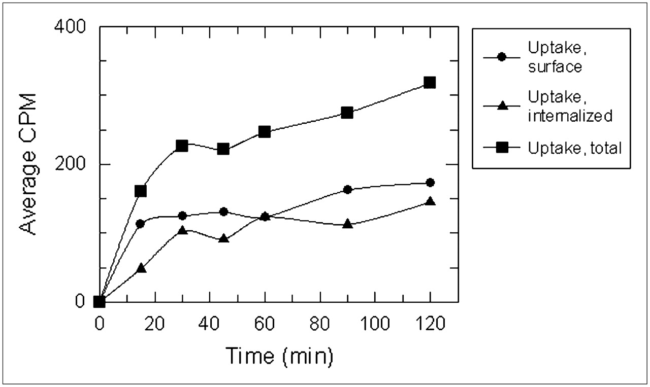
Incubation of PC-3 cells with 64Cu-CB-TE2A-8-AOC-BBN(7–14)NH2 and 64Cu-DOTA-8-AOC-BBN(7–14)NH2 is shown in Figures 3 and 4, respectively. For the 64Cu-CB-TE2A radioconjugate, the amount of internalized activity exceeded the amount of surface-associated activity. Rapid uptake of the radioconjugate was observed for the first 15 min of incubation. After 15 min of incubation, the 64Cu-CB-TE2A radioconjugate was internalized at a steadily increasing rate for the remaining time points of the internalization assay. For the 64Cu-DOTA raduiconjugate, the amount of surface-bound activity equaled or exceeded the amount of internalized activity at all time points. Similar to the 64Cu-CB-TE2A radioconjugate, internalization of the 64Cu-DOTA radioconjugate showed a steady rate of accumulation of activity in the cell over the time of the study.

Internalization of 64Cu-CB-TE2A-8-AOC-BBN(7–14)NH2 using PC-3 cells (n = 6).

Internalization of 64Cu-DOTA-8-AOC-BBN(7–14)NH2 using PC-3 cells (n = 2).
The results obtained from pharmacokinetic studies of 64Cu-DOTA-8-AOC-BBN(7–14)NH2 and 64Cu-CB-TE2A-8-AOC-BBN(7–14)NH2 in SCID mice bearing PC-3 xenografts are summarized in Tables 2 and 3, respectively. A comparison of the tumor–to–nontarget tissue ratios of the 2 radioconjugates in selected nontarget tissues is depicted in Figure 5. The pharmacokinetic values for the 64Cu-DOTA-8-AOC-BBN(7–14)NH2 radioconjugate agree well with analogous literature time points reported by Rogers et al. (9). Overall, the 64Cu-CB-TE2A-8-AOC-BBN(7–14)NH2 radioconjugate showed significantly better clearance from the mouse model than the 64Cu-DOTA-8-AOC-BBN(7–14)NH2 radioconjugate. At 24 h after injection, 98.60 ± 0.28 %ID had been excreted from the mice using the 64Cu-CB-TE2A-8-AOC-BBN(7–14)NH2 radioconjugate compared with only 67.84 ± 5.43 %ID using the 64Cu-DOTA-8-AOC-BBN(7–14)NH2 radioconjugate. Overall clearance of both 64Cu bombesin analogs proceeded mostly through the renal system, with approximately 20 %ID being excreted through the hepatobiliary system.
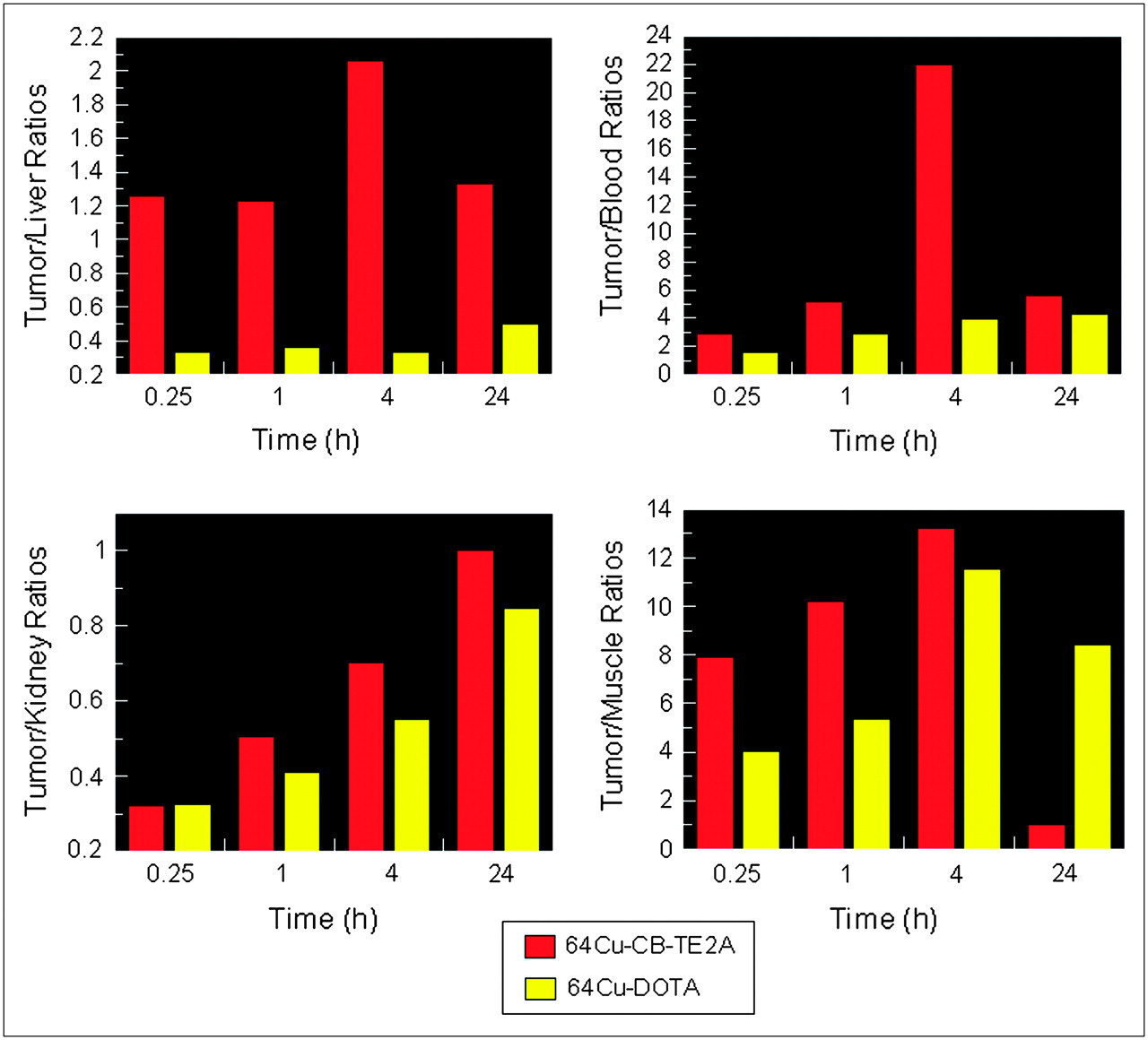
Comparison of 64Cu-CB-TE2A-8-AOC-BBN(7–14)NH2 (red) and 64Cu-DOTA-8-AOC-BBN(7–14)NH2 (yellow) using a radiolocalization index ([RI] %ID/g of tumor/%ID/g of nontarget tissue).
In Vivo Pharmacokinetic Studies of 64Cu-DOTA-8-AOC-BBN(7–14)NH2 Using SCID Mice Bearing PC-3 Xenografts
In Vivo Pharmacokinetic Studies of 64Cu-CB-TE2A-8-AOC-BBN(7–14)NH2 Using SCID Mice Bearing PC-3 Xenografts
The 64Cu-CB-TE2A-8-AOC-BBN(7–14)NH2 radioconjugate clears more efficiently from the bloodstream than the 64Cu-DOTA radioconjugate. At 24 h after injection, the retention in the blood was 0.07 ± 0.05 and 1.40 ± 0.84 %ID for the 64Cu-CB-TE2A and 64Cu-DOTA radioconjugates, respectively. For the 64Cu-DOTA-8-AOC-BBN(7–14)NH2 radioconjugate, retention of 64Cu in the liver was found to be 7.80 ± 1.51 %ID/g at 24 h after injection. The 64Cu-CB-TE2A radioconjugate shows superior clearance from the liver, with 0.21 ± 0.06 %ID/g remaining at 24 h after injection. Initial tumor uptake of the two 64Cu bombesin analogs shows statistically the same activity at 15 min after injection. However, the 64Cu-DOTA-8-AOC-BBN(7–14)NH2 radioconjugate showed significantly higher retention compared with the 64Cu-CB-TE2A-8-AOC-BBN(7–14)NH2 radioconjugate. At 24 h after injection, the tumor activity (3.88 ± 1.40 %ID/g) using the 64Cu-DOTA radioconjugate is statistically the same as the tumor uptake at 15-min, 1-h, and 4-h time points after injection. Despite the tumor retention of the 64Cu-DOTA radioconjugate, the tumor-to-liver, tumor-to-blood, and tumor-to-kidney ratios were found to be higher for the 64Cu-CB-TE2A radioconjugate at all time points due to the relative clearance of the radioconjugate from nontarget tissue. The tumor-to-muscle ratios were found to be greater for the 64Cu-CB-TE2A radioconjugate with the exception of the 24-h time point after injection. The significant retention of the 64Cu-DOTA radioconjugate and clearance of the 64Cu-CB-TE2A radioconjugate from the tumors results in a higher tumor-to-muscle ratio for the 64Cu-DOTA radioconjugate at the 24-h time point after injection. Blocking studies were performed with 100 μg of BBN coadministered with the 64Cu-CB-TE2A radioconjugate. As demonstrated with other bombesin analogs, the blocking studies for the 64Cu-CB-TE2A radioconjugate demonstrated significant BB2 receptor blocking (95%) in the BB2 receptor-rich mouse pancreas. The tumor uptake was also blocked and is consistent with blocking studies of other bombesin analogs.
microPET, fused microPET/CT, and axial images of PC-3 tumor-bearing SCID mice using 64Cu-DOTA-8-AOC-BBN(7–14)NH2 and 64Cu-CB-TE2A-8-AOC-BBN(7–14)NH2 radioconjugates are shown in Figure 6. The images of both conjugates at 20 h after injection correlate well with respective pharmacokinetic studies at 24 h after injection. For both radioconjugates, the BB2 receptor-mediated uptake in the tumors can be easily visualized in the axial images at the 20-h time point after injection. For the 64Cu-DOTA radioconjugate, substantial abdominal uptake and retention—in particular, the liver, kidneys, and gastrointestinal tract—are clearly evident. Images of the 64Cu-CB-TE2A-8-AOC-BBN(7–14)NH2 radioconjugate show substantially less abdominal activity relative to the 64Cu-DOTA radioconjugate. The most striking difference in the 2 whole-body images is the significantly reduced accumulation in the liver using the 64Cu-CB-TE2A radioconjugate.

mircoPET, fused microPET/CT, and axial images of 64Cu-DOTA-8-AOC-BBN(7–14)NH2 (A) and 64Cu-CB-TE2A-8-AOC-BBN(7–14)NH2 (B) in PC-3 tumor-bearing SCID mice at 20 h after administration. Lateral and axial projection images have been normalized to the highest pixel intensity for each respective image.
DISCUSSION
Copper radioisotopes have been shown to transchelate from chelation systems, including the DOTA and TETA tetraamine macrocycles, and bind to proteins, including serum albumin and superoxide dismutase. The reduction of Cu(II) to Cu(I) has been suggested to be the source of the kinetic instability (33,37). If this rationale is true, the chelator, on copper reduction, becomes unsuitable for the labile copper(I) species and transchelation occurs. A recent report by Weisman and coworkers investigates the electrochemical in vitro properties of tetraazamacrocycles and cross-bridge tetraazabicycles (33). Their work suggests that the CB-TE2A chelator can stabilize both copper(I) and copper(II) species—assuming copper reduction is the primary reason for transchelation—thereby significantly reducing transchelation due to copper reduction.
Under the radiolabeling conditions used in this work, the CB-TE2A conjugate was found to be more difficult to radiolabel than the DOTA conjugate. The radiochemical yield of the 64Cu-CB-TE2A-8-AOC-BBN(7–14)NH2 radioconjugate was approximately half that of the 64Cu-DOTA-8-AOC-BBN(7–14)NH2 radioconjugate. The difficulty in radiolabeling the CB-TE2A chelator has been previously noted (31,32). More vigorous labeling conditions may be required to achieve more optimal radiolabeling yields.
In vitro binding studies revealed a slightly higher binding affinity to the BB2 receptor for the Cu-CB-TE2A radioconjugate (IC50 = 0.48 ± 0.01 nM) than the Cu-DOTA radioconjugate (IC50 = 1.44 ± 0.08 nM). Internalization studies of the radioconjugates demonstrated a higher internalized–to–surface-bound ratio for the 64Cu-CB-TE2A radioconjugate. However, both conjugates demonstrated a steady increase in the concentration of activity in the cell over time. This trend is in agreement with internalization studies of the 64Cu-DOTA conjugate performed by Rogers and colleagues as well as the analogous 111In-DOTA-8-AOC-BBN(7–14)NH2 radioconjugate reported by our group (5,9).
The tumor uptake for the 64Cu-CB-TE2A and 64Cu-DOTA radioconjugates was 6.95 ± 2.27 and 4.95 ± 0.91 %ID/g, respectively, at 15 min after injection. Pharmacokinetic studies have been reported for several radiometals—111In, 177Lu, 90Y, and 149Pm—using the DOTA-8-AOC-BBN(7–14)NH2 peptide (5,7,38,39). In these studies, the radiometal was found to have high initial uptake in the tumor (e.g., 111In: 7.59 ± 2.11 %ID/g at 15 min after injection) and significant clearance of the radioactivity from the tumor over time (e.g., 111In: 1.56 ± 0.45 %ID/g at 24 h after injection). The 64Cu-CB-TE2A-8-AOC-BBN(7–14)NH2 radioconjugate demonstrated significant clearance from the tumor, with 0.28 ± 0.21 %ID/g or 4% of the 15-min value remaining at 24 h after injection. The retention profile of the 64Cu-CB-TE2A radioconjugate is consistent with previously reported radiometal complexes of DOTA-8-AOC-BBN(7–14)NH2. In contrast, 64Cu-DOTA-8-AOC-BBN(7–14)NH2 demonstrated high retention and essentially no clearance from tumor tissue, with 3.48 ± 0.90, 3.00 ± 1.10, and 3.88 ± 1.40 %ID/g at 1, 4, and 24 h after injection, respectively. The tumor tissue clearance profile of the 64Cu-DOTA radioconjugate suggests the retention is due to a nonreversible trapping mechanism, such as transchelation of the radiometal from the chelator to intracellular proteins (24,27,31).
In vivo pharmacokinetic studies show stark contrasts in the biodistribution and retention of 64Cu-DOTA-8-AOC-BBN(7–14)NH2 and 64Cu-CB-TE2A-8-AOC-BBN(7–14)NH2. The blood retention of 64Cu-DOTA-8-AOC-BBN(7–14)NH2 was 1.22 ± 0.62 %ID/g at 1 h after injection, which on average declined slightly to 0.90 ± 0.48 %ID/g at 24 h after injection. The significant blood retention is likely due to transchelation of the 64Cu radionuclide from the kinetically unstable DOTA chelation system to serum proteins such as serum albumin (25–27). The high blood activity associated with the 64Cu-DOTA-8-AOC-BBN(7–14)NH2 conjugate results in abnormally high background activities in all tissues. In contrast, the 64Cu-CB-TE2A-8-AOC-BBN(7–14)NH2 radioconjugate shows good blood clearance with 0.51 ± 0.14 and 0.05 ± 0.03 %ID/g at 1 and 24 h after injection, respectively. The significantly reduced blood retention of the 64Cu-CB-TE2A radioconjugate correlates with the enhanced kinetic stability offered by the CB-TE2A chelation system. The blood retention of the 64Cu-CB-TE2A bombesin radioconjugate is, to our knowledge, the lowest reported blood-associated activity at the 24-h time point after injection for a 64Cu bombesin analog to date.
In vivo unstable copper compounds are known to exhibit high retention in the liver. For the 64Cu-DOTA radioconjugate, 7.80 ± 1.51 %ID/g remained in the liver at 24 h after injection. The 64Cu-CB-TE2A radioconjugate demonstrated significantly lower retention at 24 h after injection, with a liver-associated activity of 0.21 ± 0.06 %ID/g. By RP-HPLC, the 64Cu-CB-TE2A radioconjugate was found to be more lipophilic than the 64Cu-DOTA radioconjugate. At 15 min after injection, the hepatobiliary clearance of the 64Cu-CB-TE2A-8-AOC-BBN(7–14)NH2 radioconjugate (18.12 ± 1.25 %ID small intestines) was found to be significantly greater than the 64Cu-DOTA-8-AOC-BBN(7–14)NH2 radioconjugate (9.58 ± 3.95 %ID small intestines). However, for 64Cu-DOTA-8-AOC-BBN(7–14)NH2, the considerable liver retention of 64Cu, likely due to transchelation, significantly reduces the amount of activity passed from the liver to the small intestines at the 15-min time point after injection.
Total excretion (urine and fecal) values for the 2 radioconjugates, 64Cu-CB-TE2A-8-AOC-BBN(7–14)NH2 and 64Cu-DOTA-8-AOC-BBN(7–14)NH2, are in sharp contrast to one another. At 24 h after injection, 98.60 ± 0.28 %ID of the 64Cu activity had been excreted (urine and feces) from the mouse using the 64Cu-CB-TE2A-8-AOC-BBN(7–14)NH2 radioconjugate. The excretion values are consistent with those of the analogous radioconjugate, 111In-DOTA-8-AOC-BBN(7–14)NH2 (87.2 ± 4.3 %ID excreted in urine at 24 h after injection). In contrast, only 67.84 ± 5.43 %ID of the 64Cu activity was excreted from the mouse by 24 h after injection using 64Cu-DOTA-8-AOC-BBN(7–14)NH2. As the only difference between the 2 copper radioconjugates is the chelation system, the excretion values further support the hypothesis that the CB-TE2A conjugate substantially stabilizes the 64Cu radionuclide in vivo, thereby significantly reducing transchelation.
microPET/CT images of both radioconjugates at 20 h after injection (Fig. 6), using PC-3 tumor-bearing SCID mice, agree well with the data obtained from pharmacokinetic studies. Because of biologic clearance properties of the radioconjugates, there was significantly less activity associated with the image of the 64Cu-CB-TE2A radioconjugate (∼370 kBq [∼10 μCi]) compared with that of the 64Cu-DOTA radioconjugate (∼7.4 MBq [∼200 μCi]). The PC-3 tumor xenografts in both mice are easily visualized in the normalized images and show similar tumor-to-background ratios. For the 64Cu-DOTA radioconjugate, the image revealed significant activity in the liver, kidneys, and gastrointestinal tract. The image using the 64Cu-CB-TE2A radioconjugate demonstrated considerably reduced activity located in the kidneys and gastrointestinal tract. Because of the markedly reduced activity in the liver using the 64Cu-CB-TE2A radioconjugate, the liver could not be easily visualized.
CONCLUSION
The pharmacokinetic and microPET/CT studies demonstrate significantly improved nontarget tissue clearance properties associated with 64Cu-CB-TE2A-8-AOC-BBN(7–14)NH2. The significant improvement in clearance is attributed to the improved in vivo stability of the 64Cu-CB-TE2A chelate complex as compared with the 64Cu-DOTA chelate complex. While the stabilization of the 64Cu radionuclide using the CB-TE2A chelation system resulted in lower retention of the radiotracer in the PC-3 tumors, the substantial decrease in nontarget tissue retention significantly increases the potential clinical applicability of 64Cu bombesin analogs for diagnostic imaging of prostate cancer. Future studies performed in our laboratory will focus on developing more hydrophilic 64Cu CB-TE2A bombesin analogs that exhibit higher renal clearance and increased tumor retention
Acknowledgments
The authors acknowledge Farah Naz, Nicholas R. Bell, Serena N. Carter, and Andrew Walters for useful discussions and their assistance with data collection and The Proteomics Center at the University of Missouri–Columbia for mass spectrometry services. This work was supported with the resources and the use of facilities at the Harry S. Truman Memorial VA Hospital. This work was also supported by grant NCI DHHS-R01-CA72942 from the National Cancer Institute, by grant ACS RSG-99-331-04-CDD from the American Cancer Society, and by grant NIH DHHS-1P50-CA13013 from the National Institutes of Health. The University of Missouri holds U.S. patents 6 200 546, 6 921 526, 7 060 247, and 7 147 838 on technology described in this article, of which Timothy J. Hoffman, Gary L. Sieckman, and Wynn A. Volkert are coinventors.
Footnotes
-
COPYRIGHT © 2007 by the Society of Nuclear Medicine, Inc.
References
- Received for publication January 9, 2007.
- Accepted for publication April 23, 2007.


Jump to section
Related Articles
Cited By...
- Improving Contrast and Detectability: Imaging with [55Co]Co-DOTATATE in Comparison with [64Cu]Cu-DOTATATE and [68Ga]Ga-DOTATATE
- Novel "Add-On" Molecule Based on Evans Blue Confers Superior Pharmacokinetics and Transforms Drugs to Theranostic Agents
- Clinical Translation of a Dual Integrin {alpha}v{beta}3- and Gastrin-Releasing Peptide Receptor-Targeting PET Radiotracer, 68Ga-BBN-RGD
- 68Ga-NOTA-Aca-BBN(7-14) PET/CT in Healthy Volunteers and Glioma Patients
- In Vivo Molecular Imaging of Thrombosis and Thrombolysis Using a Fibrin-Binding Positron Emission Tomographic Probe
- Synthesis and In Vitro and In Vivo Evaluation of Hypoxia-Enhanced 111In-Bombesin Conjugates for Prostate Cancer Imaging
- Targeted Radiotherapy of Prostate Cancer with a Gastrin-Releasing Peptide Receptor Antagonist Is Effective as Monotherapy and in Combination with Rapamycin
- Positron-emission Tomography (PET) Imaging Agents for Diagnosis of Human Prostate Cancer: Agonist vs. Antagonist Ligands
- Exploring molecular genetics of bladder cancer: lessons learned from mouse models
- Radiopeptide Imaging and Therapy in Europe
- Bombesin Antagonist-Based Radioligands for Translational Nuclear Imaging of Gastrin-Releasing Peptide Receptor-Positive Tumors
- In Vitro and In Vivo Evaluation of 64Cu-Labeled SarAr-Bombesin Analogs in Gastrin-Releasing Peptide Receptor-Expressing Prostate Cancer
- Small-Animal PET of Tumors with 64Cu-Labeled RGD-Bombesin Heterodimer
- Humanized Radioiodinated Minibody For Imaging of Prostate Stem Cell Antigen-Expressing Tumors
- International Union of Pharmacology. LXVIII. Mammalian Bombesin Receptors: Nomenclature, Distribution, Pharmacology, Signaling, and Functions in Normal and Disease States