


Abstract
Breast cancer (BC) consists of multiple subtypes defined by various molecular characteristics, for instance, estrogen receptor (ER) expression. Methods for visualizing BC include mammography, MR imaging, ultrasound, and nuclear medicine–based methods such as 99mTc-sestamibi and 18F-FDG PET, unfortunately all lacking specificity. Peptide receptor scintigraphy and peptide receptor radionuclide therapy are successfully applied for imaging and therapy of somatostatin receptor–expressing neuroendocrine tumors using somatostatin receptor radioligands. On the basis of a similar rationale, radioligands targeting the gastrin-releasing peptide receptor (GRP-R) might offer a specific method for imaging and therapy of BC. The aim of this study was to explore the application of GRP-R radioligands for imaging and therapy of BC by introducing valid preclinical in vitro and in vivo models. Methods: GRP-R expression of 50 clinical BC specimens and the correlation with ER expression was studied by in vitro autoradiography with the GRP-R agonist 111In-AMBA. GRP-R expression was also analyzed in 9 BC cell lines applying 111In-AMBA internalization assays and quantitative reverse transcriptase polymerase chain reaction. In vitro cytotoxicity of 177Lu-AMBA was determined on the GRP-R–expressing BC cell line T47D. SPECT/CT imaging and biodistribution were studied in mice with subcutaneous and orthotopic ER-positive T47D and MCF7 xenografts after injection of the GRP-R antagonist 111In-JMV4168. Results: Most of the human BC specimens (96%) and BC cell lines (6/9) were found to express GRP-R. GRP-R tumor expression was positively (P = 0.026, χ2(4) = 12,911) correlated with ER expression in the human BC specimens. Treatment of T47D cells with 10−7 M/50 MBq of 177Lu-AMBA resulted in 80% reduction of cells in vitro. Furthermore, subcutaneous and orthotopic tumors from both BC cell lines were successfully visualized in vivo by SPECT/CT using 111In-JMV4168; T47D tumors exhibited a higher uptake than MCF7 xenografts. Conclusion: Targeting GRP-R–expressing BC tumors using GRP-R radioligands is promising for nuclear imaging and therapy, especially in ER-positive BC patients.
Breast cancer (BC) is a highly heterogeneous disease characterized by multiple molecular features, such as expression of the estrogen receptor (ER), progesterone receptor, and human epidermal growth factor receptor 2 (1). On the basis of molecular characteristics, BC can be divided in 4 subgroups: luminal A and B, human epidermal growth factor receptor 2–like, and triple-negative tumors (2). The treatment and prognosis of BC are highly dependent on this classification. Mammography is the standard imaging technique used during nationwide screening of BC. Additional MR imaging or ultrasound can be performed (3). However, all 3 methods have drawbacks. Mammography lacks sensitivity and specificity, leading to false-positive and false-negative results. Especially in women with dense breast tissue, mammograms are hard to interpret (4). MR imaging and ultrasound also lack specificity. In addition, MR imaging is labor-intensive and expensive, whereas ultrasound is operator-dependent (5).
In nuclear medicine, methods to detect BC lesions and monitor response to treatment include 201TI scintigraphy, 99mTc-sestamibi or 99mTc-tetrofosmin scintigraphy, and 18F-FDG PET (6). Unfortunately, these methods also lack specificity.
Peptide receptor scintigraphy applies specific in vivo targeting of tumor lesions overexpressing a receptor of interest. This method was first described by Krenning et al. (7), using a radioactive somatostatin receptor (SSTR) ligand to image neuroendocrine tumors (NETs) overexpressing the SSTR subtype 2. To date, imaging with SSTR radioligands is routinely applied to characterize and evaluate (tumor localization, staging, monitoring) NETs (8). Furthermore, SSTR radioligands labeled with therapeutic radionuclides are successfully used for treatment of NET patients, using so-called peptide receptor radionuclide therapy (PRRT) (9).
Following the same principle, the gastrin-releasing peptide receptor (GRP-R) can be targeted. GRP-Rs are expressed on most BC cases; autoradiography studies by Reubi et al. (10) showed that 65% of the breast tumors analyzed expressed the GRP-R. Moreover, also lymph node metastases from primary tumors expressing GRP-R were positive (10–12). GRP-R binds a family of peptides, including the gastrin-releasing peptide; its C-terminal fragment neuromedin C, both endogenous in humans; and the amphibian tetradecapeptide bombesin.
The application of GRP-R radioligands, analogs of the above native peptides, might offer a sensitive and specific method for imaging of GRP-R overexpressing BC. In addition, PRRT with radiolabeled GRP-R ligands might be feasible. Recently, multiple GRP-R radioligands, receptor agonists and antagonists, have been described that can be labeled with positron and γ emitters, for example, 68Ga and 111In, for imaging and with β and α emitters, for example, 177Lu and 213Bi, for therapeutic purposes, as yet primarily aiming at application in prostate cancer (13). Preclinical studies evaluating the potential of targeting the GRP-R in BC using GRP-R radioligands are limited. The aim of this study was to further characterize the GRP-R as a potential target for the visualization of BC lesions and for PRRT. For this purpose, we analyzed GRP-R expression in human clinical BC specimens and correlated this with ER expression. In addition, we used selected GRP-R–/ER-expressing human-derived BC cells to perform in vitro cytotoxicity assays and to develop suitable xenograft-bearing animal models for preclinical imaging studies.
MATERIALS AND METHODS
Radiolabeled GRP-R Ligands
For in vitro studies, the GRP-R agonist AMBA (BioSynthema) was used (14). Because radiolabeled AMBA exhibits high uptake in the gastrointestinal tract that potentially interferes with orthotopic tumor visualization, the GRP-R antagonist JMV4168 (University of Montpellier) was used for in vivo studies (14,15). The GRP-R ligands were radiolabeled with 111In (Covidien) or 177Lu (IDB), using quenchers (10 mM methionine, 3.5 mM ascorbic acid, and 3.5 mM gentisic acid) to prevent radiolysis (16). Radiolabeling was performed for 20 min at 80°C as previously described (17), with a specific activity of 100 MBq/nmol (for in vitro studies, both 111In and 177Lu) or 150 MBq/nmol (in vivo studies). Radiometal incorporation and radiochemical purity, measured by instant thin-layer chromatography on silica gel and high-pressure liquid chromatography as previously described (16), were greater than 95% and greater than 90%, respectively.
In Vitro Autoradiography on Human BC Specimens
In vitro autoradiography was performed on 50 human BC specimens with known ER protein status. Specimens were selected from the Erasmus MC fresh frozen tissue bank. The study (MEC02·953) was approved by the Erasmus MC Medical Ethical Committee and adhered to the Code of Conduct of the Federation of Medical Scientific Societies in The Netherlands.
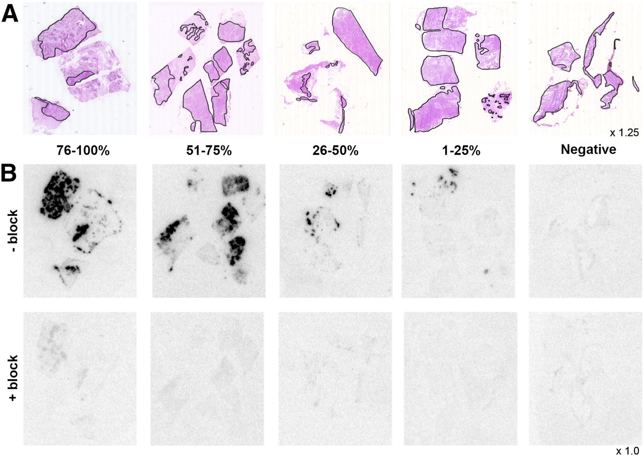
Frozen sections (10 μm) of the BC specimens were incubated with 10−9 M/0.1 MBq of 111In-AMBA, without or with 10−6 M unlabeled Tyr4-bombesin (Sigma-Aldrich), to determine nonspecific binding, for 1 h and exposed to super resolution phosphor screens (PerkinElmer) for at least 24 h and read using the Cyclone (PerkinElmer). Adjacent tissue sections were stained with hematoxylin and eosin to determine tumor content. Autoradiography results were scored visually by 3 independent observers. Among the positive tumors, a division was made between tumors that were 1%–25%, 26%–50%, 51%–75%, and 76%–100% positive.
Cell Culture, Internalization Assay, and Quantitative Reverse Transcriptase Polymerase Chain Reaction (RT-qPCR)
Nine human-derived BC cell lines, obtained from the Department of Medical Oncology, Erasmus MC, with different molecular properties (Supplemental Table 1; supplemental materials are available online at http://jnm.snmjournals.org) were screened for GRP-R expression using internalization assays and RT-qPCR. Cell lines, authenticated by short tandem repeat profiling using the Powerplex STR kit (Promega), were cultured as described by Riaz et al. (18).
In the internalization assay, cells were incubated for 1 h at 37°C with 10−9 M/0.1 MBq of 111In-AMBA (without or with 10−6 M unlabeled Tyr4-bombesin). In addition, assays were performed using 10−7 M/10 MBq, 10−8 M/1 MBq, and 10−9 M/0.1 MBq of 111In-AMBA with incubation times of 1, 2, and 4 h to select the optimal conditions for the in vitro cytotoxicity studies. The internalization assay protocol is described in the supplemental materials.
To measure GRP-R messenger RNA levels of the BC cell lines, RNA was isolated using RNA-Bee (Campro Scientific) according to the manufacturer’s instructions. Subsequently, complementary DNA synthesis and RT-qPCR were performed and normalized using the δ Cq method on the average of 2 reference genes (HMBS and HPRT1) as previously described (19). The quantification of target genes was performed using the Taqman probe–based gene expression assay, GRP-R, Hs01055872_m (Applied BioSystems/Life Technologies), according to the manufacturer’s instructions.
In Vitro Cytotoxicity Studies
Cells (12.5 × 10⁶/5 mL in a T25 culture flask, seeded 1 d before the experiment) were treated with 10−7 M/50 MBq, 5 × 10⁻⁸ M/25 MBq, or 10−8 M/5 MBq of 177Lu-AMBA in 5 mL of internalization medium for 4 h. Untreated cells, 177Lu-diethylenetriaminepentaacetic acid (DTPA)–treated, and unlabeled AMBA-treated cells served as controls. After incubation, cells were washed twice with phosphate-buffered saline (GIBCO/Life Technologies) and detached using 0.1 mM ethylenediaminetetraacetic acid. Cells were resuspended in medium, counted, and seeded in 3 wells of 12-well plates (12,500 cells/well/1 mL). Seven days after treatment, cells were fixed using 1 mL of 10% trichloroacetic acid (Sigma-Aldrich), and a sulforhodamine B colometric assay was performed to determine cell density. Results are expressed as percentages relative to untreated controls. The sulforhodamine B colometric assay protocol is described in the supplemental materials.
In Vivo Imaging, Biodistribution, and In Vitro Autoradiography of BC Xenografts
All animal studies were in agreement with the Animal Welfare Committee requirements of Eramus MC and conducted in accordance with accepted guidelines. BALB/c nu/nu female mice (6–8 wk) (Janvier), supplemented with β-estradiol (4 mg/L; Sigma-Aldrich) in drinking water, were subcutaneously (between the shoulders) and orthotopically (left fourth mammary fat pad) inoculated with 8 × 10⁶ T47D cells or 7 × 10⁶ MCF7 cells (n = 6 for each cell line).
A 40-min SPECT/CT scan was acquired at 2 time points after inoculation (time point 1 [t1], 40 ± 3 d, and time point 2 [t2], 103 ± 3 d), using the NanoSPECT/CT scanner (Bioscan) 4 h after intravenous injection of approximately 35 MBq/200 pmol of 111In-JMV4168, coinjected with 300 μg of phosphoramidon (Peptides International Inc.) to inhibit in vivo enzymatic degradation of the peptide. During the scan acquisition, the animals were anesthetized with isoflurane/O2 and body temperature was maintained. Bladder uptake was masked after reconstruction of the images. After the second scan, animals were euthanized, and tumors and organs were collected, weighed, and counted in a γ counter. Data obtained were expressed as percentage injected dose per gram of tissue (%ID/g). In addition, in vitro autoradiography was performed on the excised tumors, using 111In-JMV4168 without or with 10−6 M unlabeled Tyr4-bombesin, to demonstrate receptor specificity of the tracer. Scanning, reconstruction, and counting details are described in the supplemental materials. Supplemental Figure 1 depicts the time line of the in vivo experiments.
Statistics
Statistical analyses are described in the supplemental materials.
RESULTS
GRP-R Expression in Clinical BC Specimens
The autoradiography results of 50 human BC tissue specimens with known ER protein status were scored for GRP-R expression. Hematoxylin and eosin staining of adjacent tissue sections was used to discriminate between malignant and healthy tissue (Fig. 1A). Most (48/50 [96%]) of the BC specimens analyzed were positive for GRP-R. The majority (56%) of the samples showed greater than 75% positivity, indicating a homogeneous GRP-R expression. Of the remaining samples, 29% was 1%–25% positive, 8% was 26%–50% positive, and 6% was 51%–75% positive (Fig. 1B). In addition, a significant, positive (P = 0.026, χ2(4) = 12,911) correlation was found between ER status and extent of GRP-R expression.

(A) Hematoxylin and eosin staining of sections indicating tumor-containing regions, corresponding to autoradiography results in B. (B) Representative examples of 111In-AMBA binding to human BC specimens, without (-block) and with (+block) 10−6 M Tyr4-bombesin. Binding was GRP-R–specific because no binding was observed when the GRP-R was blocked by Tyr4-bombesin. Among GRP-R–positive tumors, a subdivision was made between tumors that were 1%–25%, 26%–50%, 51%–75%, and 76%–100% GRP-R–positive.
GRP-R Expression in Human-Derived BC Cell Lines and In Vitro Cytotoxicity Studies
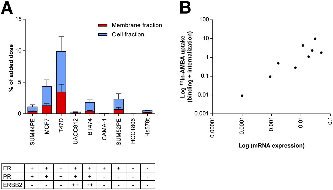
Most of the BC cell lines examined (6/9) selectively bound and internalized 111In-AMBA, although to a variable extent (Fig. 2A). This process was GRP-R–specific, because binding and internalization were significantly decreased when an excess of unlabeled GRP-R ligand was added. The extent of 111In-AMBA binding and internalization seemed higher in ER-positive BC cell lines than in the ER-negative BC cell lines. However, the observed difference was not statistically significant (P = 0.757), probably because of the lower power of the cell line study than the clinical BC specimen study. Also, no clear correlation with progesterone receptor (P = 0.209) and human epidermal growth factor receptor 2 status (P = 0.192) of the BC cell lines was found.

(A) Selective binding and internalization after 1 h of incubation with 10−9 M/0.1 MBq of 111In-AMBA, a GRP-R agonist. Nine human-derived BC cell lines with different molecular properties (18) were screened. Both membrane- and internalized/cell fraction are displayed. Results shown are average of 3 independent experiments, each performed in triplicate (mean ± SD). (B) Significant correlation (P = 0.0108, Rs = 0.8167) between GRP-R messenger RNA (mRNA) expression and level of 111In-AMBA uptake (membrane plus cell fraction).
To confirm that internalization levels correlated with GRP-R expression and were not attributed to, for example, receptor recycling efficiency, GRP-R expression was independently quantified by RT-qPCR (Supplemental Table 1). A significant positive correlation (P = 0.0108, Rs = 0.8167) was found between messenger RNA levels of GRP-R and 111In-AMBA uptake (Fig. 2B).
The ER-positive cell lines T47D and MCF7 showed the highest uptake of 111In-AMBA. T47D was therefore selected for in vitro cytotoxicity experiments, and both T47D and MCF7 were used for inoculation in mice to create relevant in vivo models.
To determine the optimal incubation time and radioligand concentration for in vitro cytotoxicity studies, taking into account potential GRP-R saturation, T47D cells were incubated with 3 different concentrations of 111In-AMBA (10−7 M/10 MBq, 10−8 M/1 MBq, and 10−9 M/0.1 MBq) for 3 different incubation times (1, 2, and 4 h). The highest absolute amount of 111In-AMBA (counts per minute) was observed after 4 h of incubation with 10−7 M 111In-AMBA, both in the membrane-bound and in the internalized fraction (Fig. 3A).
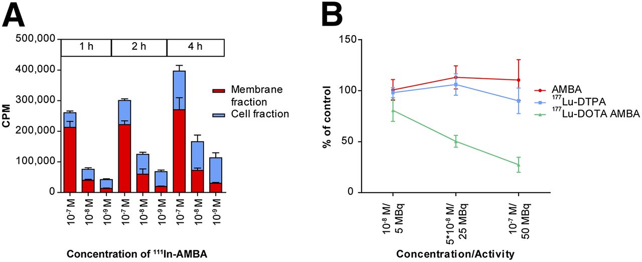
(A) Uptake of 111In-AMBA by GRP-R–expressing human-derived BC cell line T47D, at 1, 2, and 4 h after incubation with 10−7 M/10 MBq, 10−8 M/1 MBq, or 10−9 M/0.1 MBq of 111In-AMBA. Results shown are average of 2 independent experiments, each performed in triplicate (mean ± SD). (B) Treatment of T47D cells with 3 different concentrations of AMBA, 177Lu-DTPA, and 177Lu-AMBA for 4 h. Significant reduction of 80% in cell viability was reached when cells were treated with 10−7 M/50 MBq of 177Lu-AMBA, whereas no significant effects were observed when cells were similarly treated with AMBA or 177Lu-DTPA. Results shown are average of 3 independent experiments, each performed in triplicate (mean ± SD).
When T47D cells were treated for 4 h with 177Lu-AMBA, a significant reduction in cell number of 20%, 50%, and 80% was observed after incubation with 10−8 M/5 MBq, 5 × 10⁻⁸ M/25 MBq, or 10−7 M/50 MBq of 177Lu-AMBA, respectively (Fig. 3B). Treatment with the same amount of 177Lu-DTPA for 4 h, which is not actively internalized, did not inhibit cell proliferation, similarly to incubation with unlabeled AMBA. Therefore, the therapeutic effect of 177Lu-AMBA can be associated to the combination of specific GRP-R–mediated uptake and retention of 177Lu within the cells over time.
In Vivo Visualization of GRP-R Expression in BC Xenografts
We successfully obtained T47D and MCF7 xenografts in immune-deficient female mice orally supplemented with estrogen.
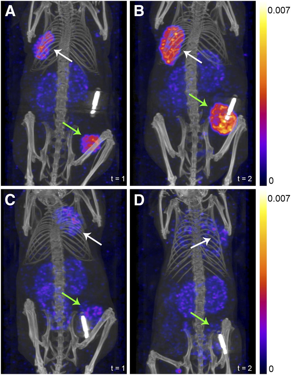
At 2 time points after tumor cell inoculation, T47D and MCF7 xenografts were visualized using in vivo SPECT/CT imaging after an injection of approximately 35 MBq/200 pmol of 111In-JMV4168 + 300 μg of phosphoramidon. T47D xenografts had a significantly (P < 0.0001) higher uptake than MCF7 xenografts (748–1,706 vs. 169–658 kBq/g for subcutaneous tumors and 1,151–2,127 vs. 189–672 kBq/g for orthotopic tumors). Therefore, T47D xenografts were visualized with higher contrast than MCF7 xenografts (Fig. 4). No significant difference in uptake between orthotopic and subcutaneous tumors of both T47D (P = 0.92) and MCF7 xenografts (P = 0.95) was determined. Biodistribution results obtained at t2 confirmed higher radiotracer uptake and a better tumor-to-kidney ratio (2.1 vs. 0.4) in T47D than MCF7 xenografts (Fig. 5). In vitro autoradiography of the excised tumors demonstrated specific binding of 111In-JMV4168 (Supplemental Fig. 2). However, the proliferation of MCF7 xenografts was faster than that of T47D xenografts; orthotopic tumors ranged from 178 to 390 mm3 for T47D xenografts and from 755 to 1,830 mm3 for MCF7 xenografts at t1. At that time point, subcutaneous T47D xenografts ranged from 11 to 630 mm3, compared with 532–2,095 mm3 for MCF7 xenografts. As yet, we cannot exclude the effect of tumor size on radiotracer uptake. Two of 6 T47D subcutaneous tumors were small (12.3 and 4.0 mm3) and were therefore excluded from the biodistribution study.

SPECT/CT scans indicating orthotopic (green arrows) and subcutaneous (white arrows) tumors of T47D and MCF7 xenograft–bearing mice at 4 h after injection of approximately 35 MBq (200 pmol) of 111In-JMV4168 + 300 μg of phosphoramidon. T47D xenografts were scanned at t1 = 43 d (A) and t2 = 100 d (B). MCF7 xenografts were scanned at t1 = 37 d (C) and t2 = 106 d (D). Bladder uptake is masked. Mice were provided with a chip for identification purposes. T47D xenografts were visualized with higher contrast than MCF7 xenografts.

Quantification of 111In-JMV4168 uptake in tumors and organs collected after last scan (t2); n = 6 for each mouse model (2 small subcutaneous T47D xenografts were excluded). Significantly (P < 0.0001) higher uptake of 111In-JMV4168 was determined in T47D than in MCF7 xenografts.
DISCUSSION
To gain more insight into the feasibility of diagnostic imaging and therapeutic applications of GRP-R radioligands in BC patients, we aimed to develop reliable preclinical models expressing the GRP-R based on well-characterized human-derived BC cell lines. We first confirmed GRP-R expression on clinical BC specimens and found that most (96%) indeed expressed the GRP-R, which was higher than the 65% of GRP-R–expressing BC specimens reported by Reubi et al. (10). In the latter study, GRP-R, analyzed by in vitro autoradiography, was more frequently and more densely expressed than any other of the investigated targets, including neuropeptide Y receptor subtype 1, SSTR, and vasoactive intestinal peptide receptor. We observed a homogeneous GRP-R expression in more than half (56%) of the tumors, supporting the conclusion that GRP-R is a suitable target for radioligands in BC patients.
Furthermore, a significant correlation was found between GRP-R and ER expression in the clinical BC specimens analyzed. ER-positive tumors were associated with higher GRP-R expression, indicating BC patients with ER-positive tumors as a potential target group for imaging or therapy with GRP-R radioligands. Our findings were in accordance with studies by Halmos et al. (12), showing a significant positive correlation between ER expression and GRP-R binding affinity examined on isolated cell membranes from BC samples. Because ER-positive tumors account for 75% of all breast tumors, GRP-R–based imaging could offer new imaging and therapeutic possibilities for most BC patients (2,20). In addition, because current treatments with antihormonal agents, such as tamoxifen and aromatase inhibitors, are ineffective in about 40% of the ER-positive metastatic BC patients (20,21), GRP-R ligands radiolabeled with particle emitters might offer new therapeutic options for these patients.
Furthermore, we found that most of the examined BC cell lines expressed the GRP-R. T47D was selected for in vitro therapy and, together with MCF7, for in vivo imaging studies.
Treatment of T47D cells with 177Lu-AMBA resulted in a significant decrease in cell viability, proving this cell line to be suitable as a model for future in vivo therapy studies in xenograft-bearing mice. An 80% decrease in cell viability was established using 10−7 M/50 MBq of 177Lu-AMBA. The uptake of 177Lu by the tumor cells appeared to be a prerequisite, because the same amounts of 177Lu-DTPA, which does not bind or internalize, showed no significant effect on cell viability after 4-h exposure. Müller et al. (22) also studied the effect of 177Lu-DTPA on cell viability, next to 177LuCl3. In line with our results, treatment with 177LuCl3, which is also internalized by cells, resulted in a larger decrease in cell viability than treatment with 177Lu-DTPA. In the study by Müller et al. (22), a decrease in cell viability was measured after treatment with 177Lu-DTPA as well, but cells were treated for 4 d with 177Lu-DTPA in contrast to the 4-h treatment in our study, which can explain the discrepancy.
On the basis of our in vitro cytotoxicity studies with 177Lu-AMBA, we expect a similar effect of GRP-R antagonists on cell viability, because binding of the radioligand to the receptor results in cell membrane–bound accumulation of 177Lu, enabling radiation-induced DNA damage and thus cytotoxicity, despite the lack of internalization. A recent study by Wild et al. (23) comparing the application of 177Lu-labeled SSTR agonist and antagonist for PRRT in NET patients showed a higher tumor dose for the SSTR antagonist than for the agonist and therefore successful therapeutic effects, providing evidence that treatment with receptor antagonists is feasible.
According to previous studies, adverse effects have been reported during the clinical evaluation of 177Lu-AMBA in prostate cancer patients (24). Such undesirable effects can be evaded with GRP-R antagonists instead of agonists (25). For this reason and to prevent interference with orthotopic tumor visualization, the GRP-R antagonist JMV4168 was used for in vivo imaging studies.
With the aim of inhibiting the in vivo enzymatic degradation of 111In-JMV4168, which may potentially interfere with GRP-R targeting and tumor uptake, 111In-JMV4168 was coinjected with the neutral endopeptidase inhibitor phosphoramidon (26). Recent studies by Nock et al. (27) have shown significantly prolonged survival of several radioligands in the circulation when phosphoramidon was administered simultaneously. In the current study, 111In-JMV4168 + phosphoramidon was successfully applied to visualize both orthotopic and subcutaneous BC tumors in mice, approximately 100 mm3 in size, indicating that GRP-R expression was sufficiently high to discriminate tumor lesions. Already approximately 1 %ID/g uptake of MCF7 xenografts resulting in approximately 200 kBq of 111In/g enabled visualization of tumor lesions, which was even higher for T47D xenografts, showing about 5 %ID/g uptake. Parry et al. (28) reported a similar tumor uptake in subcutaneous T47D xenografts using different GRP-R radioligands. Thus, these mouse models will now be used for future studies to examine the therapeutic potential of 177Lu-labeled GRP-R radioligands.
Recently several imaging studies using 99mTc-labeled bombesin analogs in BC patients were performed with promising results (29–32), identifying tumor lesions in patients suspected for BC based on mammography. In the most recent study by Shariati et al. (32), a sensitivity of 61% and specificity of 100% were reported, indicating that imaging with bombesin analogs is promising and deserves further investigation. Interestingly, 2 of these imaging studies described the identification of lymph node metastases, demonstrating the presence of the GRP-R on lymph node metastases next to primary tumor tissue (31,32). However, the capacity to visualize distant metastases still needs to be established. In a study by Bergsma et al. (33), 50% of the disseminated BC lesions studied were successfully visualized using the 68Ga-labeled GRP-R antagonist sarabesin-3 without adverse effects, once more showing the successful application of radiolabeled GRP-R antagonists in BC patients. None of these studies reported on correlation of positive scintigraphy with BC subtype and molecular markers.
Up to now, GRP-R–based PRRT has been performed targeting prostate cancer, either in xenograft-bearing animals, with GRP-R agonists or antagonists (14,34), or in patients during a pilot study using 177Lu-AMBA (24). However, GRP-R–based PRRT is not yet being applied in BC animal models or patients. When GRP-R radioligands will be used for therapeutic purposes, the physiologic uptake in the pancreas should be considered regarding safety. Yet, the radiosensitivity of the pancreas is relatively low, and the fast pancreatic washout of radiolabeled GRP-R antagonists is favorable in this respect (35).
The results obtained in the current study together with the positive first experience with 99mTc and 68Ga GRP-R radioligands to localize BC are encouraging for future application of the so-called theranostic approach in BC, using, for example DOTA-coupled receptor ligands radiolabeled with 111In and 68Ga for imaging or with 177Lu, 90Y, or 213Bi for therapeutic purposes.
CONCLUSION
We confirmed GRP-R expression in clinical BC specimens, correlating with ER expression. Hence, ER-positive BC patients are potential candidates for imaging and PRRT using GRP-R radioligands. In addition, we successfully developed BC xenograft mouse models by inoculation of the selected ER- and GRP-R–positive BC cell lines T47D and MCF7 and visualized the generated tumors using 111In-JMV4168, coinjected with phosphoramidon. These mouse models will be used to study the therapeutic effects of 177Lu-labeled GRP-R radioligands, such as 177Lu-JMV4168.
DISCLOSURE
The costs of publication of this article were defrayed in part by the payment of page charges. Therefore, and solely to indicate this fact, this article is hereby marked “advertisement” in accordance with 18 USC section 1734. This study was funded by the Erasmus MC grant “The Application of Radiolabeled Peptides in Breast Cancer.” The study was also supported by the Cancer Genomics Netherlands, funded by the Netherlands Organisation for Scientific Research. No other potential conflict of interest relevant to this article was reported.
Footnotes
Published online Mar. 19, 2015.
- © 2015 by the Society of Nuclear Medicine and Molecular Imaging, Inc.
REFERENCES
- Received for publication December 15, 2014.
- Accepted for publication March 4, 2015.


Jump to section
Related Articles
Cited By...
- Correlation of 68Ga-RM2 PET with Postsurgery Histopathology Findings in Patients with Newly Diagnosed Intermediate- or High-Risk Prostate Cancer
- Imaging the Distribution of Gastrin-Releasing Peptide Receptors in Cancer
- Expression of Gastrin-Releasing Peptide Receptor in Breast Cancer and Its Association with Pathologic, Biologic, and Clinical Parameters: A Study of 1,432 Primary Tumors
- Clinical Relevance of Targeting the Gastrin-Releasing Peptide Receptor, Somatostatin Receptor 2, or Chemokine C-X-C Motif Receptor 4 in Breast Cancer for Imaging and Therapy