


Abstract
The potential of targeted therapy with radiolabeled peptides has been reported in several clinical trials. Although there have been many improvements in dose estimation, a general and reliable dosimetric approach in peptide receptor radionuclide therapy (PRRT) is still a matter of debate. This article reviews the methods for PRRT dosimetry and the results presented in the literature. Radiopharmaceutical characteristics, data processing, dosimetric outcomes, and methods to protect critical organs are reported. The biological effective dose, based on the linear quadratic model, is also described.
The utility of peptide receptor radionuclide therapy (PRRT) has been explored in clinical trials, and new radiopharmaceuticals are under development (1–7). Table 1 presents a list of radiopeptides in use in humans and other radioligands that are under investigation in preclinical studies. Kwekkeboom et al. (5) have recently shown quite encouraging results using a somatostatin analog (177Lu-DOTATATE [177Lu-DOTA0,Tyr3,Thr8]octreotide) in patients with neuroendocrine tumors. However, this and other phase I–II studies should be confirmed in large multicentric trials to establish the role of PRRT in clinical oncology. Moreover, a large field of technologic improvement must be developed to overcome some of the problems emerging from the pioneer studies. The high dose to kidney, bone marrow, and other nontarget tissues, as well as the large variability in biodistribution and tumor uptake among patients, should be addressed. Thus, precise and reliable dosimetry of normal organs and tumor with these agents is needed, and its achievement is challenging.
Radiopeptides for Targeted Radiotherapy
Dose estimates to target organs are generally performed using the MIRD scheme (8). Cumulative activity in organs of interest can be determined by numeric or compartmental models (9) and absorbed dose calculations performed using dedicated software (OLINDA/EXM) (10,11). These programs have kinetic models and phantoms with reference parameters for patients of different size, age, and sex and also the possibility to include some patient-specific adjustments to the standard models. To improve the accuracy of activity to dose conversion, more sophisticated methods may include actual organ shape and size, inhomogeneous activity distributions, fused SPECT/CT images, and Monte Carlo codes (12). Once rough data are analyzed, the activities in normal tissues and tumor tissues must be converted into time–activity curves, and, finally, the absorbed doses must be estimated.
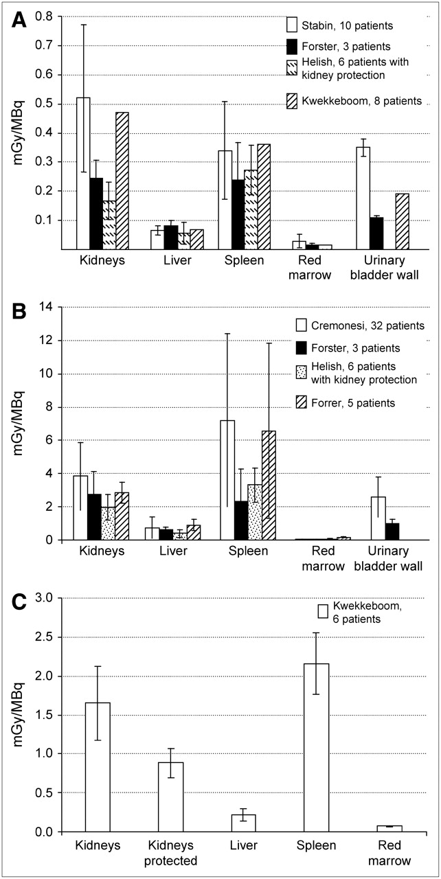
Somatotostatin analogs are certainly the most clinically used radiopeptides, and this review will address mainly the results published for 111In-DTPA-octreotide, 90Y-DOTATOC, and 177Lu-DOTATATE (Figs. 1A–1C).

Dosimetric data published for principal PRRT trials: (A) 111In-DTPA-octreotide (Stabin (13), Forster (22), Helish (23), Kwekkeboom (14)). (B) 90Y-DOTATOC (Cremonesi (15), Forster (22), Helish (23), Forrer (16)). (C) 177Lu-DOTATATE (Kwekkeboom (30)). Large ranges of variability emerge, even in large cohort of patients. This suggests that mean values of absorbed doses among patients should not be the only criterion to plan PRRT. Besides the methods used for dosimetry, interindividual differences are attributable, especially to organ functionality, metabolism, or receptor density in organs.
111IN-, 90Y-, AND 177LU-PEPTIDE DOSIMETRY
111In-Peptides
The physical properties of 111In make it suitable for both diagnostic and dosimetric purposes (Table 2). Dosimetry is facilitated by the γ-ray emission (173, 247 keV) and the relatively long half-life (2.83 d), which matches the peptide biologic half-life. Therefore, a suitable number of scintigraphic images can be obtained over >3 d (9,13) for dosimetry analysis. In principle, planar views are not ideal for dosimetry. However, serial whole-body scans might offer sufficient information on biodistribution and its variation over the time. This represents a good alternative to the more time-consuming SPECT technique, whose limited field of view usually requires 2 or more acquisitions for each time point.
Characteristics of Radionuclides for PRRT
111In-DTPA-octreotide is the commercially available 111In-labeled compound used for diagnosis and staging of somatostatin receptor–positive tumors. 111In-DOTATOC has been also used in clinical protocols (14–17) for dosimetric analysis when planning therapy.
The idea of using 111In-coupled peptides for therapy—in particular, 111In-DTPA-octreotide (18,19)—stems from the possible benefit of the Auger emission of this radionuclide. Auger electrons are high linear energy transfer particles, able to deliver high doses within a very short range (<10 μm). However, the high cytotoxic potential of the Auger electrons requires close proximity of the 111In-labeled peptide within the nucleus, preferably intercalating with the DNA chain (20).
The pharmacokinetics of 111In-DTPA-octreotide demonstrate fast blood clearance, with low exposure of the whole body (13,19). Activity is excreted through the kidneys, with a very small activity in the bowel (<2%). No specific uptake of the tracer is usually observed in bone marrow, allowing the blood-derived method to be the used for dose evaluation (0.01–0.06 mGy/MBq). The organs receiving the highest dose (Fig. 1A) are the spleen (0.10–0.85 mGy/MBq), the kidneys (0.12–0.91 mGy/MBq), and the liver (0.05–0.24 mGy/MBq). The absorbed dose to tumor, evaluated as the mean energy released to the whole tumor tissue, varies widely (0.7–30.5 mGy/MBq).
Despite some encouraging preliminary results, clinical trials with 111In-DTPA-octreotide showed only rare cases of tumor regression (4). This probably relates to the limited effect of Auger energy, likely because the nuclide is probably too far from the nucleus.
90Y-Peptides
90Y is a high-energy β−-emitter (Eave, β = 0.935 MeV), with a physical half-life (T1/2 phys = 64.1 h) compatible with the pharmacokinetics of peptides, and a long penetration range in tissue (Rmax = 11.3 mm). Thus, 90Y is particularly suitable for radionuclide therapy, considering the nonhomogeneous distribution of peptides in solid tumors (Table 2). The probability of killing most neoplastic cells is related to the so-called cross-fire effect. However, there is a relatively high radiation exposure to normal tissues, such as liver, kidneys, and spleen.
90Y-DOTATOC, 90Y-DOTATATE, and 90Y-lanreotide are the principal 90Y-labeled radiopharmaceuticals used for PRRT therapy (4,6,21). A major drawback of 90Y-labeled peptides is the lack of γ-emission, which makes it difficult to get specific dosimetry in each patient. To estimate a specific dose, alternative methods such as imaging with analogs labeled with 111In or substituting the positron emitter 86Y for 90Y may be used (15,16,22–24).
111In-Based Methods.
The first approach simulated therapy with 90Y-compunds using diagnostic activities of 111In-DTPA-octreotide (14,24). 111In-DTPA-octreotide is limited as a surrogate for 90Y-DOTATOC because of the difference in chelators and the single amino acid modification of the octreopeptide (24). A more suitable alternative uses the same peptide as the therapeutic agent labeled with 111In. If the biologic behavior of the 90Y and 111In analogs is similar in vivo, this approach is useful (6,15,17). Images derived from 111In-DOTATOC, 111In-DOTATATE, and 111In-lanreotide visibly reflect the higher in vivo affinity and specific uptake in somatostatin receptor–expressing tissues, and especially in the tumor, compared with 111In-DTPA-octreotide (5,6,14,24). To date, imaging and pharmacokinetics analysis with 111In-labeled peptides remain a feasible and reliable procedure (17,24). Moreover, the most relevant advantage of this approach is in the physical half-life of 111In, comparable with that of 90Y and with the biologic half-life of peptides, which allows derivation of the time–activity curves.
86Y-Based Methods.
Although the chemical behavior of 111In-DOTATOC is similar to that of the analog labeled with 90Y, the chemical nature of the radionuclide may affect the binding affinity for somatostatin receptors (25). To address this question, the labeling of the same compound with the positron emitter of the same element 86Y has been introduced. In particular, the biokinetics of 86Y-DOTATOC, totally preserving the chemical structure of 90Y-DOTATOC, has been investigated and is generally considered as the gold standard for mimicking therapy. Although a comparison of 111In-DOTATOC versus 86Y-DOTATOC, in the same patients, is desirable, the biokinetics of 111In-DTPA-octreotide versus 86Y-DOTATOC have been compared instead. The results indicated that although 111In-DTPA-octreotide is not the optimal surrogate for measuring dosimetry for 90Y-DOTATOC, it may yield acceptable results for certain clinical applications—in particular, for patient recruitment in clinical trials (22–24).
PET has the advantage of increased accuracy and spatial resolution (21). Nevertheless, the use of 86Y for PRRT dosimetry is presently limited by the availability of 86Y. In addition, the T1/2 phys of 14.7 h and the positron abundance of 33% limit the detailed data acquisition to <40 h (Table 2). Late activity concentrations are not experimentally obtainable and must be estimated by extrapolation, reducing the reliability of the integrated activities. This is especially true for tissues with a delayed clearance or metabolism, such as the kidneys. Additional technical problems occur because of the emission of multiple, high-energy, γ-rays in a cascade in the course of decay of 86Y (26). Extrapolation of the 86Y distribution to 90Y dosimetry requires correction for these factors, as proposed by several authors (27,28). Inadequate corrections may lead to important activity overestimations, especially in bone and bone marrow.
A summary of the dosimetric results of 90Y-DOTATOC using both 111In-DOTATOC and 86Y-DOTATOC is shown in Figure 1B. Besides the method selected, results were concordant on some essential aspects: (a) the pharmacokinetics data of 90Y-DOTATOC showed a very fast blood clearance and rapid urinary elimination; (b) the highest predicted absorbed doses were found in the spleen (range, 1.5–19.4 mGy/MBq), kidneys (range, 1.06–10.3 mGy/MBq), and liver (range, 0.1–2.6 mGy/MBq). As for the absorbed dose to the red marrow, most authors did not report any significant uptake in bone or bone marrow and used blood-derived methods. The dose values ranged from 0.01 to 0.20 mGy/MBq and from 0.04 to 0.08 mGy/MBq when evaluated by 111In-DOTATOC or 86Y-DOTATOC, respectively. Other authors observed some uptake in the red marrow by 86Y-DOTATOC images (29). Higher red marrow doses (0.11–0.23 mGy/MBq) were reported in this case, although the information provided by 86Y-images remains uncertain.
Diagnostic studies demonstrated highly variable tumor doses with both 111In-DOTATOC and 86Y-DOTATOC (15,16,23) (Table 3). Nonetheless, the efficacy of PRRT with 90Y-DOTATOC has been observed in several clinical trials. The major drawback of 90Y-peptides is that the activity administered is limited by the high renal dose, which can preclude the achievement of a prescribed tumor dose.
Tumor Dosimetry for Principal Radiopharmaceuticals Used in PRRT
177Lu-Peptides
The physical properties of 177Lu offer intermediate advantages between 90Y and 111In (Table 2). 177Lu is a β−-particle emitter (Emax = 0.50 MeV), with a long half-life (6.73 d), and a R50 (the distance within which the β-particles of 90Y transfer 50% of their energy) of approximately 1 mm. It is also a γ-emitter of low-emission abundance (113 [6%] and 208 [11%] keV). These characteristics enable imaging and therapy with the same complex and allow dosimetry to be performed before and during treatment as well. Comparison of its penetration range in tissue (Rmax = 2 mm) with 90Y indicates a lower cross-fire effect partially compensated by a higher percentage of the radiation energy absorbed in very small volumes. This makes 177Lu a good candidate nuclide for the treatment of small tumors (<2 cm) and micrometastases with 177Lu-DOTATATE.
A study comparing 177Lu-DOTATATE with 111In-DTPA-octreotide (30) demonstrated similar biologic half-lives and principal source organs for the 2 radiocompounds, with varying uptakes in organs and lesions with different expression of somatostatin receptors. These results support the use of similar methods for data collection and similar time schedules for the dosimetry of both 111In-peptides and 177Lu-peptides, although experimental data for the time–activity curves can be further prolonged in the latter case.
The dosimetric data on 177Lu–DOTATATE (Fig. 1C) are still limited (4,5,30). The blood clearance and urinary excretion are fast, similar to the other somatostatin analogs described. The dosimetry of normal organs is lower for 177Lu-DOTATATE as compared with 90Y-DOTATOC, with ranges of 1.8–2.7 mGy/MBq to the spleen, 1.0–2.2 mGy/MBq to the kidneys (lowered to 0.7–1.1 mGy/MBq with protection), and 0.1–0.3 mGy/MBq to the liver. Red marrow dose, derived by the blood approach, ranged from 0.05 to 0.08 mGy/MBq (30). Although the absorbed dose to the gonads has not been reported in the literature, a significant decrease of serum testosterone has been observed, which requires 18–24 mo for reversal (5). This effect is likely secondary to the high activity received by the urinary bladder, with consequent irradiation of the gonads from γ-rays, especially in men, or even to a possible local uptake.
Tumor Dosimetry
A high variability of intrapatient and intralesion tumor uptake has been seen in PRRT studies, which is independent of the radiopharmaceuticals used (Table 3). The wide range of tumor doses was not surprising, likely related to biologic and pathologic factors—differences in tumor volume, hypoxia, necrosis, viability, interstitial pressure, heterogeneity in binding affinity, and the receptor density.
Interestingly, a correlation between tumor dose and tumor mass reduction has been reported (24). Responding tumors could be identified as those receiving much higher doses compared with nonresponding tumors (up to 6-fold: 232 Gy vs. 37 Gy). This emphasized the challenge to deliver the highest activity to the tumor, while sparing normal tissues.
The most suitable choice of the radiopharmaceutical is crucial in therapy planning and should be performed individually, based on tumor volume and localization, adjacent tissues, and affinity for the targeting compound. As peptides are internalized, the physical characteristics of the radionuclide should be fully exploited. The lower tissue penetration range of 177Lu may reasonably exert a more favorable effect on small tumors compared with 90Y. Conversely, the cross-fire effect of 90Y may induce a superior radiation burden in larger lesions. New perspectives pursue the use of cocktails of 177Lu- and 90Y-radiopeptides, promising the treatment of different-sized lesions. Preclinical animal studies showed encouraging data, and the radiobiologic value of these cocktails will hopefully be addressed in patients in the very near future (31).
Red Marrow Dosimetry
Red marrow toxicity represents the limiting factor in numerous radionuclide therapies. Several investigators describe models for red marrow dosimetry and tolerance (10,32). Nevertheless, red marrow has a very complex structure, and the mechanisms regulating activity uptake are still not clear. Marrow radiation burden varies with the radiolabeled molecule, the specific binding, and the residual activity in the blood. Difficulties in modeling the dosimetry of red marrow are therefore comprehensible and are well illustrated in the literature (11,33,34).
The principal approaches used to evaluate the red marrow dose can be distinguished in blood-based methods and imaging-based methods.
The blood-based method was expressly investigated in the study of radiolabeled monoclonal antibodies (mAbs) in cases of no specific uptake in the red marrow (35). The activity concentration in the red marrow was linearly related to the activity concentration in the blood or in the plasma by an experimental factor (in the range of 0.2–0.4, for mAbs, likely related to their molecular weight). The possibility of implementing the blood-based method in PRRT needs an additional parameter, as peptide-bound activity in the red marrow probably distributes in a volume larger than the extracellular space. Moreover, few bone marrow samples taken in patients receiving 111In-DTPA-octreotide showed a red marrow activity concentration equal to that in plasma at the same time points (36,37). Consequently, for peptides, a factor for the concentration ratio of red marrow to blood close to 1 has been suggested.
The image-based method applies when images clearly demonstrate a specific uptake in bone or bone marrow. In this case, the activity in selected areas of known red marrow volume (e.g., sacrum, lumbar vertebrae) is quantified from images and scaled for the whole red marrow. In most studies with somatostatin analogs, no specific bone marrow localization is seen, and the blood activity is used as a surrogate indicator. Images of 111In- or 177Lu-labeled somatostatin analogs usually do not evidence any significant uptake in red marrow or in bone. Consequently, many authors prefer to extrapolate the red marrow dose from the time–activity curve in the blood, plasma, or remainder of the body (15,22). Assessment of bone marrow dose in most radionuclide therapies remains a challenge (32–34).
Other Radiopeptides of Interest for PRRT
Besides the radiopeptides already in use for therapeutic purposes, other radiocompounds are worth mentioning for their potential role in PRRT (Table 1). Among these, the development of peptides labeled with positron-emitter radionuclides is of special interest. In particular, 68Ga is a very attractive β+-emitter radionuclide because of its availability from a generator. Somatostatin and bombesin derivatives labeled with 68Ga have shown ideal characteristics, such as fast clearance and target localization in clinical studies. Recently, 68Ga-DOTATOC has been shown to provide excellent diagnostic information in patients. Although its biokinetics may not be ideal to mirror the dosimetry of therapeutic agents, it may have a main role in the prediction of the PRRT response and follow-up (38,39).
Other radiopeptides investigated in preclinical studies have given valuable results, not only for imaging, such as [18F]FP-Gluc-TOCA (40) but also for therapy, including bombesin derivatives labeled with 166Ho and 188Re (7), and bombesin and somatostatin derivatives labeled with 64Cu and 67Cu (64Cu-DOTA-[Lys3]bombesin (41), 64Cu-DOTA-PEG-BN(7–14) (42), 64Cu-TETA-octreotide (43)). 64Cu-TETA-octreotide, administered in a small number of patients with neuroendocrine tumors, showed clear lesion detection and had favorable pharmacokinetics (43). The special attention merited by Cu-labeled peptides is related to the physical characteristics of 67Cu and of 64Cu, which are especially suitable for both imaging and therapy (67Cu, γ-imaging/PRRT; 64Cu, PET imaging/PRRT).
KIDNEY DOSIMETRY: METHODS, PITFALLS, AND IMPROVEMENTS
All the radiopharmaceuticals used for PRRT have shown high renal activity concentration. This has shifted—or enlarged—the general concern of toxicity from red marrow to kidneys. Kidneys are dose-limiting organs for PRRT—in particular, with 90Y-DOTATOC (3,24,44)—making accurate renal dosimetry critical to minimize radiation nephropathy.
In most cases side effects were unforeseen or were not correlated with the administered activity or with the calculated kidney dose. In other cases, such as in therapy with 111In-DTPA-octreotide or 177Lu-DOTATATE, despite the high kidney doses (comparable with those using 90Y-peptide therapies), alteration of renal function parameters was much lower than expected. These findings were surprising, as the occurrence of nephritis was believed to be determined, in principle, by radiation dose. The alarming high doses to kidneys, and—most importantly—cases of late renal failure in patients who received kidney doses below the conventional threshold dose derived from external-beam radiotherapy (EBRT) (44,45) suggested the need to reexamine this problem.
The first evaluations of kidney dosimetry were usually based on a robust standard approach, assuming uniform activity distributions and standard masses within the MIRD scheme. However, the excessive interpatient variability of kidney absorbed doses suggested the need for more precise measurements. A first crucial improvement in renal dosimetry was obtained by the inclusion of the actual kidney masses (derived from CT) in the dosimetric estimates. Patient kidney volumes varied between 231 and 503 mL (in 25 patients), some values being very far from the reference volumes of 288 mL (male) and 264 mL (female). Consequently, kidney absorbed doses rescaled by the true masses provided values differing up to 100%. The relevance of this simple correction has been recently confirmed in a retrospective clinical study, in which the unexpected renal toxicity in a few patients could be explained by the significantly smaller kidney mass (24).
A further contribution to inaccuracy occurred because of the nonuniform radioactivity distribution in the kidney. Scintigraphic images demonstrated higher and persistent uptake in the cortex compared with the medulla. Unfortunately, the spatial resolution of SPECT or PET/CT equipment, can indicate—but not precisely establish—the fine differences among subregions. This has been confirmed by ex vivo autoradiography in patients receiving 111In-DTPA-octreotide (46). These experimental findings could open the way to more accurate renal dosimetry.
To face the problem of uneven uptake in the kidney, the MIRD Committee offered a multiregion model for a suborgan kidney dosimetry (10,47)—with cortex, medulla, pelvis, and papillae as possible source or target regions. In particular, in patients undergoing PRRT, specific kidney dose evaluations can be obtained by rescaling the actual volumes of the renal regions and by the activity assigned to cortex and medulla, evaluated, for example, by PET or SPECT images or, more accurately, by voxel-based methods (48–50). Alternatively, the experimental results of renal autoradiograms can be considered as rational for detailed kidney dosimetry (50).
In addition to the suborgan activity distribution, the physical characteristics of the radionuclide can be a major determinant of the nephrotoxic potential of the agent and, hence, of the risk–benefit balance. This has been confirmed clinically by the frequent occurrence of renal impairment in patients treated with 90Y-peptide therapy compared with the sporadic incidence observed with 111In-DTPA-octreotide and 177Lu-TATE therapies (despite mean cumulative kidney doses up to 45 Gy) (3,51). Two potential explanations are the particle range and the site of peptide accumulation: localization in the proximal tubuli, with their radioresistant cells, able to repair and regenerate, versus the glomeruli with their radiosensitive cells, not able to regenerate, can make a major difference in outcome. Therefore, the Auger electrons of 111In-peptides, and also the short-range β-particles of 177Lu-peptides, irradiate the tubular cells more selectively compared with the glomeruli. On the contrary, the long-range β-particles of 90Y-peptides may increase the toxicity due to the irradiation of the glomerular cells.
Methods for Possible Kidney Dose Reduction
It has been observed that renal uptake is not somatostatin receptor driven but related primarily to the very rapid clearance of the small radiopeptides that are filtered through the glomeruli and reabsorbed by the tubular cells (24,25,52). Preclinical and clinical studies showed that the infusion of positively charged amino acids (e.g., lysine, arginine)—before, during, and after the injection of the radioligand—is able to block the tubular peptide reabsorption process (2,25,51,53). On the basis of these findings, the efficacy and side effects of several renal protector regimens were tested. Different amounts of amino acid solutions and combinations with other positively charged molecules were studied, and the timing, the duration of the infusion, and the amino acid toxicity (mostly gastrointestinal) were analyzed (2,50,51,54–57).
The results of 86Y-DOTATOC or 111In-DOTATOC as a radiotracer to determine tracer retention in the kidneys after pretreatment with amino acids can be summarized as follows:
(a) The blood clearance and the activity elimination are not substantially modified by renal protectors; tumor uptake is not altered; and the biodistribution in source organs other than kidneys leads to minimal changes (mostly in the spleen).
(b) Protector agents maintain the typical trend of the time–activity curves in kidneys but reduce the renal uptake at all time points; and absorbed doses are consistently lowered 20%–30% (up to 40%) by amino acids or 30%–40% (up to 55%) by other positively charged molecule combinations.
(c) Both the total dose and the duration of the infusion influence the dose sparing (up to 65%, with a combination of positively charged molecules infused over 2 d after injection).
Dose and Dose Rate: Linear Quadratic (LQ) Model from EBRT to PRRT
According to the experience gained from EBRT, absorbed doses to the kidneys of 23 and 28 Gy are associated with a 5% (TD5/5 [dose for a probability of 5% injury within 5 y]) probability and a 50% (TD50/5 [dose for a probability of 50% injury within 5 y]) probability of causing deterministic late side effects within 5 y, respectively. It is also known that doses of >25 Gy may lead to acute radiation nephropathy with a latent period of 6–12 mo, whereas, at lower doses, chronic radiation nephropathy may become clinically apparent 1–5 y after irradiation (58). Unfortunately, these findings do not apply to internal radionuclide therapies. Although the general radiobiologic principles are the same, the radiation dose delivered during radionuclide therapy differs in many respects from a dose delivered by EBRT. First, the radiation dose rate in EBRT is high (1–3 Gy/min) and the total prescribed dose is delivered in some fractions (typically, 2 Gy/fraction); in contrast, the radiation dose rate in PRRT is low (<3 mGy/min) and variable, with a continuous exponential decrease related to biologic and physical decay (59,60).
The dose rate has a most important radiobiologic impact, in that it may alter the recovery from radiation damage. This has been widely investigated and demonstrated in EBRT, and it is well known that, at equal dose, fractionation of EBRT lowers toxicity. In general, early-responding tissues (most tumors) are characterized by cell-survival curves with smaller shoulders compared with late-responding tissues (60). The influence of the dose rate may be even more relevant in radionuclide therapy compared with EBRT, as the variability of both dose rate and dose distribution might strengthen the reparable sublethal damage: the lower the dose rate, the lower the damage (61). These considerations suggest that the sparing effect should benefit the tumor-to-kidney dose ratio in PRRT, as the kidney is a late-responding tissue.
Recently, some authors have used the LQ model to quantify the dose-rate sparing concepts expressly for radionuclide therapy, taking into account the different dose rate as compared with EBRT (61). The LQ model describes the biologic effect in irradiated tissue by the surviving fraction (S) of cells that received a radiation dose D: where αD accounts for the double-strand breaks induced by a single ionizing event, and the quadratic component βD2 accounts for the cell kill by multiple sublethal events.
The biological effective dose (BED = −1/α ln S) represents the dose producing the same biological effect obtained under different irradiation conditions. The α/β ratio relates the intrinsic radiosensitivity (α) and the potential sparing capacity (β) for a specified tissue or effect.
In general, tissues with low α/β values (normal tissues, α/β = 2–5 Gy; kidneys, α/β ∼2.4 Gy (46)) have more time to recover from sublethal damages and are more influenced by the dose rate compared with tissues with high α/β values (tumor tissues, α/β = 5–25 Gy).
On considering radionuclide therapy, the effect of the repair potential of the dose rate and of the delivery of the dose—protracted and possibly divided in cycles—must be included. Therefore, an additional parameter has been introduced in the LQ equation to finally provide a revised expression for the BED estimate (61,62): where Di is the dose delivered per cycle i, T1/2rep is the repair half-time of sublethal damage, and T1/2eff is the effective half-life of the radiopharmaceutical in the specific tissue.
This refined LQ model has been suggested with the intent of increasing the dose–response correlation in radionuclide therapy and applied, in particular, to PRRT, focusing on the kidney. Some results recently presented in the literature are briefly reported.
In a retrospective analysis on patients who received 90Y-DOTATOC therapy, dosimetry of the kidneys was reevaluated by the inclusion of some patient-specific adjustments. The contribution of this new approach was investigated for a possible dose–effect correlation for kidneys (50). In particular, according to the LQ model, the BED for kidneys was determined for each patient, considering the parameters derived from the specific biokinetics of 90Y-DOTATOC (T1/2eff = 30 h) and from the literature (T1/2rep = 2.8 h; α/β = 2.6 Gy) (54,62). The results of this study (50) strongly indicated that improvements in dosimetry reflect improved prediction of radiation effects. Accounting for the total activity administered in patients, the kidney absorbed dose range resulted in 19.4–39.6 Gy (when considering the actual kidney mass and the activity localized in the cortex), whereas the BED range was 27.7–59.3 Gy. Significantly, a correlation was found between BED and renal impairment (evaluated by means of loss of creatinine clearance per year), as opposed to the absorbed dose values alone, when evaluated by the standard or the refined methods as well. As a further relevant issue, it also emerged that patients with high BED and more serious kidney side effects received the treatment in a low number of cycles. These findings are consistent with other preliminary results reported in the literature (2,54,56), similarly suggesting an improved repair possibility for kidney tissues in the case of a higher number of cycles and a slow infusion of the radiocompounds over 1 h. Therefore, on the basis of the typical different sensitivities of most tumor tissues and kidney tissues to the dose rate and the number of cycles, treatment protocols based on multiple cycles could represent a powerful strategy to lower toxicity and, possibly, to improve the therapeutic outcome. Total administered activities could be increased accordingly.
CONCLUSION
Patient variability requires careful individual dosimetry to determine the activity to be administered in PRRT. However, the value of the kidney absorbed dose may not be sufficient to accurately predict the likelihood of renal toxicity for individual patients. Other factors contribute to the biologic effectiveness, including the tissue radiosensitivity, dose rate, detailed intraorgan activity distribution, and cycle therapy scheme. New multiregional and radiobiologic models improve the prediction of dosimetry—in particular, the renal side effects. Moreover, the biologic equivalent dose approach strongly supports the need for clinical randomized trials, which could definitively compare the therapeutic efficacy of equal therapeutic activities administered in multiple cycles versus a single or very few cycles.
Acknowledgments
The authors thank Prof. Giuliano Mariani and Dr. Marco Chinol for constructive discussion and Deborah Console for typing the manuscript. We are especially grateful to Prof. Mike Stabin and Prof. William Strauss for their support and valuable comments.
Footnotes
-
↵* NOTE: FOR CE CREDIT, YOU CAN ACCESS THIS ACTIVITY THROUGH THE SNM WEB SITE (http://www.snm.org/ce_online) THROUGH SEPTEMBER 2007.
-
COPYRIGHT © 2006 by the Society of Nuclear Medicine, Inc.
References
- Received for publication February 6, 2006.
- Accepted for publication June 12, 2006.


Jump to section
Related Articles
Cited By...
- Can 177Lu-DOTATATE Kidney Absorbed Doses be Predicted from Pretherapy SSTR PET? Findings from Multicenter Data
- Absorbed Dose-Response Relationship in Patients with Gastroenteropancreatic Neuroendocrine Tumors Treated with [177Lu]Lu-DOTATATE: One Step Closer to Personalized Medicine
- Comparison of 3 Different Therapeutic Particles in Radioembolization of Locally Advanced Intrahepatic Cholangiocarcinoma
- Radiotheranostic Agent 64Cu-cyclam-RAFT-c(-RGDfK-)4 for Management of Peritoneal Metastasis in Ovarian Cancer
- mTOR inhibitors as radiosensitizers in neuroendocrine neoplasms
- {alpha}V{beta}3 Integrin-Targeted Radionuclide Therapy with 64Cu-cyclam-RAFT-c(-RGDfK-)4
- Dose Response of Pancreatic Neuroendocrine Tumors Treated with Peptide Receptor Radionuclide Therapy Using 177Lu-DOTATATE
- 90Y Radioembolization After Radiation Exposure from Peptide Receptor Radionuclide Therapy
- 4-Step Renal Dosimetry Dependent on Cortex Geometry Applied to 90Y Peptide Receptor Radiotherapy: Evaluation Using a Fillable Kidney Phantom Imaged by 90Y PET
- Intraindividual Comparison of Selective Arterial versus Venous 68Ga-DOTATOC PET/CT in Patients with Gastroenteropancreatic Neuroendocrine Tumors
- MIRD Pamphlet No. 20: The Effect of Model Assumptions on Kidney Dosimetry and Response--Implications for Radionuclide Therapy
- Intraoperative Avidination for Radionuclide Therapy: A Prospective New Development to Accelerate Radiotherapy in Breast Cancer