


Article Figures & Data
Figures
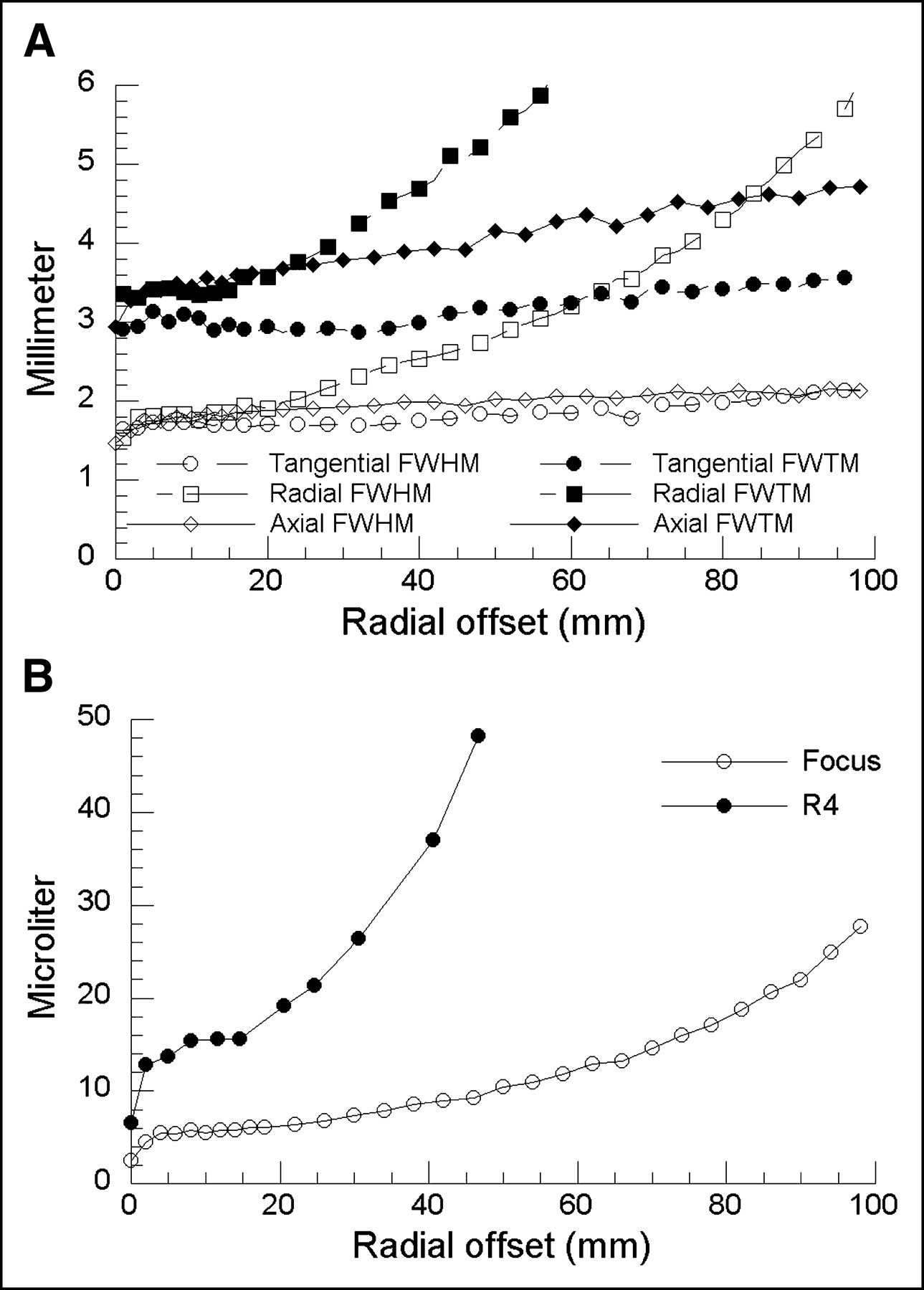
- FIGURE 1.
Reconstructed image resolution of microPET Focus system as function of radial offset from CFOV. (A) FWHM and FWTM of radial, tangential, and axial image resolutions were calculated by linear extrapolation of corresponding profiles extracted from point source images. (B) Volumetric resolution of the Focus compared with the R4.
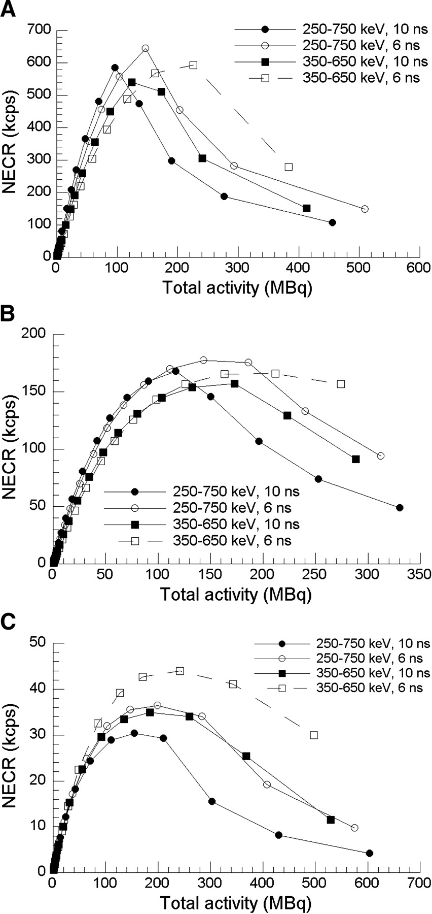
- FIGURE 2.
NEC rate of mouse (A), rat (B), and monkey (C) phantoms. Peak NEC rate of 645 kcps is reached with setting of 250–750 keV and 6 ns for mouse phantom. With rat phantom, peak NEC rate is 177 kcps with 250–750 keV and 6 ns. With monkey phantom, peak NEC rate is 44 kcps with 350–650 keV and 6 ns. NECR = NEC rate.
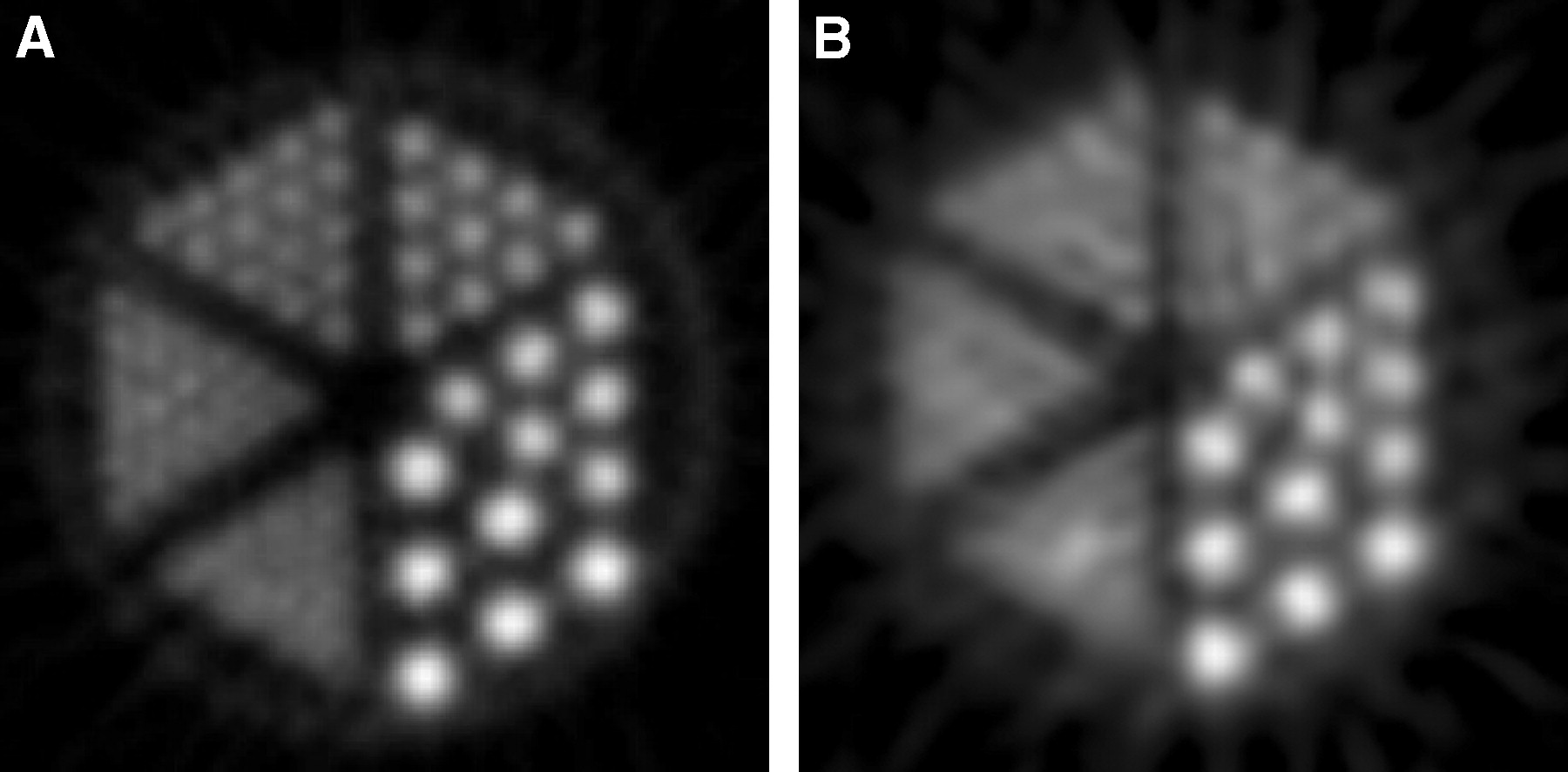
- FIGURE 3.
(A) Emission image measured by the Focus. (B) Same phantom as in A scanned by the R4. Diameter of rods was 0.80, 1.00, 1.25, 1.50, 2.00, and 2.50 mm, respectively. Center-to-center distance between adjacent rods was 2 times the rod diameter.
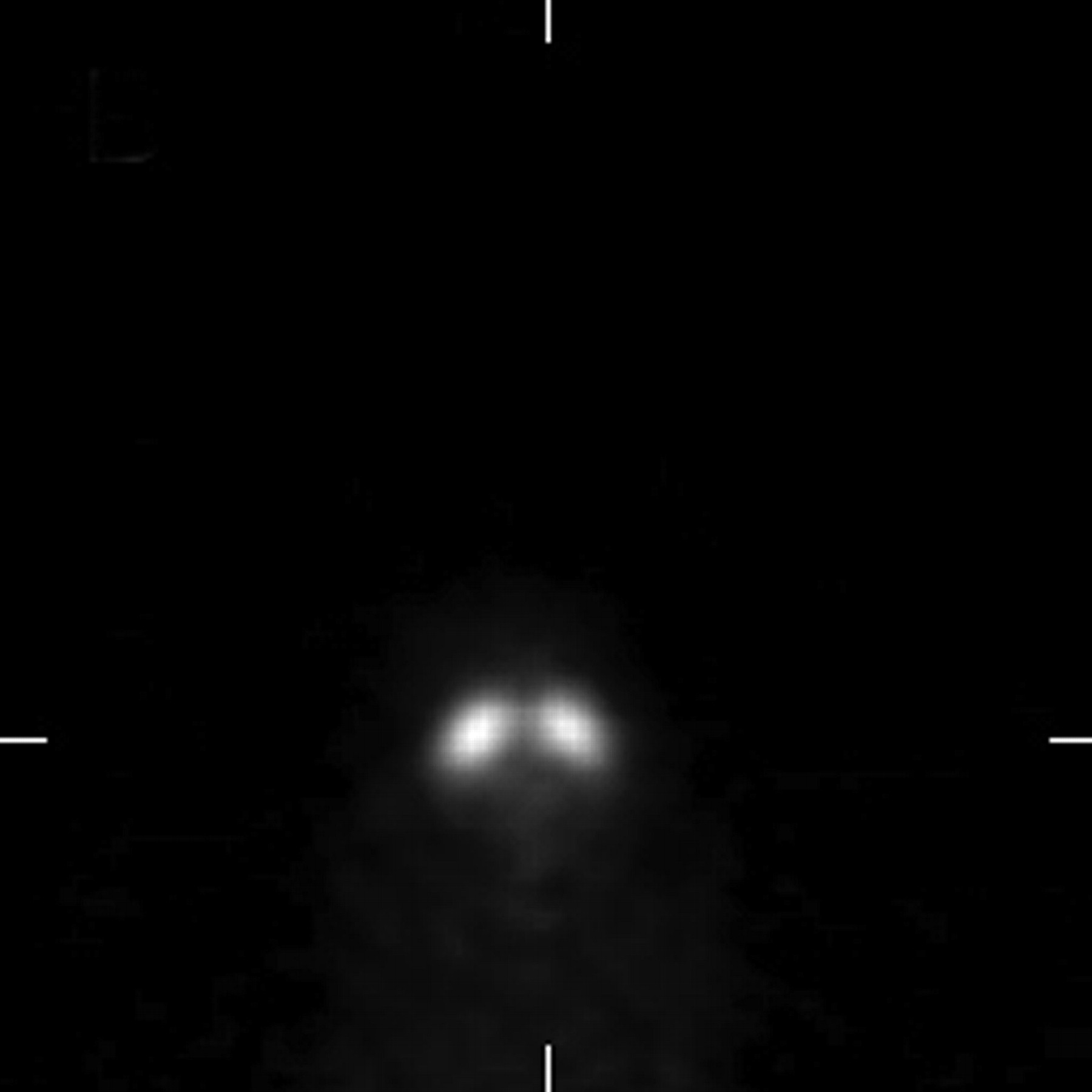
- FIGURE 4.
Coronal image of 18.7-g mouse injected with 17.8 MBq (0.48 mCi) of 11C-CFT. Image corresponds to 50-min acquisition starting at 70 min after injection by summing the last 10 frames of 2-h dynamic study.
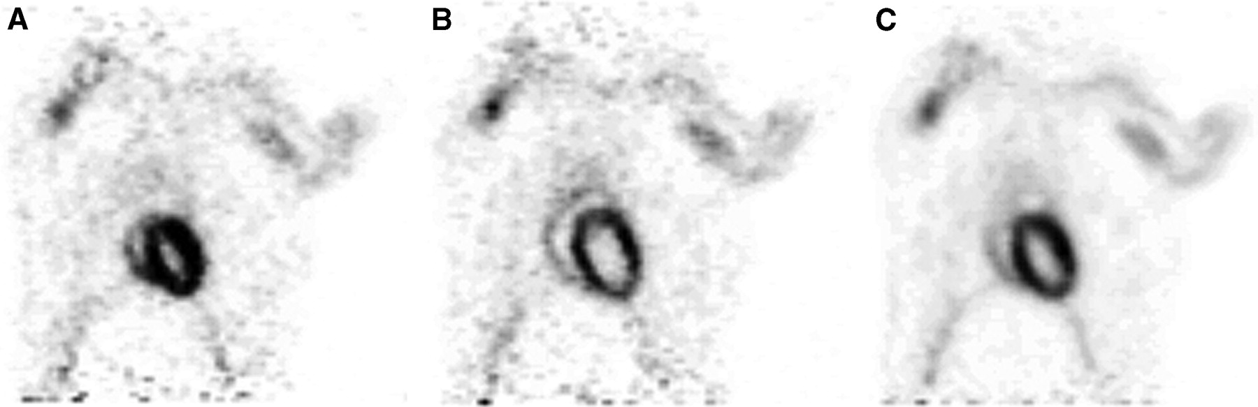
- FIGURE 5.
Cardiac-gated study of 363-g Sprague–Dawley rat injected with 35.7 MBq (0.97 mCi) of 18F-FDG. Acquisition time was 30 min started 110 min after injection. Coronal image of heart: (A) gated at end-systole, (B) gated at end-diastole, and (C) nongated.
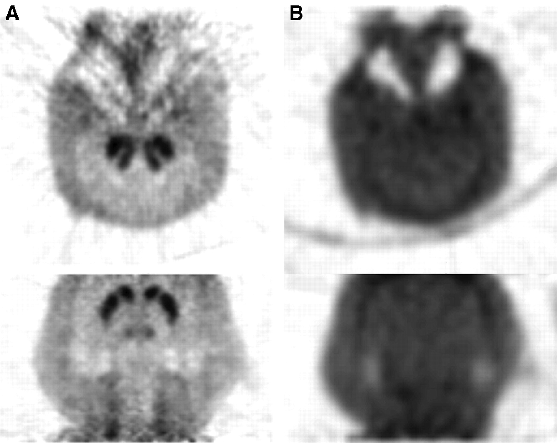
- FIGURE 6.
Two-hour dynamic imaging of 5.4-kg macaque monkey injected with 166 MBq (4.5 mCi) of 18F-FDOPA. (A) Transverse (top) and coronal (bottom) views of emission images correspond to summed frames of 15–30 (40–120 min after injection). (B) Transmission images of monkey head of same transverse and coronal slices.


Tables
Category Parameter Concorde Focus Concorde P4 Concorde R4 Prototype microPET microPET II Detector Crystal material LSO LSO LSO LSO LSO Crystal size (mm) 1.51 × 1.51 × 10.00 2.2 × 2.2 × 10.0 2.2 × 2.2 × 10.0 2 × 2 × 10 0.975 × 0.975 × 12.500 Crystal pitch (mm) 1.59 2.45 2.45 2.25 1.15 Crystal array 144 (12 × 12) 64 (8 × 8) 64 (8 × 8) 64 (8 × 8) 196 (14 × 14) Photomultiplier tube Hamamatsu R5900-C12 Hamamatsu R5900-C12 Hamamatsu R5900-C12 Philips XP1722, 64 channel Hamamatsu H7546-M64 System No. of detectors 168 168 96 30 90 No. of crystals 24,192 10,752 6,144 1,920 17,640 No. of rings 48 32 32 8 42 No. of crystals/ring 504 336 192 240 420 Ring diameter (cm) 25.8 26.1 14.8 17.2 16.0 Gantry aperture (cm) 22.0 22.0 13.0 16.0 15.3 Axial FOV (cm) 7.6 7.8 7.8 1.8 4.9 Transaxial FOV (cm) 19.00 19.00 9.40 11.25 8.00 Dataset No. of sinograms 3D 2,304 1,024 1,024 64 1,764 2D 95 63 63 Not used 83 Sinogram size 288 × 252 192 × 168 84 × 96 100 × 120 140 × 210 Dataset size (MB) 3D 638 126 33 1.54 207 2D 26.3 7.8 2.0 Not used 9.7 Sampling distance (mm) 0.795 1.225 1.225 1.125 0.575 Phantom type Energy window (keV) Timing window (ns) Scatter fraction (%) Mouse body (3-cm Ø, 7-cm L, volume = 49.5 mL) 250–750 10 19.0 6 19.0 350–650 10 12.8 6 12.7 Rat body (6-cm Ø, 15-cm L, volume = 424 mL) 250–750 10 36.5 6 36.4 350–650 10 27.6 6 27.4 Monkey body (10.1-cm Ø, 40-cm L, volume = 3,205 mL) 250–750 10 57.6 6 58.1 350–650 10 40.9 6 40.9 Ø = diameter; L = length.
In this issue


Jump to section
Related Articles
Cited By...
- Metabolic resistance of A{beta}3pE-42, a target epitope of the anti-Alzheimer therapeutic antibody, donanemab
- Submillimeter-Resolution PET for High-Sensitivity Mouse Brain Imaging
- Evaluation of Candidate Theranostics for 227Th/89Zr Paired Radioimmunotherapy of Lymphoma
- High-resolution positron emission microscopy of patient-derived tumor organoids
- Performance Evaluation of a High-Resolution Nonhuman Primate PET/CT System
- Reproducibility and Comparability of Preclinical PET Imaging Data: A Multicenter Small-Animal PET Study
- Cell-Proliferation Imaging for Monitoring Response to CDK4/6 Inhibition Combined with Endocrine-Therapy in Breast Cancer: Comparison of [18F]FLT and [18F]ISO-1 PET/CT
- Noninvasive Interrogation of DLL3 Expression in Metastatic Small Cell Lung Cancer
- Preclinical Evaluation and Quantification of 18F-FPEB as a Radioligand for PET Imaging of the Metabotropic Glutamate Receptor 5
- Imaging the Norepinephrine Transporter in Neuroblastoma: A Comparison of [18F]-MFBG and 123I-MIBG
- Evaluation of Frame-Based and Event-by-Event Motion-Correction Methods for Awake Monkey Brain PET Imaging
- Performance Evaluation of the Small-Animal nanoScan PET/MRI System
- Quantitative PET Imaging Detects Early Metabolic Remodeling in a Mouse Model of Pressure-Overload Left Ventricular Hypertrophy In Vivo
- VECTor: A Preclinical Imaging System for Simultaneous Submillimeter SPECT and PET
- In Vivo Measurement of the Affinity and Density of Metabotropic Glutamate Receptor Subtype 1 in Rat Brain Using 18F-FITM in Small-Animal PET
- Small-Animal PET: What Is It, and Why Do We Need It?
- NEMA NU 4-2008 Comparison of Preclinical PET Imaging Systems
- Assessment of PET Tracer Uptake in Hormone-Independent and Hormone-Dependent Xenograft Prostate Cancer Mouse Models
- Gastric Cancer Growth Control by BEZ235 In Vivo Does Not Correlate with PI3K/mTOR Target Inhibition but with [18F]FLT Uptake
- Whiskers Area as Extracerebral Reference Tissue for Quantification of Rat Brain Metabolism Using 18F-FDG PET: Application to Focal Cerebral Ischemia
- Simplified Quantification of Myocardial Flow Reserve with flurpiridaz F 18: Validation with Microspheres in a Pig Model
- In Vivo Positron Emission Tomographic Imaging of Glial Responses to Amyloid-{beta} and Tau Pathologies in Mouse Models of Alzheimer's Disease and Related Disorders
- Dynamic PET Denoising with HYPR Processing
- Establishment of In Vivo Brain Imaging Method in Conscious Mice
- Delivery of Na/I Symporter Gene into Skeletal Muscle Using Nanobubbles and Ultrasound: Visualization of Gene Expression by PET
- Efficacy of PHA-848125, a Cyclin-Dependent Kinase Inhibitor, on the K-RasG12DLA2 Lung Adenocarcinoma Transgenic Mouse Model: Evaluation by Multimodality Imaging
- NEMA NU4-2008 Image Quality Performance Report for the microPET Focus 120 and for Various Transmission and Reconstruction Methods
- In Vivo Metabolic Phenotyping of Myocardial Substrate Metabolism in Rodents: Differential Efficacy of Metformin and Rosiglitazone Monotherapy
- Quantification of Cerebral Glucose Metabolic Rate in Mice Using 18F-FDG and Small-Animal PET
- A Novel Technology for the Imaging of Acidic Prostate Tumors by Positron Emission Tomography
- Recent Advances in Small-Animal Cardiovascular Imaging
- In Vivo Biodistribution, PET Imaging, and Tumor Accumulation of 86Y- and 111In-Antimindin/RG-1, Engineered Antibody Fragments in LNCaP Tumor-Bearing Nude Mice
- Performance Evaluation of the Inveon Dedicated PET Preclinical Tomograph Based on the NEMA NU-4 Standards
- Neuroimaging and Physiological Evidence for Involvement of Glutamatergic Transmission in Regulation of the Striatal Dopaminergic System
- Derivation of a Compartmental Model for Quantifying 64Cu-DOTA-RGD Kinetics in Tumor-Bearing Mice
- In Vitro and In Vivo Evaluations of a Hydrophilic 64Cu-Bis(Thiosemicarbazonato)-Glucose Conjugate for Hypoxia Imaging
- Impact of Contamination from Scattered Photons in Singles-Mode Transmission Data on Quantitative Small-Animal PET Imaging
- Engineered Antibody Fragments with Infinite Affinity as Reporter Genes for PET Imaging
- Micro Insert: A Prototype Full-Ring PET Device for Improving the Image Resolution of a Small-Animal PET Scanner
- Time Course of Alterations in Myocardial Glucose Utilization in the Zucker Diabetic Fatty Rat with Correlation to Gene Expression of Glucose Transporters: A Small-Animal PET Investigation
- PET/MRI Dual-Modality Tumor Imaging Using Arginine-Glycine-Aspartic (RGD)-Conjugated Radiolabeled Iron Oxide Nanoparticles
- A Prototype PET Scanner with DOI-Encoding Detectors
- Changes in Tumor Metabolism as Readout for Mammalian Target of Rapamycin Kinase Inhibition by Rapamycin in Glioblastoma
- Initial Characterization of an 18F-Labeled Myocardial Perfusion Tracer
- Virtual-Pinhole PET
- 1-11C-Acetate as a PET Radiopharmaceutical for Imaging Fatty Acid Synthase Expression in Prostate Cancer
- A Feasibility Study of a Prototype PET Insert Device to Convert a General-Purpose Animal PET Scanner to Higher Resolution
- Longitudinal, Quantitative Assessment of Amyloid, Neuroinflammation, and Anti-Amyloid Treatment in a Living Mouse Model of Alzheimer's Disease Enabled by Positron Emission Tomography
- Performance Measurement of the microPET Focus 120 Scanner
- Fabrication and Characterization of a 0.5-mm Lutetium Oxyorthosilicate Detector Array for High-Resolution PET Applications
- Synthesis and Biologic Evaluation of 64Cu-Labeled Rhenium-Cyclized {alpha}-MSH Peptide Analog Using a Cross-Bridged Cyclam Chelator
- 64Cu-Azabicyclo[3.2.2]Nonane Thiosemicarbazone Complexes: Radiopharmaceuticals for PET of Topoisomerase II Expression in Tumors
- Performance Evaluation of the GE Healthcare eXplore VISTA Dual-Ring Small-Animal PET Scanner
- Monitoring Tumor Glucose Utilization by Positron Emission Tomography for the Prediction of Treatment Response to Epidermal Growth Factor Receptor Kinase Inhibitors.
- Noninvasive Measurement of Cardiovascular Function in Mice with High-Temporal-Resolution Small-Animal PET
- Performance Test of an LSO-APD Detector in a 7-T MRI Scanner for Simultaneous PET/MRI
- Assessment of Myocardial Metabolism in Diabetic Rats Using Small-Animal PET: A Feasibility Study
- Assessment of Myocardial Blood Flow Using 15O-Water and 1-11C-Acetate in Rats with Small-Animal PET
- Reproducibility of 3'-Deoxy-3'-18F-Fluorothymidine MicroPET Studies in Tumor Xenografts in Mice