
Main menu

  • Home
  • Content
    • Current
    • Ahead of print
    • Past Issues
    • JNM Supplement
    • SNMMI Annual Meeting Abstracts
    • Continuing Education
    • JNM Podcasts
  • Subscriptions
    • Subscribers
    • Institutional and Non-member
    • Rates
    • Journal Claims
    • Corporate & Special Sales
  • Authors
    • Submit to JNM
    • Information for Authors
    • Assignment of Copyright
    • AQARA requirements
  • Info
    • Reviewers
    • Permissions
    • Advertisers
  • About
    • About Us
    • Editorial Board
    • Contact Information
  • More
    • Alerts
    • Feedback
    • Help
    • SNMMI Journals
  • SNMMI
    • JNM
    • JNMT
    • SNMMI Journals
    • SNMMI

User menu

  • Subscribe
  • My alerts
  • Log in
  • My Cart

Search

  • Advanced search
Journal of Nuclear Medicine
  • SNMMI
    • JNM
    • JNMT
    • SNMMI Journals
    • SNMMI
  • Subscribe
  • My alerts
  • Log in
  • My Cart
Journal of Nuclear Medicine

Advanced Search

  • Home
  • Content
    • Current
    • Ahead of print
    • Past Issues
    • JNM Supplement
    • SNMMI Annual Meeting Abstracts
    • Continuing Education
    • JNM Podcasts
  • Subscriptions
    • Subscribers
    • Institutional and Non-member
    • Rates
    • Journal Claims
    • Corporate & Special Sales
  • Authors
    • Submit to JNM
    • Information for Authors
    • Assignment of Copyright
    • AQARA requirements
  • Info
    • Reviewers
    • Permissions
    • Advertisers
  • About
    • About Us
    • Editorial Board
    • Contact Information
  • More
    • Alerts
    • Feedback
    • Help
    • SNMMI Journals
  • View or Listen to JNM Podcast
  • Visit JNM on Facebook
  • Join JNM on LinkedIn
  • Follow JNM on Twitter
  • Subscribe to our RSS feeds
OtherBASIC SCIENCE INVESTIGATIONS

Experimental and Clinical Evaluation of Iterative Reconstruction (OSEM) in Dynamic PET: Quantitative Characteristics and Effects on Kinetic Modeling

Ronald Boellaard, Arthur van Lingen and Adriaan A. Lammertsma
Journal of Nuclear Medicine May 2001, 42 (5) 808-817;
Ronald Boellaard
  • Find this author on Google Scholar
  • Find this author on PubMed
  • Search for this author on this site
Arthur van Lingen
  • Find this author on Google Scholar
  • Find this author on PubMed
  • Search for this author on this site
Adriaan A. Lammertsma
  • Find this author on Google Scholar
  • Find this author on PubMed
  • Search for this author on this site
  • Article
  • Figures & Data
  • Info & Metrics
  • PDF
Loading

Article Figures & Data

Figures

  • Tables
  • FIGURE 1.
    • Download figure
    • Open in new tab
    • Download powerpoint
    FIGURE 1.

    Image noise (COV) as function of emission scan NEC (NECEx) for Tx of 2, 5, and 30 min for FBP reconstruction (A) and OSEM 2 × 12 reconstruction (B). Results obtained with other transmission scan durations have been omitted for clarity.
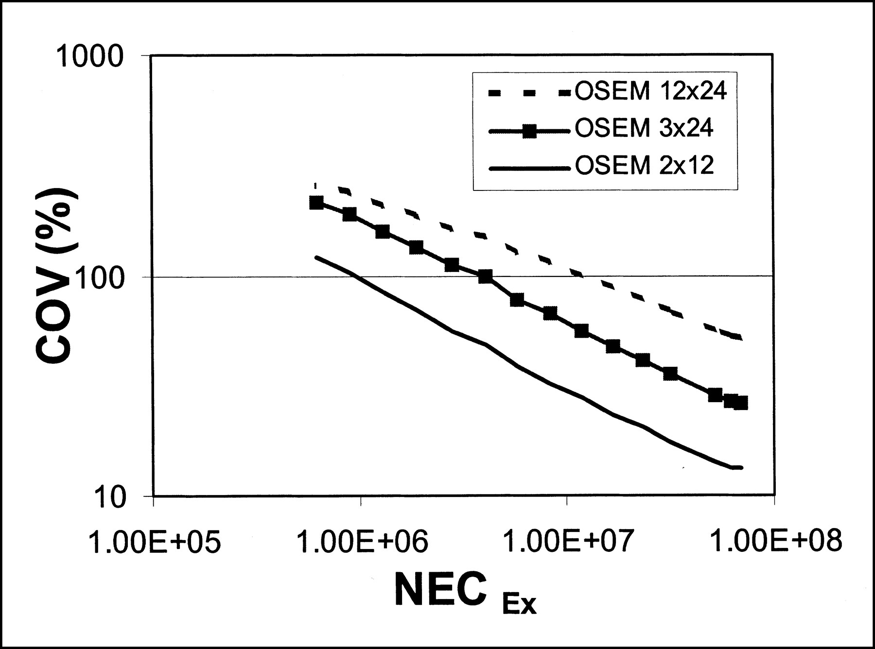
  • FIGURE 2.
    • Download figure
    • Open in new tab
    • Download powerpoint
    FIGURE 2.

    Image noise (COV) as function of emission scan NEC (NECEx) obtained with 2 × 12, 3 × 24, and 12 × 24 iterations of OSEM.

  • FIGURE 3.
    • Download figure
    • Open in new tab
    • Download powerpoint
    FIGURE 3.

    (A) Image noise (COV) as function of emission scan NEC (NECEx) obtained with 5-cm-diameter and 20-cm-diameter phantoms. (B) Ratio of image noise within insert region (COVinsert) of NEMA phantom between OSEM 2 × 12 and FBP as function of image contrast, defined as ratio of activity concentration (AC) between insert and background (BG) regions.

  • FIGURE 4.
    • Download figure
    • Open in new tab
    • Download powerpoint
    FIGURE 4.

    Bias as function of image contrast, defined as ratio of activity concentration (AC) between insert and background regions (BG). Note that image contrast <1 corresponds to cold spots in hot background.
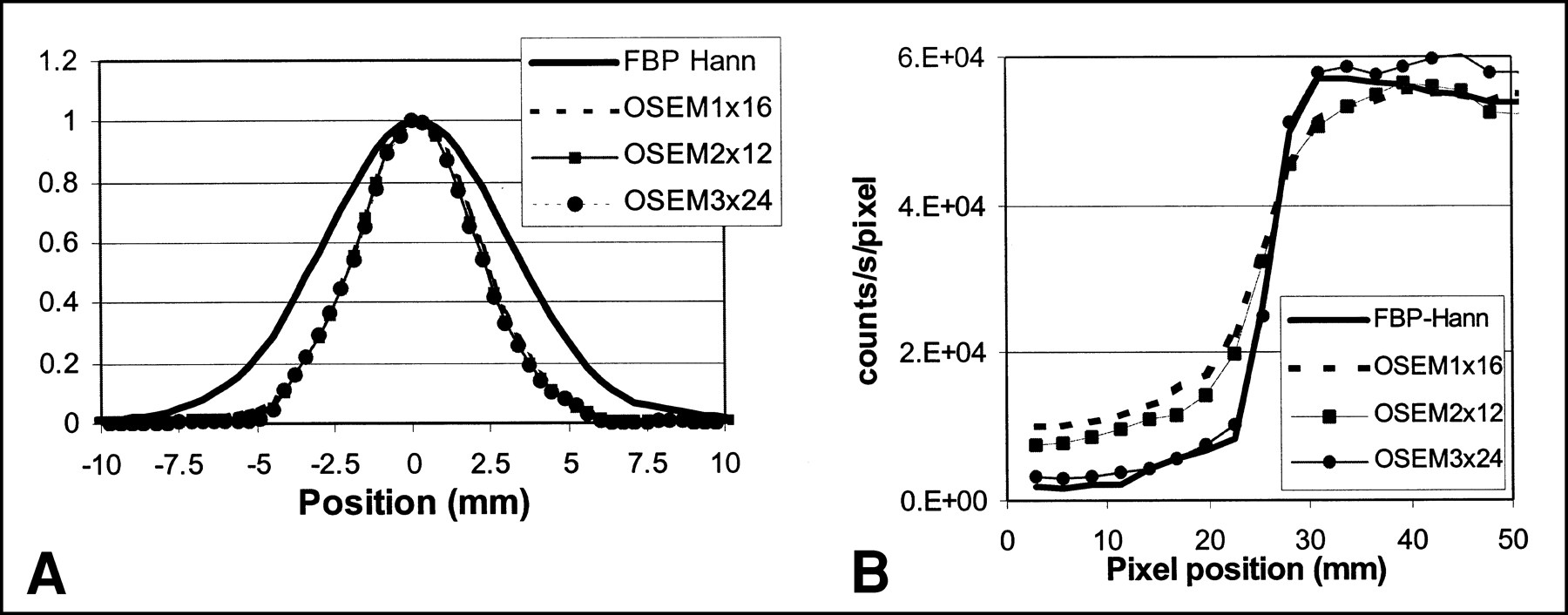
  • FIGURE 5.
    • Download figure
    • Open in new tab
    • Download powerpoint
    FIGURE 5.

    (A) Line spread functions for FBP, OSEM 1 × 16, OSEM 2 × 12, and OSEM 3 × 24. (B) Activity profiles across edge of cold insert and warm background of NEMA phantom for FBP, OSEM 1 × 16, OSEM 2 × 12, and OSEM 3 × 24.
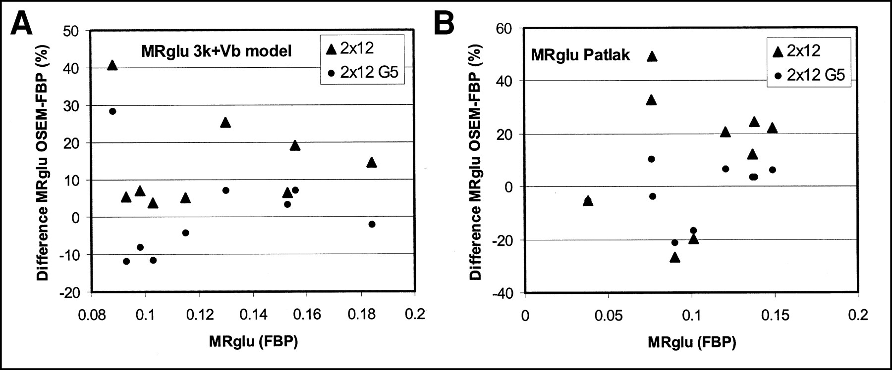
  • FIGURE 6.
    • Download figure
    • Open in new tab
    • Download powerpoint
    FIGURE 6.

    Percentage difference of lung tumor MRglu values (μmol/mL/min) between OSEM and FBP data as function of MRglu values derived from FBP data using standard two-tissue compartment model with blood volume (Vb) parameter (A) and Patlak analysis (B). Data were obtained for nine lung tumors.
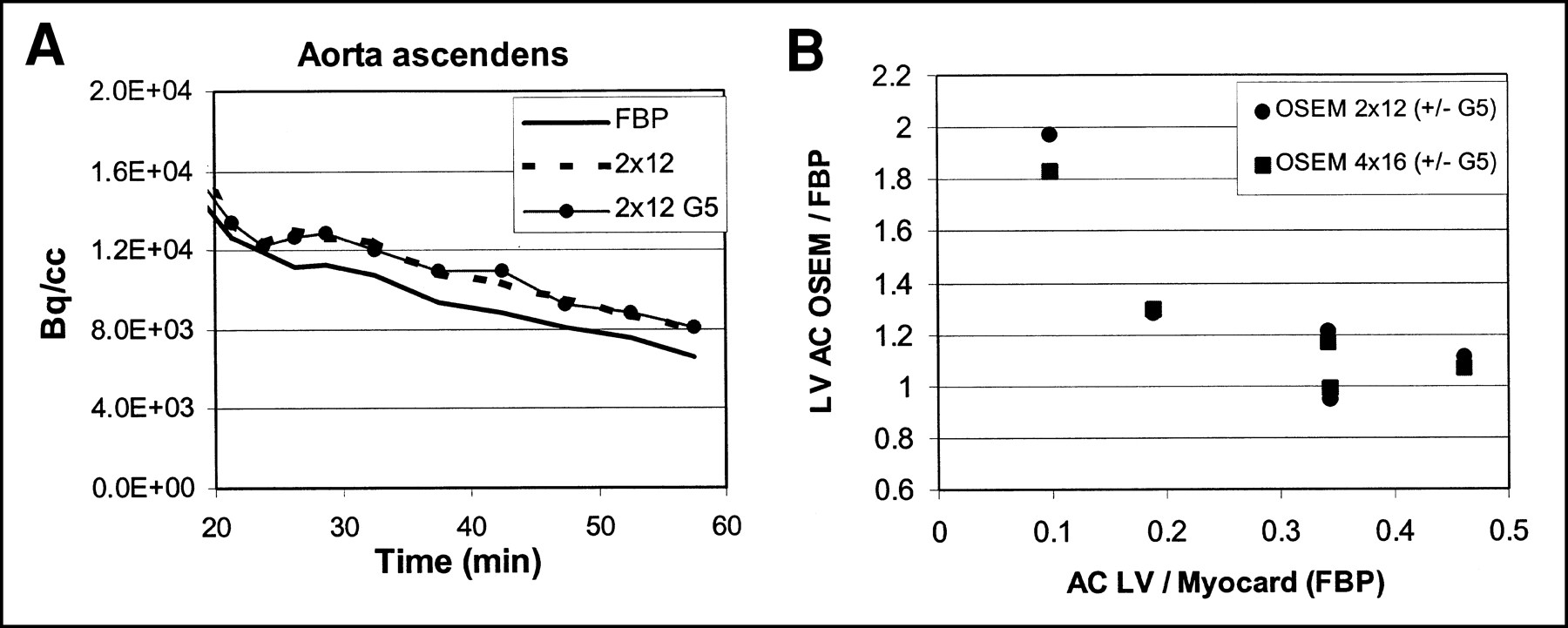
  • FIGURE 7.
    • Download figure
    • Open in new tab
    • Download powerpoint
    FIGURE 7.

    (A) Example of time–activity curve from ROI on aorta ascendens for last 35 min of FDG scan. (B) Bias of left ventricular (LV) activity concentration (AC) as function of image contrast for OSEM 2 × 12 with and without smoothing and OSEM 4 × 16 with and without smoothing.

Tables

  • Figures
    • View popup
    TABLE 1

    Values for C1, C2, and Ratio C2/C1 for Various Reconstruction Methods

    ReconstructionC1C2C2/C1
    FBP307860.28
    OSEM 2 × 12553550.10
    OSEM 3 × 242,4601850.075
    OSEM 12 × 243.1 × 1054.6 × 1030.015
    • Values calculated with Equation 1.

    • View popup
    TABLE 2

    Values of C1 and PL of Equation 4 for Various OSEM Iterations

    ReconstructionC1PL
    OSEM 2 × 125530.50
    OSEM 3 × 242,4600.43
    OSEM 12 × 243.1 × 1050.35
    • View popup
    TABLE 3

    Ratio of ROI Values (OSEM/FBP) for Interval 45–60 Minutes of FDG Scan

    TissueROI2 × 124 × 16G5
    2 × 124 × 16
    MyocardiumShort axis, template ROI1.101.081.081.05
    Axial slices, isocontour ROI1.131.211.011.02
    Tumor2.5- to 5-cm diameter1.14—0.99—
    BrainGray matter0.981.040.940.99
    • G5 = with smoothing.

    • Data are given for OSEM 2 × 12, 4 × 16 with and without 5-mm FWHM Gaussian smoothing of reconstructed image. Uncertainty of these ratios is 2% (1 SD) or less.

    • View popup
    TABLE 4

    Ratio of MRglu (OSEM/FBP) for Various OSEM Reconstruction Techniques

    TissueROI2 × 124 × 16G5
    2 × 124 × 16
    MyocardiumShort axis, template ROI1.111.091.081.05
    Axial slices, isocontour ROI1.091.151.021.02
    Tumor2.5- to 5-cm diameter1.14—1.01—
    BrainGray matter0.971.020.940.98
    • G5 = with smoothing.

    • Data are given for OSEM 2 × 12, 4 × 16 with and without 5-mm FWHM Gaussian smoothing of reconstructed image. Uncertainty of these ratios is 3% (1 SD) or less for brain and myocardium. For tumors, larger variation of this ratio was observed, as shown in Figure 6.

PreviousNext
Back to top

In this issue

Journal of Nuclear Medicine
Vol. 42, Issue 5
May 1, 2001
  • Table of Contents
  • Index by author
Print
Download PDF
Article Alerts
Sign In to Email Alerts with your Email Address
Email Article

Thank you for your interest in spreading the word on Journal of Nuclear Medicine.

NOTE: We only request your email address so that the person you are recommending the page to knows that you wanted them to see it, and that it is not junk mail. We do not capture any email address.

Enter multiple addresses on separate lines or separate them with commas.
Experimental and Clinical Evaluation of Iterative Reconstruction (OSEM) in Dynamic PET: Quantitative Characteristics and Effects on Kinetic Modeling
(Your Name) has sent you a message from Journal of Nuclear Medicine
(Your Name) thought you would like to see the Journal of Nuclear Medicine web site.
Citation Tools
Experimental and Clinical Evaluation of Iterative Reconstruction (OSEM) in Dynamic PET: Quantitative Characteristics and Effects on Kinetic Modeling
Ronald Boellaard, Arthur van Lingen, Adriaan A. Lammertsma
Journal of Nuclear Medicine May 2001, 42 (5) 808-817;

Citation Manager Formats

  • BibTeX
  • Bookends
  • EasyBib
  • EndNote (tagged)
  • EndNote 8 (xml)
  • Medlars
  • Mendeley
  • Papers
  • RefWorks Tagged
  • Ref Manager
  • RIS
  • Zotero
Share
Experimental and Clinical Evaluation of Iterative Reconstruction (OSEM) in Dynamic PET: Quantitative Characteristics and Effects on Kinetic Modeling
Ronald Boellaard, Arthur van Lingen, Adriaan A. Lammertsma
Journal of Nuclear Medicine May 2001, 42 (5) 808-817;
Twitter logo Facebook logo LinkedIn logo Mendeley logo
  • Tweet Widget
  • Facebook Like
  • Google Plus One
Bookmark this article

Jump to section

  • Article
    • Abstract
    • MATERIALS AND METHODS
    • RESULTS
    • DISCUSSION
    • CONCLUSION
    • Acknowledgments
    • Footnotes
    • REFERENCES
  • Figures & Data
  • Info & Metrics
  • PDF

Related Articles

  • No related articles found.
  • PubMed
  • Google Scholar

Cited By...

  • Consensus Recommendations on the Use of 18F-FDG PET/CT in Lung Disease
  • Forward to the Past: The Case for Quantitative PET Imaging
  • A Monte Carlo Study of the Dependence of Early Frame Sampling on Uncertainty and Bias in Pharmacokinetic Parameters from Dynamic PET
  • Using the NEMA NU 4 PET Image Quality Phantom in Multipinhole Small-Animal SPECT
  • Effects of Image Characteristics on Performance of Tumor Delineation Methods: A Test-Retest Assessment
  • Optimization of the Injected Activity in Dynamic 3D PET: A Generalized Approach Using Patient-Specific NECs as Demonstrated by a Series of 15O-H2O Scans
  • HRRT Versus HR+ Human Brain PET Studies: An Interscanner Test-Retest Study
  • Accuracy of 3-Dimensional Reconstruction Algorithms for the High-Resolution Research Tomograph
  • Effect of Reconstruction Algorithms on Myocardial Blood Flow Measurement with 13N-Ammonia PET
  • 18F-Labeled Bombesin Analogs for Targeting GRP Receptor-Expressing Prostate Cancer
  • Prognostic Relevance of Response Evaluation Using [18F]-2-Fluoro-2-Deoxy-D-Glucose Positron Emission Tomography in Patients With Locally Advanced Non-Small-Cell Lung Cancer
  • PET/CT: Challenge for Nuclear Cardiology
  • Delayed Contrast Enhancement and Perfusable Tissue Index in Hypertrophic Cardiomyopathy: Comparison Between Cardiac MRI and PET
  • Influence of Reconstruction Iterations on 18F-FDG PET/CT Standardized Uptake Values
  • Quantitative Comparison of Analytic and Iterative Reconstruction Methods in 2- and 3-Dimensional Dynamic Cardiac 18F-FDG PET
  • Number of Iterations When Comparing MLEM/OSEM with FBP
  • Effects of Noise, Image Resolution, and ROI Definition on the Accuracy of Standard Uptake Values: A Simulation Study
  • Perfusable Tissue Index as a Potential Marker of Fibrosis in Patients with Idiopathic Dilated Cardiomyopathy
  • CT Attenuation Correction for Myocardial Perfusion Quantification Using a PET/CT Hybrid Scanner
  • Clinical Implications of Different Image Reconstruction Parameters for Interpretation of Whole-Body PET Studies in Cancer Patients
  • Postinjection Transmission Scanning in Myocardial 18F-FDG PET Studies Using Both Filtered Backprojection and Iterative Reconstruction
  • PET Instrumentation and Reconstruction Algorithms in Whole-Body Applications
  • Methods to Monitor Response to Chemotherapy in Non-Small Cell Lung Cancer with 18F-FDG PET
  • Measurement of Perfusion in Stage IIIA-N2 Non-Small Cell Lung Cancer Using H215O and Positron Emission Tomography
  • Google Scholar

More in this TOC Section

  • PET/MRI of Hypoxic Atherosclerosis Using 64Cu-ATSM in a Rabbit Model
  • Tumor Uptake of Anti-CD20 Fabs Depends on Tumor Perfusion
  • How Sensitive Is the Upper Gastrointestinal Tract to 90Y Radioembolization? A Histologic and Dosimetric Analysis in a Porcine Model
Show more Basic Science Investigations

Similar Articles

SNMMI

© 2025 SNMMI

Powered by HighWire