


Abstract
The aim of this study was to evaluate the diagnostic value of a new somatostatin analog, 68Ga-labeled 1,4,7,10-tetraazacyclododecane-N,N′,N″,N″′-tetraacetic acid-d-Phe1-Tyr3-octreotide (68Ga-DOTA-TOC), for PET in patients with known or suspected neuroendocrine tumors. PET was compared with conventional scintigraphy and dedicated CT. Methods: Eighty-four patients (48 men, 36 women; age range, 28–79 y; mean age ± SD, 58.2 ± 12.2 y) were prospectively studied. For analysis, patients were divided into 3 groups: detection of unknown primary tumor in the presence of clinical or biochemical suspicion of neuroendocrine malignancy (n = 13 patients), initial tumor staging (n = 36 patients), and follow-up after therapy (n = 35 patients). Each patient received 100–150 MBq 68Ga-DOTA-TOC. Imaging results of PET were compared with 99mTc-labeled hydrazinonicotinyl-Tyr3-octreotide (99mTc-HYNIC-TOC) and 111In-DOTA-TOC. CT was also performed on every patient using a multidetector scanner. Each imaging modality was interpreted separately by observers who were unaware of imaging findings before comparison with PET. The gold standard for defining true-positive (TP), true-negative (TN), false-positive (FP), and false-negative (FN) results was based on all available histologic, imaging, and follow-up findings. Results: PET was TP in 69 patients, TN in 12 patients, FP in 1 patient, and FN in 2 patients, indicating a sensitivity of 97%, a specificity of 92%, and an accuracy of 96%. The FP finding was caused by enhanced tracer accumulation in the pancreatic head, and the FN results were obtained in patients with a tumor of the gastrointestinal tract displaying liver metastases. 68Ga-DOTA-TOC showed higher diagnostic efficacy compared with SPECT (TP in 37 patients, TN in 12 patients, FP in 1 patient, and FN in 34 patients) and diagnostic CT (TP in 41 patients, TN in 12 patients, FP in 5 patients, and FN in 26 patients). This difference was of statistical significance (P < 0.001). However, the combined use of PET and CT showed the highest overall accuracy. Conclusion: 68Ga-DOTA-TOC PET shows a significantly higher detection rate compared with conventional somatostatin receptor scintigraphy and diagnostic CT with clinical impact in a considerable number of patients.
Neuroendocrine tumors (NET) are a heterogeneous group of neoplasms that originate from the neural crest. These tumors are characterized by their ability to overexpress somatostatin (SST) receptors in most cells deriving from so-called neuroendocrine dispersed cells (1). The main primary sites are the gastrointestinal tract and the lung (2,3), but NET can also originate from various other sites, such as the head and neck region or the prostate.
Scintigraphy with radiolabeled SST analogs, first with an 123I label (4) and subsequently with an 111In (4,5) and 99mTc label (6), has proven useful in diagnosing these tumors. This method also shows the content of SST receptors that might indicate efficacy for treatment with octreotide or other SST analogs (1). Although SST receptor scintigraphy (SRS) shows high efficacy for whole-body imaging, there are some limitations in organs with higher physiologic uptake—for example, liver (7,8)—and in terms of detection of smaller lesions due to the detection limits of SPECT for the mentioned radiotracers. 18F-FDG PET scanning is another widely accepted imaging approach in clinical oncology. Although 18F-FDG PET shows high spatial resolution, unlike for many other malignancies, it is not indicated primarily for NET because of its poor sensitivity to detect tumors with low metabolic activity and slow growth (9).
On the other hand, morphologically orientated imaging techniques, such as contrast-enhanced multidetector CT, permit rapid volumetric acquisition and dynamic analysis of the contrast agent, which provides higher image resolution and gives information about the vascular phase for detection of even small-sized lesions of neuroendocrine origin (10). However, these methods sometimes lack specificity, as conclusions regarding malignant involvement of organ structures are based only on size criteria and the contrast enhancement pattern (11).
Initial patient studies have demonstrated the capability of PET technology using 68Ga-labeled 1,4,7,10-tetraazacyclododecane-N,N′,N′′,N′′′-tetraacetic acid-d-Phe1-Tyr3-octreotide (68Ga-DOTA-TOC) (12,13). This method clearly offers higher resolution and improved pharmacokinetcs compared with SRS, with promising results in the detection of SST receptor-expressing tumors.
The aim of the present study was to provide data on diagnostic efficacy of the new radiopharmaceutical 68Ga-DOTA-TOC for PET in a larger series of patients with known or suspected NET. The study included comparison with SPECT and CT. Patient and site-related differences of the 3 imaging modalities were analyzed in a head-to-head comparison by means of image fusion.
MATERIALS AND METHODS
Patients
From September 2004 to April 2006, 84 consecutive patients (48 men, 36 women; age range, 28–79 y; mean age ± SD, 58.2 ± 12.2 y) were enrolled in this prospective phase IIb study. For analysis, the patients who were investigated were divided into 3 groups (14): The first group consisted of patients who underwent imaging for the initial detection and localization of suspected NET and potential metastases in the presence of clinical or biochemical suspicion (detection; n = 13). Patients with histologically proven neuroendocrine malignancies were enrolled for staging purposes in the second group (staging; n = 36). In the third group, patients were referred during posttherapy follow-up (follow-up; n = 35) to exclude or to detect tumor recurrence. In the last 2 groups, at least 1 tumor manifestation was histologically confirmed in patients with multiple metastases. The patient characteristics are summarized in Table 1.
Patient Characteristics
Four patients had hypoglycemia with symptoms of both neuroglycopenia and catecholamine response. During symptoms, blood glucose levels were <40 mg/dL. Therefore, a pancreatic islet cell tumor was considered in these patients.
NET are generally differentiated between those producing hormone-related symptoms (e.g., flush or diarrhea) and those presenting without any hormonal symptoms. Accordingly, 27 patients with clinical and biochemical signs for a secreting tumor and 57 patients with a nonfunctional tumor were included.
In patients who were referred for restaging during follow-up (n = 35), various therapeutic procedures were performed before inclusion. Most of these patients were treated by surgery (n = 29), some of them without further treatment (n = 6). Additional drug therapy was administered to 23 patients. Seven patients were treated with chemotherapy, and 16 patients were treated with long-acting SST analogs alone or in combination with interferon-α.
Written informed consent was obtained from all patients before being included in the study, and the study was approved by the local ethics committee.
PET
Preparation of 68Ga-DOTA-TOC.
68Ga-DOTA-TOC was prepared using a modification of the method described by Breeman et al. (15). Briefly, a TiO2-based commercially available 68Ge/68Ga generator (Cyclotron Inc.) was eluted with 0.1N hydrochloric acid, and a 1.2-mL fraction was added to 20–40 μg of DOTA-TOC; pH was adjusted to 3.5–4.0 by adding 1 mol/L sodium acetate solution, which was followed by heating to 100°C for 7 min. The reaction solution was passed over a C18 cartridge (Sep-Pak; Waters), washed with 4 mL of water, and finally eluted with 0.5 mL 95% ethanol, which was followed by saline through a 0.2-μm sterile filter. Radiochemical purity, as determined by instant thin-layer chromatography and high-performance liquid chromatography, exceeded 95% in all cases.
Data Acquisition and Processing.
Data acquisition was performed by means of a dedicated PET scanner (GE Advance) with 15-cm axial field of view (FOV) and 55-cm transaxial FOV. Patients were imaged in 2-dimensional mode using septa. the duration of acquisition was 5 min per bed position (axial FOV) in emission mode. For the evaluation of the best imaging time, 3 emission image sets were acquired at 20, 60, and 100 min after injection. Because of scan time limitations, the first and last acquisitions scanned only the torso (4 bed positions), whereas the acquisition at 1 h after injection was performed as a whole-body scan (from head to middle of the upper leg, usually 7 bed positions). Attenuation correction was performed by means of transmission data (68Ga pin source, 3 min per bed position). Image reconstruction was performed with the system's implementation of the ordered-subsets expectation maximization iterative algorithm, using segmented attenuation correction and model-based scatter correction. The settings for iterative reconstruction were 2 iterations and 26 subsets, with 4-mm full width at half maximum (FWHM) interupdate filtering and 6-mm FWHM after filtering. The attenuation correction settings were set to segmented correction with 10-mm smoothing. No axial smoothing was performed for either emission or transmission data. For the first 8 patients, average tissue standardized uptake values (SUVs) (SUVbw, units of g/mL; bw indicates body weight) have been determined by means of manually drawn regions of interest delineating the respective tissue thresholded to 50% of the maximum uptake in that tissue.
SRS
99mTc-Labeled hydrazinonicotinyl-Tyr3-octreotide (99mTc-HYNIC-TOC) was prepared using a kit formulation as recently described (16). Each patient received a mean activity of 400 MBq (intravenously) of the tracer. Whole-body imaging was performed at 2 and 4 h after injection using a dual-detector VertexPlus scintillation camera (Philips), which was followed by SPECT (6). The scan speed for whole-body imaging was 10 cm/min when using the 99mTc-labeled derivative. The camera was equipped with a low-energy, all-purpose, parallel-hole collimator (window setting, 140 keV; width, 10%; 180° rotation detector head; 64 projections; 128 × 128 matrix; 40-s acquisition time per projection). The SPECT image data were reconstructed by standard filtered backprojection using a Butterworth filter.
DOTA-TOC was radiolabeled as reported elsewhere (17). 111In-DOTA-TOC whole-body scintigraphy in anterior and posterior views was performed at 4, 24, and 48 h after a single-dose injection of 150 MBq of the 111In-labeled radiopharmaceutical. Scintigraphic acquisitions were obtained with the same double-head γ-camera as described (ADAC; VertexPlus), equipped with a medium-energy, parallel-hole collimator (window setting, 172 and 246 keV; window width, 20%). The scan speed for whole-body imaging was 5 cm/min using the 111In-labeled radiopharmaceutical. SPECT was acquired after 24-h whole-body imaging using the same reconstruction algorithm as mentioned earlier.
Sixty-six patients were investigated with only 1 tracer: 33 patients with 99mTc-HYNIC-TOC and 33 with 111In-DOTA-TOC. In 18 patients, both radiopharmaceuticals were used for comparison with PET and CT.
CT and Image Fusion Procedure
Helical CT scans of the thorax and the abdomen with a slice thickness of 2.5 mm were obtained with the HiSpeed CT/i Advantage scanner (GE Healthcare). Typically, 150 mL (twice the weight of the patient in kilograms) iopromidum contrast media (Ultravist 370; Schering) were administered at 5 mL/s, with scan delays of approximately 30 s for the late arterial phase and 70 s for the portal phase.
For image fusion, the PET, SPECT, and CT scans were performed sequentially using an individualized vacuum mattress with external markers attached to it. For every image acquisition (PET, SPECT, and CT), the patient was repositioned into the vacuum mattress (11). The image fusion procedure was used for anatomic delineation of abnormal findings in SPECT and PET.
Interpretation and Data Evaluation
PET and SPECT studies were interpreted independently by 2 experienced nuclear medicine physicians. Corresponding studies were compared lesion by lesion for final analysis and ruled as matching or mismatching by the 2 nuclear medicine specialists. If the result of the 2 viewers was discordant, a third reader—who acted as referee—was consulted. They were aware of the patients' clinical history, which was provided by the referring physician but were unaware of any result of other imaging modalities. The criteria for image interpretation of PET and SPECT are summarized in Table 2. As a measure for diagnostic yield, the number of lesions that could be identified clearly as single foci was determined. Lesions within the liver were rated as 1 organ metastasis, considering the irregular configuration and confluence of some lesions, so that an individual metastasis frequently was not delineated. A lesion-by-lesion analysis was performed for all other tumor foci. Concordant findings on nuclear medicine techniques (PET and SPECT) and CT meant that both techniques (PET or SPECT and CT) were consistent with malignancy. In the case of discrepancies with regard to nuclear medicine and CT findings, further assessment of abnormal foci was mandatory—that is, by histologic proof or follow-up controls with CT or MRI after 3 mo and, if necessary, after 6 mo. If malignant evolution on follow-up or progression on therapy was observed, these suggestive findings were considered malignant for the final decision. Those patients with no abnormal findings were monitored over a period of at least 6 mo with repeated CT or MR scans before a scan result was considered true-negative (TN).
Criteria for Visual Study Interpretation
CT and, if necessary, MR scans were interpreted by experienced radiologists who were unaware of the scintigraphic results. A positive diagnosis was based on the specific appearance of malignant disease derived from NET as reported elsewhere (18). Thus far, abnormal findings were assessed histologically in 13 patients. Repeated clinical examinations with CT were performed in 73 cases and with MR in 22 cases. More details are given in Table 1. Attention has been directed toward unproven findings of the scans.
Statistical Analysis
The results of the 3 imaging modalities (PET, SPECT, and CT) were classified as true-positive (TP), TN, false-positive (FP), or false-negative (FN) according to the reference standard, as described earlier. The χ2 test for independence, or the Fisher exact test when appropriate, was used to evaluate differences in lesion detectability when subgroups of the patients being investigated were statistically compared (111In-DOTA-TOC vs. 99mTc-HYNIC-TOC and secreting vs. nonsecreting tumors). The McNemar test of correlated properties was used to statistically compare the imaging results of 68Ga-DOTA-TOC PET with SPECT and diagnostic CT. Analysis was done on a lesion basis and on a patient basis. All P values < 0.05 were considered significant. Cohen's κ-statistic with 95% confidence intervals was calculated to show the degree of association between the techniques. The function of uptake over time was assessed using linear regression analysis.
For evaluation of the clinical value of 68Ga-DOTA-TOC PET in comparison with the other imaging modalities, organ systems were assessed for recognition of any lesion in the tissue. The focus of interest was related to unknown tumor lesions arising in organ systems, unaware of malignant involvement from other imaging techniques (e.g., bone), with clinically relevant information in terms of further patient management.
RESULTS
Biodistribution of 68Ga-DOTA-TOC
Uptake of 68Ga-DOTA-TOC was routinely found in neuroendocrine tissue (pituitary and adrenal glands) and in 57 patients (67.8%) in the pancreatic head without known pathology. Additionally, the spleen and the urinary excretion system also showed enhanced tracer accumulation. Homogeneous uptake was observed in the liver and in 34 patients (40.4%) in the thyroid gland. Because of the excellent tumor-to-organ contrast, NET lesions could be easily identified by visual analysis (Fig. 1). However, anatomic delineation of abnormal findings was difficult without image fusion because of highly specific tracer uptake. Intravenous injection of 68Ga-DOTA-TOC was well tolerated in all patients, and no side effects were observed in any patients after tracer injection. On the basis of an initial evaluation of 8 patients, the optimal time of acquisition was determined to be 100 min after injection. In 1 of these 8 patients, 2 additional thoracic findings were observed when comparing late acquisition 100 min after injection with the earlier acquisition (patient 12). The acquisition protocol was adapted after this initial series with only 1 single whole-body scanning at 100 min after injection. Linear regression analysis of tumor uptake values shows a significant increase in SUVs of 14% (60 min vs. 20 min, R2 = 0.96), 9% (100 vs. 60 min, R2 = 0.97), and 24% (100 vs. 20 min, R2 = 0.89). Interpatient SUVs at different times are given in Table 3.
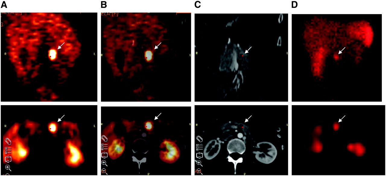
A 28-y-old female was referred for primary diagnosis of a NET because of elevated tumor markers in serum. PET (A) clearly depicted an abnormal focus in upper abdomen (arrow). This lesion could be delineated in the pancreas after image fusion with CT (B). There was also increased contrast medium enhancement in the margin when using helical CT (C). SPECT with 99mTc-HYNIC-TOC was also positive for this tumor in upper abdomen (D). This positive finding was confirmed by histopathology revealing a NET with 1 cm in diameter. (Top) Coronal views; (bottom) axial views.
SUVs at Different Time Points
Analysis on Patient Basis
Results from all 84 patients studied are summarized in Table 4. Among the 84 patients, 68Ga-DOTA-TOC PET was TP in 69 (82.1%), TN in 12 (14.3%), FP in 1 (1.2%), and FN in 2 (2.4%) patients, indicating a sensitivity of 97% (69/71 patients), a specificity of 92% (12/13 patients), and an accuracy of 96% (81/84 patients) on a patient basis. An analysis per patient comparing the scan results of PET with SPECT and with diagnostic CT emphasizes the improved diagnostic efficacy of 68Ga-DOTA-TOC, with a P value of < 0.001 using the McNemar test (Table 5). Cohen's κ-statistic of 0.3 showed only fair association between the techniques.
Results of PET vs. SPECT and CT: Analysis per Patient
Comparison of 3 Imaging Modalities: PET, SPECT, and CT
Analysis on Lesion Basis
68Ga-DOTA-TOC PET studies detected 375 abnormal findings in 70 patients, of which 374 were TP and 1 was FP. This FP finding was found in patient 30, who had clinical symptoms suggestive of a secreting NET and elevated tumor markers (chromogranin A [CgA] of 34.6 U/L and neuron-specific enolase [NSE] of 26.7 μg/L) and who presented with enhanced tracer uptake in the pancreatic head. Surgical exploration was negative, and histology and further follow-up controls did not confirm this finding to be malignant. Overall, 23 abnormal findings in 22 patients were considered malignant in the pancreas. Fourteen of those were found in the pancreatic head, with 1 FP finding.
68Ga-DOTA-TOC was FN in 2 patients. A 47-y-old woman was referred for initial staging of a NET unknown primary (patient 36). PET and SPECT were negative for multiple liver metastases that were histologically confirmed by biopsy. Both nuclear medicine techniques were also negative for histologically confirmed small liver metastases in the other patient, a 67-y-old man (patient 78). This patient was referred for follow-up after surgery and chemotherapy of a rectal tumor. In both patients, diagnostic CT revealed a TP scan result.
68Ga-DOTA-TOC and Functional Status of NET
The fraction of patients with clinical and biochemical features of a NET consisted of 18 TP, 8 TN, and 1 FP results, whereas in the group of patients with nonfunctioning tumors, 51 TP, 4 TN, and 2 FN results were observed. When comparing both groups, no statistically significant difference was found for PET (P = 0.96). Both patients with the FN scan result did not show any functional activity of the tumor, whereas the FP result was observed in a patient with elevated CgA level and persisting diarrhea, suggestive of a hormone-active tumor, as mentioned earlier.
PET Versus Scintigraphy (SPECT) and Diagnostic CT
All 3 modalities (PET, SPECT, and CT) showed an equivalent scan result in 39 patients (46.4%), including 27 TP and 12 TN results (Fig. 1).
Discrepancies between PET and SPECT were found in 32 patients (38%), all of whom were TP with PET and FN with SPECT. In this patient group, liver metastases were missed in 10 cases. Twenty-two additional small lymph node metastases also were not detected with SPECT in 15 patients. In 2 patients with carcinoid tumors, small peritoneal deposits escaped detection with SPECT. Furthermore, 32 bone metastases were not delineated by conventional scintigraphy but were positive with 68Ga-DOTA-TOC PET. Discrepancies between PET and CT were found in 34 patients (40.5%), of whom there were 2 TP, 1 TN, 5 FP, and 26 FN findings with CT. FP findings with CT were caused by suggestive small nodular lung lesions in 2 patients and in 2 additional cases by enlarged lymph nodes. One 55-y-old male patient was referred for initial detection of a NET in the case of elevated CgA levels (patient 52). Abdominal CT visualized a lesion in the wall of the jejunum with a diameter of 1.4 cm. The contrast medium showed enhanced uptake of a primary NET. However, PET and SPECT were negative. Surgical exploration revealed a benign leiomyoma, which was proven by histology. Site-related differences are illustrated in Table 6.
Site-Related Findings
Eighteen patients were investigated with both SPECT tracers, yielding a comparable scan result. The 99mTc-labeled compound was TP in 18 patients, TN in 11, FP in 1, and FN in 21 patients. When using 111In-DOTA-TOC, the scan result was TP in 29 patients, TN in 1, and FN in 21 patients. No statistically significant difference was observed between the 2 groups (P = 0.84).
Clinically Valuable Information Obtained by PET
In 18 patients (21.4%), 68Ga-DOTA-TOC provided further clinically relevant information in comparison with diagnostic CT alone, including 9 patients with unknown bone metastases (Fig. 2). The primary tumor or residual tumor at the primary site was demonstrated in 5 patients with 68Ga-DOTA-TOC PET but escaped detection by CT. A 61-y-old woman (patient 65) was referred after treatment of a pulmonary carcinoid tumor for follow-up. Diagnostic CT was negative, but PET revealed small metastatic lesions in the myocardium and in the pancreas, with focally enhanced tracer accumulation. Multiple liver metastases were known in a 47-y-old woman (patient 80) who was investigated during follow-up after surgical resection of a small bowel carcinoid. 68Ga-DOTA-TOC additionally showed a small lesion in the right breast initially not found with the other 2 modalities (Figure 3). This lesion with a diameter of 7–4 mm and 3 other metastases in the liver were surgically removed. In 2 patients, small liver metastases were not shown with diagnostic CT and SPECT (Fig. 4).

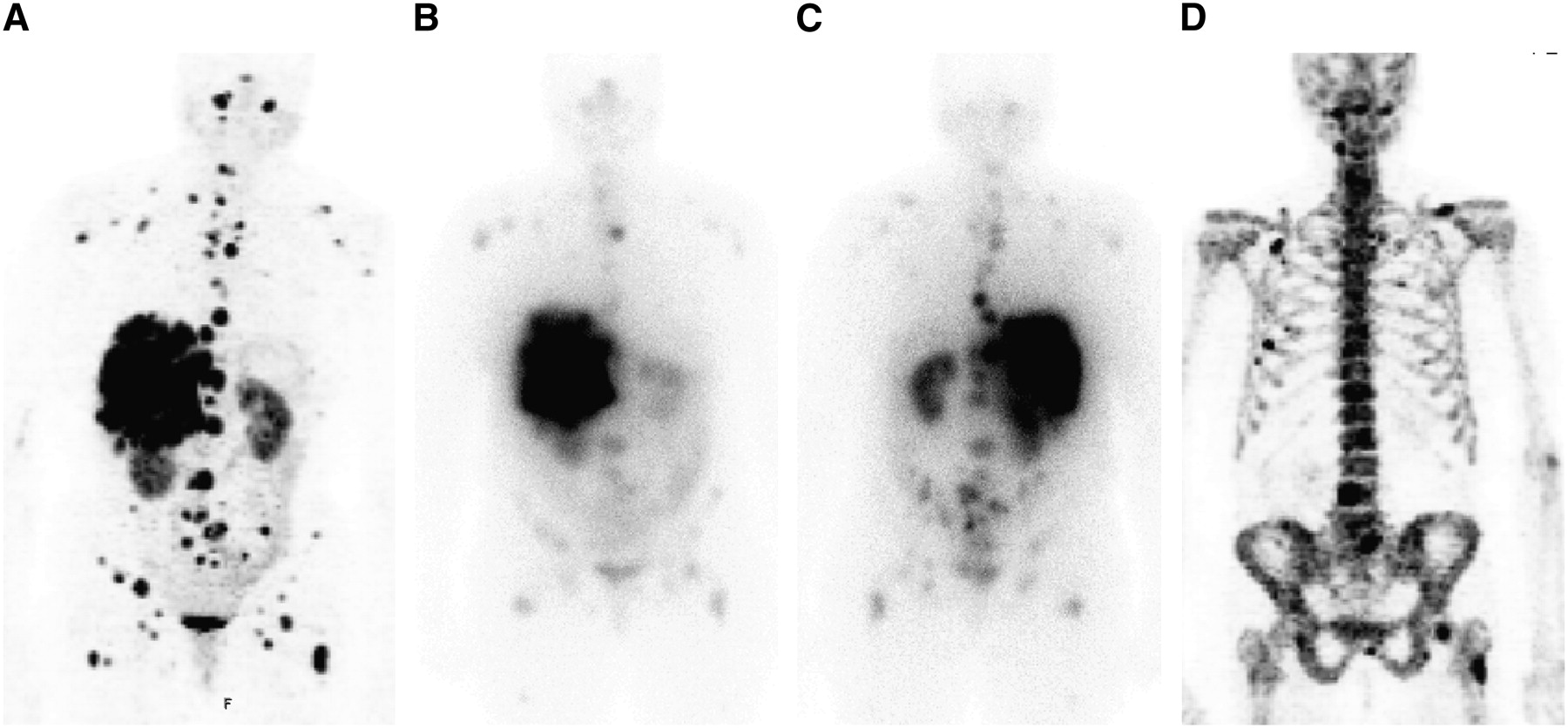
A 56-y-old woman with multiple liver and lymph node metastases was referred for restaging after surgery and chemotherapy. CT presented these tumor lesions; however, it was negative for bone lesions. Beside the visceral metastases, some additional osteoblastic and osteolytic bone metastases were clearly depicted with 68Ga-DOTA-TOC (A). Only some of these bone metastases were delineated by conventional scintigraphy (B, anterior view; C, posterior view). Osteoblastic bone lesions were confirmed by 18F-Na-fluoride PET (D). Retrospective CT analysis after image fusion revealed some of these bone metastases.

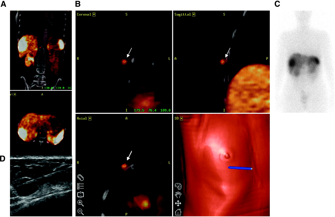
A 47-y-old female patient was referred for scanning after resection of a carcinoid of the ileum. Multiple liver metastases were known (A). Additionally, 68Ga-DOTA-TOC showed a small lesion in right breast (arrows) (B). This finding was initially not detected with CT or scintigraphy (C). Ultrasound-guided fine-needle biopsy confirmed a metastasis in soft tissue derived from the NET with 7- to 4-mm diameter (D). This tumor lesion and 3 liver metastases were consecutively surgically removed.

A 62-y-old male patient was investigated after resection of a small bowel carcinoid. 68Ga-DOTA-TOC PET displayed multiple small liver metastases (A). These liver lesions were negative with the other 2 modalities, CT and scintigraphy (B) including SPECT (C). Ultrasonography (D) and further follow-up controls confirmed these lesions. Diameters of metastases were in the range of 1 cm. Positive PET finding initiated treatment with [177Lu-DOTA0,Tyr3,Thr8]octreotide (177Lu-DOTA-TATE).
Compared with scintigraphy, 68Ga-DOTA-TOC PET provided further valuable clinical information in 12 patients (14.3%). Three patients have just been mentioned. Unknown bone metastases were shown in 5 patients. Surgical intervention was omitted in 3 patients because widespread disease was detected by 68Ga-DOTA-TOC, showing additional unknown distant tumor lesions. One 34-y-old male patient was investigated after chemotherapy and chemoembolization of metastatic lesions in the liver of a NET unknown origin. PET additionally showed the primary tumor in the pancreatic head and local lymph node metastases (patient 34).
DISCUSSION
SRS has gained widespread acceptance as the imaging method of choice in NET patients, showing high sensitivity and good specificity for detection of the primary tumor and secondary lesions (4,14,19–21). However, because of low spatial resolution, this technique has a poor capability to detect lesions with smaller size and lower receptor density. 68Ga-DOTA-TOC has emerged as a new PET tracer showing better results compared with conventional nuclear medicine examinations in a small group of patients (12,13). Initial results were confirmed by our prospective study in a larger number of patients with statistically significant higher diagnostic accuracy compared with conventional SRS as well as diagnostic CT. In 25% (21/84) of patients with NET, 68Ga-DOTA-TOC provided additional information that was obtained with none of the other imaging procedures.
The better imaging properties are based on the higher spatial resolution of PET and on some beneficial pharmacokinetic properties of 68Ga-DOTA-TOC (22) but also need an optimal acquisition protocol. Therefore, in 8 patients images were acquired at different times to evaluate the optimal time for acquisition, which turned out to be 100 min after injection by calculation of SUVs. SUVs were not used for diagnostic purposes, especially as the threshold and averaging method applied in this article is too complex for routine clinical use. Nevertheless, we do not rule out that simple maximum SUVs might be feasible as a clinical tool, taking into account our results.
The difference in detection rate was most pronounced for bone metastases–that is, of all 116 PET-positive lesions, SPECT delineated 84 (72.5%) lesions and CT delineated only 58 lesions (50%). These additional findings have prompted therapeutic interventions in some patients but also have a prognostic implication because unknown distant bone metastases are considered as a negative prognostic factor, possibly requiring a more aggressive treatment regime (23,24). On the other hand, some limitations can be found in the detection of liver metastases using 68Ga-DOTA-TOC, as it is also known for SPECT (7,8). Radiologic techniques are found to be valuable for evaluation of this organ, in which metastases are frequently found in NET patients (25,26). In the present study the combined use of PET and CT also showed the highest overall accuracy for diagnosis of liver metastases, as CT provided complementary information in those 2 patients who were negative with PET. Diagnostic CT additionally reveals the individual anatomy, assisting in delineation of abnormal findings, which was very important in many patients when using 68Ga-DOTA-TOC. On the other hand, tumor deposits—for example, bone metastases—frequently escaped detection by initial CT evaluation. Some of these lesions, however, were consecutively identified after image fusion in the CT scan guided by the findings of the PET scan. This implies that the PET scan is an excellent method for screening of tumor lesions followed by a more directed CT.
The very specific binding of 68Ga-DOTA-TOC may lead to overinterpretation of tracer accumulation. Therefore, interpretation should be done cautiously in organs showing physiologically enhanced tracer uptake. The only FP case, for instance, was found in a patient with clinical features suggestive of a NET presenting focally enhanced tracer uptake in the pancreatic head mimicking the tumor.
One limitation of this study is based on the use of 2 different compounds for conventional scintigraphy, 99mTc-HYNIC-TOC and 111In-DOTA-TOC. However, it has been shown for both radiopharmaceuticals that the detection capability for NET is comparable with 111In-DTPA-d-Phe1-octreotide (where DTPA is diethylenetriaminepentaacetic acid) (6,17,27). Equivalent scan results were also obtained with both tracers in some patients, and no statistical difference was observed when 99mTc-HYNIC-TOC was compared with 111In-DOTA-TOC. Therefore, conventional scintigraphy, including SPECT acquisition, was confined to 1 group for head-to-head comparison with PET.
11C-5-Hydroxytryptophan and 18F-fluoro-l-3,4-dihydroxyphenylalanine are substrates of the intermediary metabolic pathway in terms of the APUD concept (where APUD is amine precursor uptake and decarboxylation). Promising results have been obtained with both radiopharmaceuticals in patients with NET, exceeding the detection rate of SPECT and CT (28,29). A limitation of this concept seems to be that nonfunctioning tumors may be difficult to detect, as accumulation reflects the secretion pattern of peptide hormones (28). Furthermore, a decision on treatment using 90Y-DOTA-TOC or [177Lu-DOTA0,Tyr3,Thr8]octreotide (177Lu-DOTA-TATE) cannot be made on the basis of the uptake behavior in tumor lesions. In contrast, several patients were successfully treated with radiopeptide therapy because of a positive pretherapeutic scan result with 68Ga-DOTA-TOC. With regard to patient convenience, it should be stressed that the whole investigation can be performed within 2 h, thereby creating lower radiation burden compared with some other nuclear medicine techniques as indicated by preclinical (30) and clinical studies (12,13).
The use of a generator for a short-lived radionuclide such as 68Ga provides the basis for convenient, easy use of this radionuclide. Labeling of DOTA-derivatized peptides is straightforward and can be performed in a very short time (<30 min). This guarantees a high flexibility and good availability of this radiopharmaceutical in clinical routine in contrast to 11C-labeled compounds, requiring access to an on-site cyclotron unit, or some 18F-labeled derivatives, such as N-(1-deoxy-d-fructosyl)-N-(2-18F-fluoroproionyl)-Lys0,Tyr3-octreotate (Gluc-Lys(18F-FP)-TOCA) (31), requiring multistep synthesis with several purification steps.
CONCLUSION
Somatostatin receptor PET with 68Ga-DOTA-TOC is superior for the detection of NET compared with SPECT and diagnostic CT in various clinical situations (initial diagnosis, staging, and follow-up). The higher sensitivity for tumor detection has clinical impact in a considerable number of patients, especially when compared with CT. However, the best results are to be achieved by the combination of PET and CT. It also indicates receptor expression for targeted radiopeptide therapy.
Footnotes
-
COPYRIGHT © 2007 by the Society of Nuclear Medicine, Inc.
References
- Received for publication August 13, 2006.
- Accepted for publication November 2, 2006.


Jump to section
Related Articles
Cited By...
- SNMMI Procedure Standard/EANM Practice Guideline for SSTR PET: Imaging Neuroendocrine Tumors
- Importance of PET with 68Ga-Labeled Somatostatin Analogs (perspective on "68Ga-DOTA-Tyr3-Octreotide PET in Neuroendocrine Tumors: Comparison with Somatostatin Receptor Scintigraphy and CT" J Nucl Med. 2007;48:508-518)
- Prognostic Value of 18F-FDG PET/CT in a Large Cohort of Patients with Advanced Metastatic Neuroendocrine Neoplasms Treated with Peptide Receptor Radionuclide Therapy
- Safety, tolerability and clinical implementation of 'ready-to-use 68gallium-DOTA0-Tyr3-octreotide (68Ga-DOTATOC) (SomaKIT TOC) for injection in patients diagnosed with gastroenteropancreatic neuroendocrine tumours (GEP-NETs)
- Improving Contrast and Detectability: Imaging with [55Co]Co-DOTATATE in Comparison with [64Cu]Cu-DOTATATE and [68Ga]Ga-DOTATATE
- 111In-Pentetreotide Scintigraphy Versus 68Ga-DOTATATE PET: Impact on Krenning Scores and Effect of Tumor Burden
- Multimodality imaging in carcinoid heart disease
- Twelve-Year Follow-up After Peptide Receptor Radionuclide Therapy
- CMKLR1-targeting peptide tracers for PET/MR imaging of breast cancer
- Sensitivity Comparison of 68Ga-OPS202 and 68Ga-DOTATOC PET/CT in Patients with Gastroenteropancreatic Neuroendocrine Tumors: A Prospective Phase II Imaging Study
- Safety, Biodistribution, and Radiation Dosimetry of 68Ga-OPS202 in Patients with Gastroenteropancreatic Neuroendocrine Tumors: A Prospective Phase I Imaging Study
- Current Concepts in 68Ga-DOTATATE Imaging of Neuroendocrine Neoplasms: Interpretation, Biodistribution, Dosimetry, and Molecular Strategies
- Somatostatin Receptor 2-Targeting Compounds
- 68Ga-DOTATOC Imaging of Neuroendocrine Tumors: A Systematic Review and Metaanalysis
- Localization of Unknown Primary Site with 68Ga-DOTATOC PET/CT in Patients with Metastatic Neuroendocrine Tumor
- Parametric Net Influx Rate Images of 68Ga-DOTATOC and 68Ga-DOTATATE: Quantitative Accuracy and Improved Image Contrast
- The Impact of Somatostatin Receptor-Directed PET/CT on the Management of Patients with Neuroendocrine Tumor: A Systematic Review and Meta-Analysis
- MIB-1 Index-Stratified Assessment of Dual-Tracer PET/CT with 68Ga-DOTATATE and 18F-FDG and Multimodality Anatomic Imaging in Metastatic Neuroendocrine Tumors of Unknown Primary in a PRRT Workup Setting
- Head-to-Head Comparison of 64Cu-DOTATATE and 68Ga-DOTATOC PET/CT: A Prospective Study of 59 Patients with Neuroendocrine Tumors
- 68Ga-DOTATATE PET/CT Interobserver Agreement for Neuroendocrine Tumor Assessment: Results of a Prospective Study on 50 Patients
- Comparison of the Impact of 68Ga-DOTATATE and 18F-FDG PET/CT on Clinical Management in Patients with Neuroendocrine Tumors
- 68Ga-DOTATOC PET/CT in Patients with Iodine- and 18F-FDG-Negative Differentiated Thyroid Carcinoma and Elevated Serum Thyroglobulin
- A Delphic consensus assessment: imaging and biomarkers in gastroenteropancreatic neuroendocrine tumor disease management
- Clinical Translation of a Click-Labeled 18F-Octreotate Radioligand for Imaging Neuroendocrine Tumors
- Potential value of EUS in pancreatic surveillance of VHL patients
- Safety and Efficacy of 68Ga-DOTATATE PET/CT for Diagnosis, Staging, and Treatment Management of Neuroendocrine Tumors
- Evaluation of the Efficacy of Targeted Imaging Agents
- Prospective Study of 68Ga-DOTATATE Positron Emission Tomography/Computed Tomography for Detecting Gastro-Entero-Pancreatic Neuroendocrine Tumors and Unknown Primary Sites
- Significance of a Single-Time-Point Somatostatin Receptor SPECT/Multiphase CT Protocol in the Diagnostic Work-up of Gastroenteropancreatic Neuroendocrine Neoplasms
- Appendiceal neuroendocrine neoplasms: diagnosis and management
- Prognostic Value of 68Ga-DOTANOC PET/CT SUVmax in Patients with Neuroendocrine Tumors of the Pancreas
- 64Cu-DOTATATE PET for Neuroendocrine Tumors: A Prospective Head-to-Head Comparison with 111In-DTPA-Octreotide in 112 Patients
- Increased 68Ga-DOTATATE Uptake in PET Imaging Discriminates Meningioma and Tumor-Free Tissue
- Can Complementary 68Ga-DOTATATE and 18F-FDG PET/CT Establish the Missing Link Between Histopathology and Therapeutic Approach in Gastroenteropancreatic Neuroendocrine Tumors?
- 68Ga-DOTATATE PET/CT, 99mTc-HYNIC-Octreotide SPECT/CT, and Whole-Body MR Imaging in Detection of Neuroendocrine Tumors: A Prospective Trial
- Preclinical Evaluation of a High-Affinity 18F-Trifluoroborate Octreotate Derivative for Somatostatin Receptor Imaging
- PET/MR in Oncology: Non-18F-FDG Tracers for Routine Applications
- Evaluating digestive neuroendocrine tumor progression and therapeutic responses in the era of targeted therapies: state of the art
- 18F-Fluorodihydroxyphenylalanine PET/CT in Patients with Neuroendocrine Tumors of Unknown Origin: Relation to Tumor Origin and Differentiation
- Promises of Cyclotron-Produced 44Sc as a Diagnostic Match for Trivalent {beta}--Emitters: In Vitro and In Vivo Study of a 44Sc-DOTA-Folate Conjugate
- Comparison of Response Evaluation in Patients with Gastroenteropancreatic and Thoracic Neuroendocrine Tumors After Treatment with [177Lu-DOTA0,Tyr3]Octreotate
- Comparison of 68Ga-DOTANOC and 68Ga-DOTATATE PET/CT Within Patients with Gastroenteropancreatic Neuroendocrine Tumors
- The Role of 68Ga-DOTATATE PET/CT in Suspected Neuroendocrine Tumors
- Tumor Response Assessment to Treatment with [177Lu-DOTA0,Tyr3]Octreotate in Patients with Gastroenteropancreatic and Bronchial Neuroendocrine Tumors: Differential Response of Bone Versus Soft-Tissue Lesions
- Unexpected Sensitivity of sst2 Antagonists to N-Terminal Radiometal Modifications
- A Man with Abdominal Pain: Enough Evidence for Surgery?
- Nuclear medicine imaging of neuroendocrine tumours
- Clinical PET of Neuroendocrine Tumors Using 64Cu-DOTATATE: First-in-Humans Study
- Guidelines for the management of gastroenteropancreatic neuroendocrine (including carcinoid) tumours (NETs)
- 68Ga-DOTATOC Versus 68Ga-DOTATATE PET/CT in Functional Imaging of Neuroendocrine Tumors
- The SNM Practice Guideline for Somatostatin Receptor Scintigraphy 2.0
- Radiopeptide Imaging and Therapy in Europe
- Novel SDHD Gene Mutation (H102R) in a Patient With Metastatic Cervical Paraganglioma Effectively Treated by Peptide Receptor Radionuclide Therapy
- Treatment with Octreotide Does Not Reduce Tumor Uptake of 68Ga-DOTATATE as Measured by PET/CT in Patients with Neuroendocrine Tumors
- Nuclear medicine techniques for the imaging and treatment of neuroendocrine tumours
- 177Lu-DOTATATE Molecular Radiotherapy for Childhood Neuroblastoma
- Incidence of Increased 68Ga-DOTANOC Uptake in the Pancreatic Head in a Large Series of Extrapancreatic NET Patients Studied with Sequential PET/CT
- Somatostatin Receptors as Targets for Nuclear Medicine Imaging and Radionuclide Treatment
- 68Ga-DOTATOC PET/CT of Neuroendocrine Tumors: Spotlight on the CT Phases of a Triple-Phase Protocol
- Expression of somatostatin receptors, dopamine D2 receptors, noradrenaline transporters, and vesicular monoamine transporters in 52 pheochromocytomas and paragangliomas
- Diagnostics of Neuroendocrine Tumours
- Exendin-4-Based Radiopharmaceuticals for Glucagonlike Peptide-1 Receptor PET/CT and SPECT/CT
- The Role of 68Ga-DOTATATE PET in Patients with Neuroendocrine Tumors and Negative or Equivocal Findings on 111In-DTPA-Octreotide Scintigraphy
- 68Ga-DOTANOC PET/CT Clinical Impact in Patients with Neuroendocrine Tumors
- Somatostatin receptor-based imaging and therapy of gastroenteropancreatic neuroendocrine tumors
- Features of Carcinoid Heart Disease Identified by 2- and 3-Dimensional Echocardiography and Cardiac MRI
- A Comparison of 68Ga-DOTATATE and 18F-FDG PET/CT in Pulmonary Neuroendocrine Tumors
- 68Ga-DOTA-Tyr3-Octreotide PET for Assessing Response to Somatostatin-Receptor-Mediated Radionuclide Therapy
- Bone Metastases in Patients with Neuroendocrine Tumor: 68Ga-DOTA-Tyr3-Octreotide PET in Comparison to CT and Bone Scintigraphy
- 99mTc-HYNIC-TOC Scintigraphy Is Superior to 131I-MIBG Imaging in the Evaluation of Extraadrenal Pheochromocytoma
- The clinical value of [18F]fluoro-dihydroxyphenylalanine positron emission tomography in primary diagnosis, staging, and restaging of neuroendocrine tumors
- New Technologies for Human Cancer Imaging
- Tumor Receptor Imaging
- Utility of Radiolabeled Somatostatin Receptor Analogues for Staging/Restaging and Treatment of Somatostatin Receptor-Positive Pediatric Tumors
- Somatostatin Receptor Imaging in Patients with Neuroendocrine Tumors: Not Only SPECT?