


Abstract
Despite many advances in the past years, the treatment of metastatic prostate cancer still remains challenging. In recent years, prostate-specific membrane antigen (PSMA) inhibitors were intensively studied to develop low-molecular-weight ligands for imaging prostate cancer lesions by PET or SPECT. However, the endoradiotherapeutic use of these compounds requires optimization with regard to the radionuclide-chelating agent and the linker moiety between chelator and pharmacophore, which influence the overall pharmacokinetic properties of the resulting radioligand. In an effort to realize both detection and optimal treatment of prostate cancer, a tailor-made novel naphthyl-containing DOTA-conjugated PSMA inhibitor has been developed. Methods: The peptidomimetic structure was synthesized by solid-phase peptide chemistry and characterized using reversed-phase high-performance liquid chromatography and matrix-assisted laser desorption/ionization mass spectrometry. Subsequent 67/68Ga and 177Lu labeling resulted in radiochemical yields of greater than 97% or greater than 99%, respectively. Competitive binding and internalization experiments were performed using the PSMA-positive LNCaP cell line. The in vivo biodistribution and dynamic small-animal PET imaging studies were investigated in BALB/c nu/nu mice bearing LNCaP xenografts. Results: The chemically modified PSMA inhibitor PSMA-617 demonstrated high radiolytic stability for at least 72 h. A high inhibition potency (equilibrium dissociation constant [Ki] = 2.34 ± 2.94 nM on LNCaP; Ki = 0.37 ± 0.21 nM enzymatically determined) and highly efficient internalization into LNCaP cells were demonstrated. The small-animal PET measurements showed high tumor-to-background contrasts as early as 1 h after injection. Organ distribution revealed specific uptake in LNCaP tumors and in the kidneys 1 h after injection. With regard to therapeutic use, the compound exhibited a rapid clearance from the kidneys from 113.3 ± 24.4 at 1 h to 2.13 ± 1.36 percentage injected dose per gram at 24 h. The favorable pharmacokinetics of the molecule led to tumor-to-background ratios of 1,058 (tumor to blood) and 529 (tumor to muscle), respectively, 24 h after injection. Conclusion: The tailor-made DOTA-conjugated PSMA inhibitor PSMA-617 presented here is sustainably refined and advanced with respect to its tumor-targeting and pharmacokinetic properties by systematic chemical modification of the linker region. Therefore, this radiotracer is suitable for a first-in-human theranostic application and may help to improve the clinical management of prostate cancer in the future.
In western societies, prostate cancer (PCa) continues to be the most common cancer in elderly men and the third most frequent cause of cancer-related mortality (1). Consequently, there is a high clinical demand for more effective treatment options in the case of metastatic and hormone-refractory PCa. Specific cancer targeting based on low-molecular-weight radioligands may offer a more accurate and rapid visualization, improved staging, and highly effective endoradiotherapy.
On the basis of small molecules, promising PET radiotracers have been investigated in the last couple of years for the imaging of PCa such as 18F-fluoro- or 11C-choline (2,3), 18F-fluoro- or 11C-acetate, 11C-methionine, and peptidyl radiotracers based on the gastrin-releasing peptide receptor and the prostate-specific membrane antigen (PSMA) (4–6).
Because PSMA is strongly expressed in PCa and upregulated in poorly differentiated, metastatic, and hormone-refractory carcinomas (7,8), it represents a highly attractive target in nuclear medicine, potentially meeting the clinical requirements for an effective therapy of metastatic PCa. In particular, urea-based peptidomimetic inhibitors of PSMA were investigated mainly for diagnosis and shown to image advantageously PSMA-expressing PCa (9–11). Because of low expression levels in healthy tissue, however, PSMA has additionally the potential for high-dose endoradiotherapy, with minimized radioactivity-related side effects.
An in vivo theranostic approach combines the potential of both diagnosis and therapy in one and the same targeting molecule by labeling with either a diagnostic or a suitable therapeutic radionuclide. The β emitters such as 90Y, 131I, and 177Lu are appropriate candidates for current systemic radionuclide therapy. Both 131I and 177Lu emit γ radiation in addition to its β– particle, whereas 90Y is a pure β– emitter (12). Recently, the promising therapeutic low-molecular-weight compound 131I-MIP-1095 was clinically investigated. In this study, 28 men with metastatic castration-resistant PCa were treated with 131I-MIP-1095 (mean injected activity, 4.8 GBq). The treatment has shown a significant impact on the tumor lesions and prostate-specific antigen (PSA) values and resulted in a reduction of bone pain. However, because of the high fraction of γ radiation, patients were obliged to stay in the hospital for around 1 wk. In addition, mild hematologic toxicities were observed (13). Endoradiotherapy with the aforementioned radiometals, however, bear a high potential to improve the clinical situation. For example, 177Lu presents a lower proportion of γ radiation, which would result in a reduced stay in the hospital and lower hemotoxicity in comparison to 131I (12).
Thus, a novel theranostic compound was designed consisting of 3 components: the pharmacophore glutamate-urea-lysine; the chelator DOTA, able to complex both 68Ga and 177Lu; and a linker connecting these 2 entities. The linker turned out to be crucial for high imaging contrasts (14) because the chemical structure determines the internalization potency of the PSMA inhibitors (15). Besides the targeting properties, modifications of the linker might also influence the pharmacokinetic properties of peptidomimetic PSMA inhibitors and, therefore, improve their therapeutic potential (16). This study presents the preclinical evaluation of a tailor-made linker-optimized theranostic PSMA inhibitor with considerably optimized pharmacokinetic and targeting properties. Finally, the first individual clinical diagnostic experience with this novel radiotracer is shown, underlining its clinical potential.
MATERIALS AND METHODS
All chemicals, reagents, and solvents for the synthesis and analysis of the compound were analytic grade (for radiosynthesis ultrapure and metal-free); purchased from Sigma Aldrich, Merck, Iris Biotech, or CheMatech; and used without further purification.
The synthesis of PSMA-617 is summarized in Figure 1. The peptidomimetic glutamate-urea-lysine binding motif (steps 1–6) and the linker were synthesized by solid-phase peptide chemistry as previously described (17).
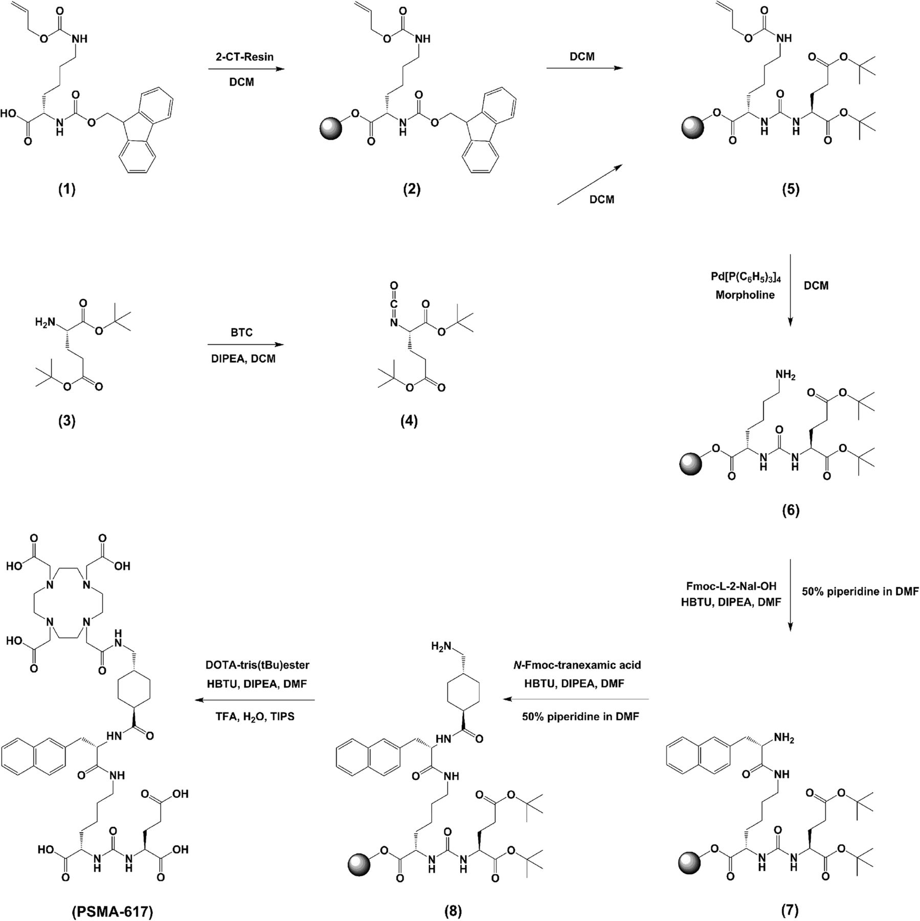
Synthesis of DOTA-conjugated PSMA inhibitor PSMA-617.
The conjugation of the chelator was performed using DOTA-tris(tBu)ester (the supplemental materials provide detailed synthesis information; supplemental materials are available at http://jnm.snmjournals.org). The final product (PSMA-617) was cleaved from the resin and deprotected.
The compound (∼42% yield) was analyzed and purified by reversed-phase high-performance liquid chromatography (RP-HPLC) and matrix-assisted laser desorption/ionization mass spectrometry (MALDI-MS) (the supplemental materials provide detailed method information).
68Ga (half-life [T1/2], 68 min; maximum energy of positrons [β+], 1.9 MeV [88%]) was gained from a 68Ge/68Ga generator based on a pyrogallol resin support (18) as [68Ga]GaCl3 in 0.1 M HCl. 67Ga (T1/2, 3.26 d; energy of photons [γ]/roentgen radiation [x], 94 keV [40%], 184 keV [24%], 296 keV [22%]) was purchased from MDS Nordion as [67Ga]GaCl3 in 0.1 M HCl. 177Lu (T1/2, 6.71 d; maximum energy of electrons [β−], 497 keV [79%]; energy of photons [γ]/roentgen radiation [x], 113 keV [6%], 208 keV [11%]) was obtained from PerkinElmer as [177Lu]LuCl3 in 0.05 M HCl.
Typically, 80–95 μL of 2.4 M HEPES (4-(2-hydroxyethyl)-1-piperazineethanesulfonic acid; pH 7.5) were mixed with 20 μl of [68Ga]Ga3+ eluate (∼70 MBq) or 5 μL of [67Ga]GaCl3 (∼50 MBq) and adjusted to pH 4.0 with 10%–30% NaOH or 0.1 M HCl, respectively. Subsequently, 5 μL of 0.1–1 mM PSMA-617 (0.5–5 nmol) in 0.1 M HEPES were added. The reaction mixture was incubated at 95°C for 15 min. Radiolabeling was performed without any separation of labeled and nonlabeled compound. The specific activity for [68Ga]PSMA-617 was in the range of 14–140 GBq/μmol and for [67Ga]PSMA-617 in the range of 10–100 GBq/μmol. For 177Lu labeling, 112 μL of sodium acetate (0.4 M; pH 5.0), 5 μL of [177Lu]LuCl3 (∼20 MBq), and 5 μL of 0.1–1 mM PSMA-617 (0.5–5 nmol) in 0.1 M HEPES were mixed and incubated for 20 min at 95°C. The specific activity was in the range of 4–40 GBq/μmol. The radiochemical yield was determined by analytic RP-HPLC and reversed-phase thin-layer chromatography (RP-TLC) on silica gel plates (60 RP-18F254S; Merck) with 0.1 M sodium citrate as a mobile phase.
The radiochemical stability of 67/68Ga-labeled PSMA-617 in phosphate-buffered saline (PBS) and human serum up to 72 h at 37°C was tested by RP-TLC. The lipophilicity was determined via the distribution of 67/68Ga-labeled PSMA-617 in the 2-phase system n-octanol and HEPES buffer. The serum protein binding was analyzed by gel filtration (the supplemental materials provide detailed method information).
Both in vitro and in vivo experiments were performed using the PSMA-positive LNCaP cell line (androgen-sensitive human lymph node metastatic lesion of prostatic adenocarcinoma, CRL-1740 [American Type Culture Collection]). The cells were supplemented with 10% fetal calf serum and l-glutamine and incubated at 37°C in an environment of humidified air containing 5% CO2. For in vivo experiments, 8-wk-old BALB/c nu/nu mice were subcutaneously inoculated into the right trunk with 5 × 10⁶ LNCaP cells in 50% Matrigel(Discovery Labware). When the size of the tumor was approximately 1 cm3, the radiolabeled compound was injected via the tail vein (∼30 MBq, 0.5 nmol for small-animal PET imaging; ∼1 MBq, 0.06 nmol for organ distribution).
The binding affinity of PSMA-617 was assayed by enzyme-based (NAALADase) and cell-based competitive assays. Data obtained from both experiments were fitted using a nonlinear regression algorithm (GraphPad Prism 5; GraphPad Software) to obtain the 50% inhibitory concentration.
The NAALADase assay is based on a competitive reaction of recombinant human PSMA (R&D Systems) with nonlabeled PSMA-617 and was performed as previously described (17) (the supplemental materials provide detailed method information).
Meanwhile, PSMA-617 became commercially available (CAS no. 9933, PSMA-617; ABX Advanced Biochemical Compounds, chemical characteristics were proved to be identical) and was additionally used as radioligand in the internalization assay. The assay was performed as described previously (19) (the supplemental materials provide detailed method information).
For small-animal PET imaging with 68Ga-labeled PSMA-617, a 50-min dynamic scan and a static scan from 100 to 120 min after injection were obtained (the supplemental materials provide detailed method information).
For organ distribution, the animals were sacrificed after indicated time points (from 1 to 24 h). The distributed radioactivity (67/68Ga or 177Lu, respectively) was measured in all dissected organs and in blood using a γ counter. The values are expressed as percentage injected dose per gram (%ID/g).
The first clinical diagnostic study with [68Ga]Ga-PSMA-617 PET/CT (1 h after injection) was performed as previously published (20–22). The administered mass of [68Ga]Ga-PSMA-617 was 2 μg. The administered activity was 288 MBq. There were no adverse or other clinically detectable pharmacologic effects. No significant changes in vital signs were observed. The outcome from this study will be described in more detail in a further publication.
RESULTS
The chemical analysis is summarized in Table 1. PSMA-617 was stable for at least 6 mo as a lyophilized fluffy white powder and also in a solution of dimethylsulfoxid-d6 or hexadeuterodimethyl sulfoxide, respectively, both at −20°C, as shown by RP-HPLC and MALDI-MS. A precursor amount of 0.5 nmol was labeled with radiogallium, with a radiochemical yield of greater than 90% in 15 min at 95°C, for both 67Ga or 68Ga. Small-animal PET imaging was performed after purification by means of solid-phase extraction using SepPak C18 cartridges (Waters). A higher amount of precursor (5 nmol) resulted in a radiochemical yield of greater than 97%. Radiolabeling with 177Lu gained a radiochemical yield of greater than 99% at low amounts of precursor (0.5 μg, 0.5 nmol).
Analytic Data of Compound PSMA-617
For determination of the radiolytic stability, PSMA-617 was radiolabeled with 68Ga or 67Ga and incubated at 37°C for 1 or 24 h, respectively, both in PBS and in human serum. [68Ga]Ga-PSMA-617 showed a high stability after 1 h in PBS and in human serum, respectively, as indicated by TLC. After 24 h, values demonstrated 1% of free activity in PBS and less than 4% in human serum. Long-term stability with 177Lu did not reveal any free activity after 1 and 24 h, neither in PBS nor in serum. After 48 h, less than 0.4% and after 72 h less than 0.6% of free 177Lu was measured in serum and less than 0.2% and less than 0.6% in PBS, respectively. The stability of the compound was also confirmed via gel filtration with a Sephadex column (Merck). This run did not demonstrate any transfer of activity to human serum proteins after 1 h of incubation at 37°C.
PSMA-617 revealed nanomolar affinity for PSMA on LNCaP cells (equilibrium dissociation constant [Ki] = 2.34 ± 2.94 nM; n = 7). In addition, the inhibition potency was determined for the natGa- and natLu-labeled PSMA-617. No significant difference in binding affinity was observed (Ki = 6.40 ± 1.02 nM for natGa complex; Ki = 6.91 ± 1.32 nM for natLu complex). The binding affinity determined by the enzyme-based NAALADase assay was subnanomolar (Ki = 0.37 ± 0.21 nM; n = 2). 68Ga-labeled PSMA-617 was specifically internalized up to 17.67 ± 4.34 percentage injected activity/106 LNCaP cells (n = 3) and 177Lu-labeled PSMA-617 up to 17.51 ± 3.99 percentage injected activity/106 LNCaP cells (n = 3), both at 37°C.
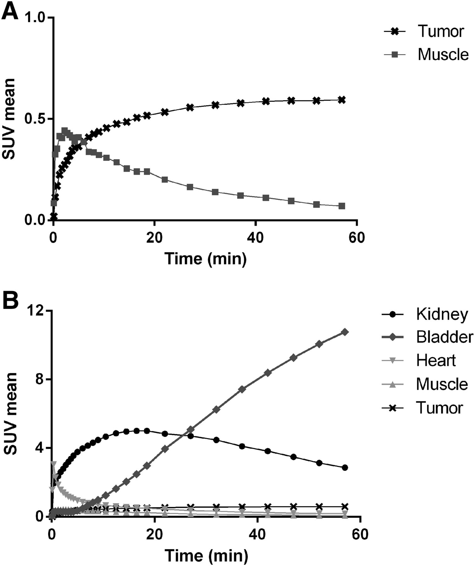
The time–activity curves obtained from dynamic PET showed a high tumor-to-muscle ratio (8.5) at 1 h after injection (Fig. 2A) and fast clearance from the kidneys, followed by a rapid accumulation of radioactivity in the bladder (Fig. 2B).

Time–activity-curves for tumor and background (A) and for relevant organs (B) up to 1 h after injection of 0.5 nmol of 68Ga-labeled PSMA-617. Data are mean standardized uptake value (SUVmean).
The small-animal PET slices (Fig. 3) reveal an increasing tumor uptake up to 2 h after injection and a rapid elimination of radioactivity from other organs, muscles, and blood. The rapid kidney excretion is also demonstrated by the maximum-intensity-projection scans (Fig. 4), whereby tumor uptake is maintained (Fig. 2).

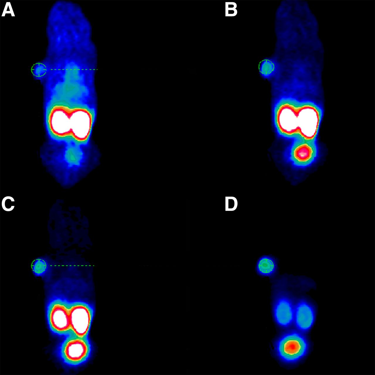
Whole-body coronal slices from small-animal PET imaging of athymic male nude mouse bearing LNCaP tumor xenografts. Tumor-targeting efficacy and pharmacokinetic properties were evaluated by injection of 0.5 nmol of 68Ga-labeled PSMA-617 (∼30 MBq) with following scans at 0–20 min (A), 20–40 min (B), 40–60 min (C), and 120 min (D) after injection.
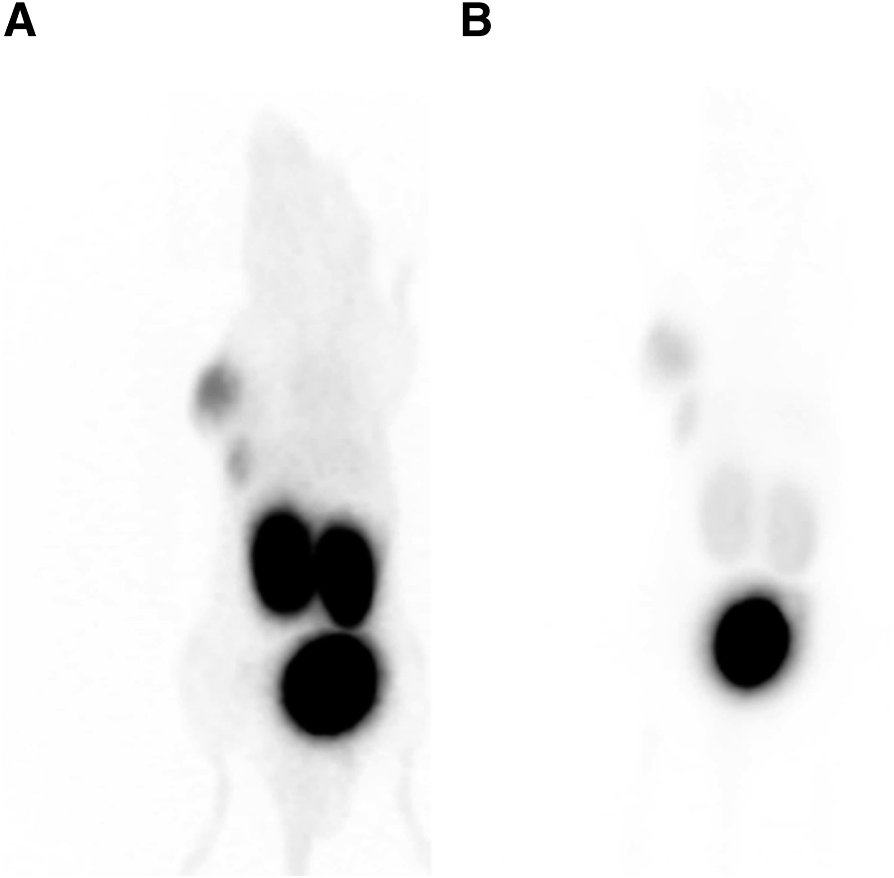
Whole-body coronal scans as maximum-intensity projections at 60 (A) and 120 min (B) after injection of 68Ga-labeled PSMA-617.
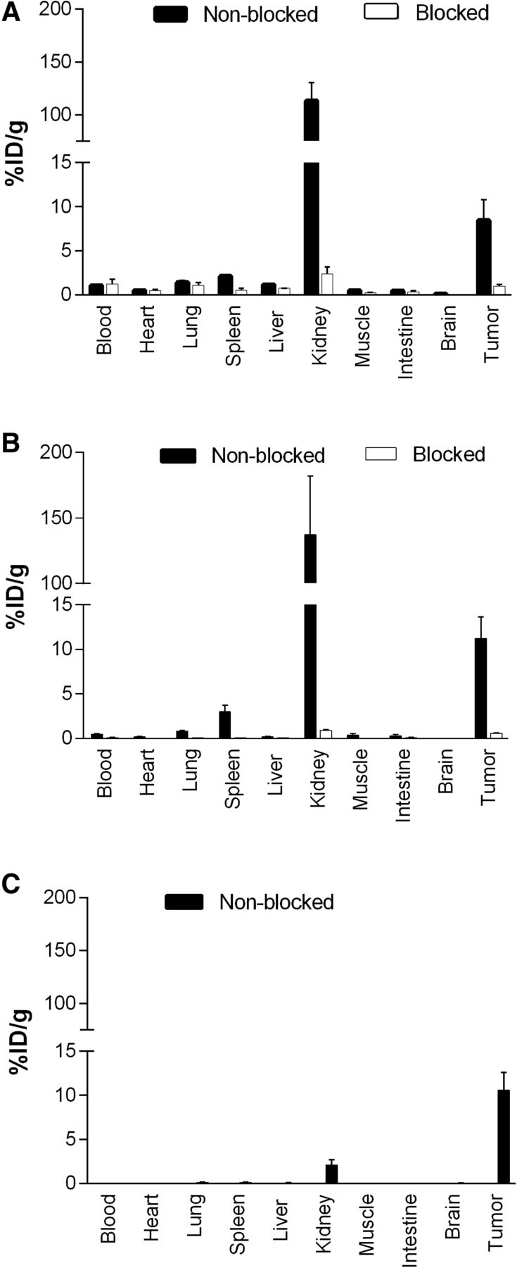
Organ distribution with 68Ga-labeled PSMA-617 after 1 h (n = 3; Fig. 5A) revealed a high specific uptake in LNCaP tumors (8.47 ± 4.09 %ID/g; 0.98 ± 0.32 %ID/g by coinjection of 2-PMPA) and in the kidneys (113.3 ± 24.4 %ID/g). The high uptake in the kidneys was nearly completely blocked (2.38 ± 1.40 %ID/g) by coinjection of 2 mg of 2-PMPA per kilogram. Other organs such as the liver (1.17 ± 0.10 %ID/g), lung (1.41 ± 0.41 %ID/g), and spleen (2.13 ± 0.16 %ID/g) showed rather low uptake and no blocking effect (data not shown), with the exception of the spleen (0.52 ± 0.36 %ID/g). Tumor-to-background ratios were 7.8 (tumor to blood) and 17.1 (tumor to muscle) at 1 h after injection.

Organ distribution of 0.06 nmol of [68Ga]Ga-PSMA-617 at 1 h after injection (A) and 0.06 nmol of [177Lu]Lu-PSMA-617 at 1 (B) and 24 h after injection (C). Specificity was demonstrated by coinjection of 2 mg of 2-PMPA per kilogram of body weight. Uptake in murine kidneys and in tumor proved to be PSMA-specific. Data are %ID/g of tissue ± SD (n = 3).
As compared with the 68Ga-labeled version, the organ distribution with 177Lu-labeled PSMA-617 (n = 3; Fig. 5B) showed a similar uptake in the LNCaP tumors (11.20 ± 4.17 %ID/g; 0.64 ± 0.07 %ID/g by coinjection of 2-PMPA) and in the kidneys (137.2 ± 77.8 %ID/g; 0.85 ± 0.22 %ID/g by coinjection of 2-PMPA [2 mg/kg]). The uptake of radioactivity in the lung (0.78 ± 0.17 %ID/g; not significant; P = 0.06) and spleen (2.98 ± 1.32 %ID/g; not significant; P = 0.25) demonstrated values similar to 68Ga-labeled PSMA-617. The liver uptake was found to be statistically different (0.22 ± 0.08 %ID/g; P < 0.01). Tumor-to-background ratios determined 1 h after injection showed slightly higher values (tumor to blood, 22.1; tumor to muscle, 25.6) than previous organ distribution with 68Ga-labeled PSMA-617.
Organ distribution with [177Lu]Lu-PSMA-617 (n = 3; Fig. 5C) showed that the high initial kidney uptake was almost completely cleared (2.13 ± 1.36 %ID/g) within 24 h whereas the tumor uptake remained high or even tended to slightly increase (10.58 ± 4.50 %ID/g; not significant; P = 0.55). Other organs such as the liver (0.08 ± 0.03 %ID/g), lung (0.11 ± 0.13 %ID/g), and spleen (0.13 ± 0.05 %ID/g) demonstrated low uptake at 24 h after injection. The favorable pharmacokinetics led to very high tumor-to-background ratios (tumor to blood, 1,058; tumor to muscle, 529) at 24 h after injection.
A side-by-side comparison of [68Ga/177Lu]Ga/Lu-PSMA-617 (177Lu for 24-h biodistribution) with the [67/68Ga]Ga-HBED-CC–conjugated (HBED-CC is N,N′-bis[2-hydroxy-5-(carboxyethyl)benzyl]ethylenediamine-N,N′-diacetic acid) PSMA inhibitor PSMA-11 (67Ga for 24-h biodistribution) (17) is presented in Tables 2 and 3 and in Figure 6. Major differences were observed in the spleen (2.13 ± 0.16 and 17.88 ± 2.87 %ID/g; P < 0.01), the kidneys (2.13 ± 1.36 and 187.4 ± 25.3 %ID/g; P < 0.01), and the tumor (10.58 ± 4.50 and 3.20 ± 2.89 %ID/g; P = 0.07) at 24 h after injection. As compared with 1 h after injection, the kidney uptake of [67Ga]Ga-PSMA-11 was not significantly reduced at 24 h after injection whereas [177Lu]Lu-PSMA-617 was nearly completely cleared.
PSMA Inhibition Potency (Ki) of 68Ga-Labeled PSMA-617 and PSMA-11
Cell Surface Binding and Internalization of PSMA-617 and PSMA-11

Organ distribution expressed as %ID/g of tissue ± SD (n = 3) of 0.06 nmol of [68Ga]Ga-PSMA-617 and [68Ga]Ga-PSMA-11 at 1 h after injection (A) and [177Lu]Lu-PSMA-617 and [67Ga]Ga-PSMA-11 at 24 h after injection (B).
Figure 7 shows the encouraging first introductory human [68Ga]Ga-PSMA-617 PET/CT imaging of a patient with a PSA level of 20 ng/mL presenting with multiple abdominal metastases. The lesions were clearly visualized with high tumor uptake and high tumor-to-background ratios already 1 h after intravenous injection.
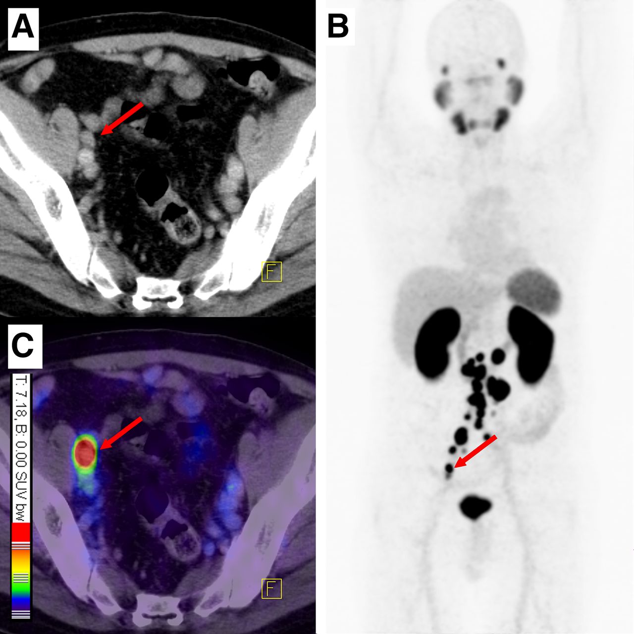
[68Ga]Ga-PSMA-617 PET/CT (1 h after injection) demonstrating a first patient with multiple lymph node metastases. Red arrows point to representative lesion with maximum standardized uptake value (SUV) of 36.5 and a tumor-to-background ratio of 52.1 at 1 h after injection. (A) Contrast-enhanced CT. (B) Maximum-intensity projection of PET scan 1 h after injection. (C) Fusion of PET scan and contrast-enhanced CT.
DISCUSSION
Once a metastatic PCa becomes hormone-refractory there are only a few therapy options, with rather poor clinical success, left. According to the current medical guidelines, antimitotic chemotherapy with docetaxel is typically recommended. However, the overall benefit for the patient is typically poor because of the reported side effects and rather short time to progression, with a reported improvement in overall survival of less than 2.5 mo (23).
As a consequence, there is a high clinical demand for more effective—that is, targeted—systemic therapy strategies. Targeted endoradiotherapy offers the possibility to treat the lesions in a specific and tumor-selective manner by addressing cell surface receptors mainly expressed on malignant cells. Because PSMA is highly expressed on the surface of metastatic and hormone-refractory PCa cells and—besides the kidneys and salivary glands—nearly no expression in healthy tissue is found, a highly effective treatment can be expected using radiolabeled urea-based small-molecule inhibitors of PSMA. Besides many encouraging reports about the diagnostic clinical use of urea-based PSMA inhibitors, Zechmann et al. have recently shown promising therapeutic effects with 131I-labeled MIP-1095 in patients with hormone-refractory PCa. The radiotracer has shown significant lesion uptake in all patients. The PSA values decreased in 60.7% of men treated by more than 50%, and 84.6% of men with bone pain showed complete or moderate reduction of pain (13).
Despite these highly encouraging preliminary clinical results, further developments are necessary to optimize the effectivity of treatment and to minimize the reported side effects. The combination of the commonly used chelating agent DOTA with the PSMA-targeting inhibitors opens the possibility of using the same vector molecule for imaging and therapeutic purposes as DOTA effectively forms complexes with diagnostic (68Ga) and therapeutic radiometals (90Y or 177Lu). The half-life of 68Ga matches the pharmacokinetics of low-molecular-weight molecules with relatively fast diffusion, target localization, and blood clearance (24). Therapeutic radionuclides such as 177Lu allow scintigraphy and subsequent dosimetry with the same compound. In comparison to 131I, 177Lu presents a lower proportion of γ radiation, resulting in a considerably reduced hospitalization, which goes along with improved radiation protection and lowered hemotoxicity (25).
In general, theranostics based on radiometals offer targeted and personalized treatment for the patient while minimizing the damage of healthy tissues. From an economic point of view, in vivo theranostics yield an improved outcome for cancer patients, clinical trials can be designed more effectively and efficiently, and finally, individual costs for cancer treatment will be reduced.
Recently, 68Ga-labeled Glu-urea-Lys-(Ahx)-HBED-CC ([68Ga]Ga-PSMA-11) proved to be a successful tracer for PET imaging of recurrent PCa (17). It was reported that the chelator HBED-CC seemed to interact advantageously with the lipophilic part of the PSMA binding pocket (17,22). However, because of the high selectivity of the complexing agent HBED-CC for 68Ga, the radiotracer is not suitable for radiolabeling with therapeutic radiometals such as 177Lu or 90Y. The here aimed linker region is designed to elucidate the structure–activity relationships and to mimic the proven additional biologic interactions of HBED-CC with the binding pocket. Thus, an ideal linker length (26), polarity (27), size, flexibility (28), presence of aromatic groups (26), or hydrophobic functionality distal to the glutamic acid moiety (29) represents an effective strategy to design highly potent PSMA inhibitor–based radiotracers.
On the basis of these considerations, a DOTA conjugate of a PSMA inhibitor with optimized targeting properties has been designed in this study. The resulting [68Ga]Ga-PSMA-617 exhibits high binding affinity for PSMA and is effectively internalized. Thus, the biologic properties of the novel tracer were significantly improved in comparison with [68Ga]Ga-PSMA-11 (17). Moreover, dynamic small-animal PET imaging and organ distribution of [68Ga]Ga-PSMA-617 showed an early enrichment in the bladder (6 min after injection), and also the maximum kidney uptake was reached as early as 15 min after injection and diminished substantially already after 20 min, whereas [68Ga]Ga-PSMA-617 was further accumulated and retained in the tumor. The favorable pharmacokinetics led to high tumor-to-background ratios (tumor to blood, 1,058; tumor to muscle, 529) with the 177Lu-labeled version 24 h after injection. In contrast to the clinical PET tracer [68Ga]Ga-PSMA-11 (21), the initial high kidney uptake was nearly completely cleared 24 h after injection.
Taken together, the studies of PSMA-617 presented here disclose that the presence of the naphthylic linker has a significant impact on the tumor-targeting and biologic activity as well as on imaging contrast and pharmacokinetics, properties which are crucial for both high imaging quality and efficient targeted endoradiotherapy. With regard to therapeutic use, the high binding affinity and internalization, prolonged tumor uptake, rapid kidney clearance, and high tumor-to-background ratio give clear clinical advantages for PSMA-617, compared with previously published DOTA-based PSMA inhibitors (11,30). In comparison to PSMA-11 (21), PSMA-617 seems to be more attractive for endoradiotherapy because of its higher tumor uptake at later time points, lower spleen accumulation, and the highly efficient clearance from the kidneys.
The first individual clinical diagnostic experience with 68Ga-labeled PSMA-617 is comparable with the recently introduced exclusive PET tracer [68Ga]Ga-PSMA-11. The fast kidney clearance shown with [177Lu]Lu-PSMA-617 encourages us to transfer the compound into the clinical scenario with the aim of a first individual endoradiotherapeutic study. In this connection, a comprehensive first-in-human clinical evaluation of [177Lu]Lu-PSMA-617 is under way (31).
CONCLUSION
Theranostic radiotracers provide patients with targeted and personalized treatment options, thereby improving the current clinical treatment options at the same time. The compound PSMA-617 exhibits high PSMA-specific tumor uptake, rapid background clearance, and fast kidney excretion, which gives clear clinical advantages for both high imaging quality and efficient endoradiotherapy of recurrent PCa.
DISCLOSURE
The costs of publication of this article were defrayed in part by the payment of page charges. Therefore, and solely to indicate this fact, this article is hereby marked “advertisement” in accordance with 18 USC section 1734. This project was supported by a PhD stipend, project no. 101247, provided by the Helmholtz International Graduate School for Cancer Research, a grant from the DFG (Deutsche Forschungsgemeinschaft) (ED234/2-1), and a grant from the Klaus Tschira Foundation. No other potential conflict of interest relevant to this article was reported.
Acknowledgments
Animal experiments complied with the current laws of the Federal Republic of Germany. In vivo studies were kindly performed by Ursula Schierbaum and Karin Leotta (both German Cancer Center Research, Heidelberg, Germany).
Footnotes
Published online Apr. 16, 2015.
- © 2015 by the Society of Nuclear Medicine and Molecular Imaging, Inc.
REFERENCES
- Received for publication August 21, 2014.
- Accepted for publication February 25, 2015.


Jump to section
Related Articles
Cited By...
- Preclinical Evaluation of 177Lu-rhPSMA-10.1, a Radiopharmaceutical for Prostate Cancer: Biodistribution and Therapeutic Efficacy
- Value of [68Ga]Ga-NYM046 PET/CT, in Comparison with 18F-FDG PET/CT, for Diagnosis of Clear Cell Renal Cell Carcinoma
- Limitations of the radiotheranostic concept in neuroendocrine tumors due to lineage-dependent somatostatin receptor expression on hematopoietic stem and progenitor cells
- Single Chelator-Minibody Theranostic Agents for 89Zr PET Imaging and 177Lu Radiopharmaceutical Therapy of PSMA-Expressing Prostate Cancer
- A Single-Arm, Low-Dose, Prospective Study of 177Lu-EB-PSMA Radioligand Therapy in Patients with Metastatic Castration-Resistant Prostate Cancer
- Synthesis and Preclinical Evaluation of 177Lu-Labeled Radiohybrid PSMA Ligands for Endoradiotherapy of Prostate Cancer
- Antitumor efficacy of 90Y-NM600 targeted radionuclide therapy and PD-1 blockade is limited by regulatory T cells in murine prostate tumors
- The History of Prostate-Specific Membrane Antigen as a Theranostic Target in Prostate Cancer: The Cornerstone Role of the Prostate Cancer Foundation
- Safety of PSMA-Targeted Molecular Radioligand Therapy with 177Lu-PSMA-617: Results from the Prospective Multicenter Phase 2 Trial RESIST-PC (NCT03042312)
- Rational Linker Design to Accelerate Excretion and Reduce Background Uptake of Peptidomimetic PSMA-Targeting Hybrid Molecules
- Nuclear Medicine Beyond VISION
- Radiolabeling of PSMA-617 with 89Zr: A Novel Use of DMSO for Radiochemical Yield Enhancement and Preliminary Small-Animal PET Results
- Cytoplasmic Localization of Prostate-Specific Membrane Antigen Inhibitors May Confer Advantages for Targeted Cancer Therapies
- 177Lu-Labeled Albumin-Binder-Conjugated PSMA-Targeting Agents with Extremely High Tumor Uptake and Enhanced Tumor-to-Kidney Absorbed Dose Ratio
- PET Imaging Quantifying 68Ga-PSMA-11 Uptake in Metastatic Colorectal Cancer
- 68Ga-PSMA PET/CT Combined with PET/Ultrasound-Guided Prostate Biopsy Can Diagnose Clinically Significant Prostate Cancer in Men with Previous Negative Biopsy Results
- Investigating PSMA-Targeted Radioligand Therapy Efficacy as a Function of Cellular PSMA Levels and Intratumoral PSMA Heterogeneity
- 3-Year Freedom from Progression After 68Ga-PSMA PET/CT-Triaged Management in Men with Biochemical Recurrence After Radical Prostatectomy: Results of a Prospective Multicenter Trial
- Development of Novel PSMA Ligands for Imaging and Therapy with Copper Isotopes
- 177Lu-PSMA-617 Radioligand Therapy in Metastatic Castration-Resistant Prostate Cancer Patients with a Single Functioning Kidney
- Preclinical Evaluation and Pilot Clinical Study of Al18F-PSMA-BCH for Prostate Cancer PET Imaging
- One-Step 18F-Labeling and Preclinical Evaluation of Prostate-Specific Membrane Antigen Trifluoroborate Probes for Cancer Imaging
- Synthesis and Preclinical Characterization of the PSMA-Targeted Hybrid Tracer PSMA-I&F for Nuclear and Fluorescence Imaging of Prostate Cancer
- Targeted {alpha}-Therapy of Metastatic Castration-Resistant Prostate Cancer with 225Ac-PSMA-617: Swimmer-Plot Analysis Suggests Efficacy Regarding Duration of Tumor Control
- PSMA-11-Derived Dual-Labeled PSMA Inhibitors for Preoperative PET Imaging and Precise Fluorescence-Guided Surgery of Prostate Cancer
- Repeated 177Lu-Labeled PSMA-617 Radioligand Therapy Using Treatment Activities of Up to 9.3 GBq
- Anatomic and Molecular Imaging in Prostate Cancer
- Establishing 177Lu-PSMA-617 Radioligand Therapy in a Syngeneic Model of Murine Prostate Cancer
- Targeted {alpha}-Therapy of Metastatic Castration-Resistant Prostate Cancer with 225Ac-PSMA-617: Dosimetry Estimate and Empiric Dose Finding
- Glu-Ureido-Based Inhibitors of Prostate-Specific Membrane Antigen: Lessons Learned During the Development of a Novel Class of Low-Molecular-Weight Theranostic Radiotracers
- Acceleration of PSMA-Targeted Theranostics to the Clinic: Can Common Sense Prevail?
- 68Ga-THP-PSMA: A PET Imaging Agent for Prostate Cancer Offering Rapid, Room-Temperature, 1-Step Kit-Based Radiolabeling
- 177Lu-PSMA Radioligand Therapy for Prostate Cancer
- Preclinical Evaluation of 18F-PSMA-1007, a New Prostate-Specific Membrane Antigen Ligand for Prostate Cancer Imaging
- German Multicenter Study Investigating 177Lu-PSMA-617 Radioligand Therapy in Advanced Prostate Cancer Patients
- 225Ac-PSMA-617 for PSMA-Targeted {alpha}-Radiation Therapy of Metastatic Castration-Resistant Prostate Cancer
- The Rise of PSMA Ligands for Diagnosis and Therapy of Prostate Cancer
- PSMA-Targeted Radionuclide Therapy of Metastatic Castration-Resistant Prostate Cancer with 177Lu-Labeled PSMA-617
- New Strategies in Prostate Cancer: Prostate-Specific Membrane Antigen (PSMA) Ligands for Diagnosis and Therapy
- The Theranostic PSMA Ligand PSMA-617 in the Diagnosis of Prostate Cancer by PET/CT: Biodistribution in Humans, Radiation Dosimetry, and First Evaluation of Tumor Lesions