
Main menu

  • Home
  • Content
    • Current
    • Ahead of print
    • Past Issues
    • JNM Supplement
    • SNMMI Annual Meeting Abstracts
    • Continuing Education
    • JNM Podcasts
  • Subscriptions
    • Subscribers
    • Institutional and Non-member
    • Rates
    • Journal Claims
    • Corporate & Special Sales
  • Authors
    • Submit to JNM
    • Information for Authors
    • Assignment of Copyright
    • AQARA requirements
  • Info
    • Reviewers
    • Permissions
    • Advertisers
  • About
    • About Us
    • Editorial Board
    • Contact Information
  • More
    • Alerts
    • Feedback
    • Help
    • SNMMI Journals
  • SNMMI
    • JNM
    • JNMT
    • SNMMI Journals
    • SNMMI

User menu

  • Subscribe
  • My alerts
  • Log in
  • Log out
  • My Cart

Search

  • Advanced search
Journal of Nuclear Medicine
  • SNMMI
    • JNM
    • JNMT
    • SNMMI Journals
    • SNMMI
  • Subscribe
  • My alerts
  • Log in
  • Log out
  • My Cart
Journal of Nuclear Medicine

Advanced Search

  • Home
  • Content
    • Current
    • Ahead of print
    • Past Issues
    • JNM Supplement
    • SNMMI Annual Meeting Abstracts
    • Continuing Education
    • JNM Podcasts
  • Subscriptions
    • Subscribers
    • Institutional and Non-member
    • Rates
    • Journal Claims
    • Corporate & Special Sales
  • Authors
    • Submit to JNM
    • Information for Authors
    • Assignment of Copyright
    • AQARA requirements
  • Info
    • Reviewers
    • Permissions
    • Advertisers
  • About
    • About Us
    • Editorial Board
    • Contact Information
  • More
    • Alerts
    • Feedback
    • Help
    • SNMMI Journals
  • View or Listen to JNM Podcast
  • Visit JNM on Facebook
  • Join JNM on LinkedIn
  • Follow JNM on Twitter
  • Subscribe to our RSS feeds
Research ArticleOncology

PET Imaging Quantifying 68Ga-PSMA-11 Uptake in Metastatic Colorectal Cancer

Tahleesa J. Cuda, Andrew D. Riddell, Cheng Liu, Vicki L. Whitehall, Jennifer Borowsky, David K. Wyld, Matthew E. Burge, Elizabeth Ahern, Alison Griffin, Nicholas J.R. Lyons, Stephen E. Rose, David A. Clark, Andrew R.L. Stevenson, John D. Hooper, Simon Puttick and Paul A. Thomas
Journal of Nuclear Medicine November 2020, 61 (11) 1576-1579; DOI: https://doi.org/10.2967/jnumed.119.233312
Tahleesa J. Cuda
1University of Queensland, Brisbane, Australia
2Metro North Hospital and Health Service, Brisbane, Australia
  • Find this author on Google Scholar
  • Find this author on PubMed
  • Search for this author on this site
Andrew D. Riddell
1University of Queensland, Brisbane, Australia
2Metro North Hospital and Health Service, Brisbane, Australia
  • Find this author on Google Scholar
  • Find this author on PubMed
  • Search for this author on this site
Cheng Liu
1University of Queensland, Brisbane, Australia
3Envoi Specialist Pathologists, Herston, Australia
4QIMR Berghofer Medical Research Institute, Herston, Australia
  • Find this author on Google Scholar
  • Find this author on PubMed
  • Search for this author on this site
Vicki L. Whitehall
1University of Queensland, Brisbane, Australia
4QIMR Berghofer Medical Research Institute, Herston, Australia
  • Find this author on Google Scholar
  • Find this author on PubMed
  • Search for this author on this site
Jennifer Borowsky
1University of Queensland, Brisbane, Australia
2Metro North Hospital and Health Service, Brisbane, Australia
4QIMR Berghofer Medical Research Institute, Herston, Australia
  • Find this author on Google Scholar
  • Find this author on PubMed
  • Search for this author on this site
David K. Wyld
1University of Queensland, Brisbane, Australia
2Metro North Hospital and Health Service, Brisbane, Australia
  • Find this author on Google Scholar
  • Find this author on PubMed
  • Search for this author on this site
Matthew E. Burge
1University of Queensland, Brisbane, Australia
2Metro North Hospital and Health Service, Brisbane, Australia
  • Find this author on Google Scholar
  • Find this author on PubMed
  • Search for this author on this site
Elizabeth Ahern
1University of Queensland, Brisbane, Australia
2Metro North Hospital and Health Service, Brisbane, Australia
4QIMR Berghofer Medical Research Institute, Herston, Australia
  • Find this author on Google Scholar
  • Find this author on PubMed
  • Search for this author on this site
Alison Griffin
4QIMR Berghofer Medical Research Institute, Herston, Australia
  • Find this author on Google Scholar
  • Find this author on PubMed
  • Search for this author on this site
Nicholas J.R. Lyons
1University of Queensland, Brisbane, Australia
2Metro North Hospital and Health Service, Brisbane, Australia
  • Find this author on Google Scholar
  • Find this author on PubMed
  • Search for this author on this site
Stephen E. Rose
5CSIRO, Herston, Australia; and
  • Find this author on Google Scholar
  • Find this author on PubMed
  • Search for this author on this site
David A. Clark
1University of Queensland, Brisbane, Australia
2Metro North Hospital and Health Service, Brisbane, Australia
  • Find this author on Google Scholar
  • Find this author on PubMed
  • Search for this author on this site
Andrew R.L. Stevenson
1University of Queensland, Brisbane, Australia
2Metro North Hospital and Health Service, Brisbane, Australia
  • Find this author on Google Scholar
  • Find this author on PubMed
  • Search for this author on this site
John D. Hooper
1University of Queensland, Brisbane, Australia
  • Find this author on Google Scholar
  • Find this author on PubMed
  • Search for this author on this site
Simon Puttick
5CSIRO, Herston, Australia; and
  • Find this author on Google Scholar
  • Find this author on PubMed
  • Search for this author on this site
Paul A. Thomas
1University of Queensland, Brisbane, Australia
2Metro North Hospital and Health Service, Brisbane, Australia
6Herston Imaging Research Facility, Herston, Australia
  • Find this author on Google Scholar
  • Find this author on PubMed
  • Search for this author on this site
  • Article
  • Figures & Data
  • Supplemental
  • Info & Metrics
  • PDF
Loading

Abstract

At diagnosis, 22% of colorectal cancer (CRC) patients have metastases, and 50% later develop metastasis. Peptide receptor radionuclide therapy (PRRT), such as 177Lu-PSMA-617, is used to treat metastatic prostate cancer. 177Lu-PSMA-617 targets prostate-specific membrane antigen (PSMA), a cell-surface protein enriched in prostate cancer and the neovasculature of other solid tumors, including CRC. We performed 68Ga-PSMA-11 PET/CT imaging of 10 patients with metastatic CRC to assess metastasis avidity. Eight patients had lesions lacking avidity, and 2 had solitary metastases exhibiting very low avidity. Despite expression of PSMA in CRC neovasculature, none of the patients exhibited tumor avidity sufficient to be considered for 177Lu-PSMA-617 PRRT.

  • metastatic colorectal cancer
  • peptide receptor radionuclide therapy
  • PET
  • theranostics

Colorectal cancer (CRC) is the fourth most common cause of cancer-related death (1). At diagnosis, 22% of patients have metastases, and 50% develop metastasis during their lifetime (1).

Theranostics uses tumor-selective ligands conjugated to radionuclides and cytotoxic agents for, respectively, cancer imaging and treatment (2). By targeting tumor cell-surface antigens, these agents are delivered selectively to malignancies (2). Using a diagnostic positron-emitting radionuclide and PET, tumor burden is quantified and response to therapy predicted on the basis of tumor avidity (2). In peptide receptor radionuclide therapy (PRRT), therapeutic α- or β-emitting radionuclides, conjugated to the same PET imaging target, induce DNA damage and cell death (2). PRRT is a mainstay treatment for neuroendocrine tumors and is emerging for metastatic prostate cancer (PC) (2).

The PRRT target prostate-specific membrane antigen (PSMA) is enriched in metastatic PC and has low expression in normal tissues (3,4). It is also elevated on the endothelial cells of certain solid tumors, including CRC, where 75%–80% of primary tumors and metastases express PSMA, which correlates with poor outcome (5,6). PSMA-11, a high-specificity and high-affinity ligand for PSMA that incorporates a radiometal chelator (7), is applied for PET imaging of metastatic PC using 68Ga-PSMA-11 (8) and PRRT using 177Lu-PSMA-617 (9).

Several case reports note CRC avidity during 68Ga-PSMA-11 imaging for metastatic PC, potentially supporting PSMA-targeted PRRT in advanced CRC (10,11). Responding to a recent call for prospective studies in place of incidental case reports or series (12), we assessed metastatic CRC avidity for 68Ga-PSMA-11 to determine whether the avidity meets the criteria for 177Lu-PSMA-617 PRRT.

MATERIALS AND METHODS

Patients

Inclusion and exclusion criteria (Supplemental Table 1; supplemental materials are available at http://jnm.snmjournals.org) and sample size (n = 10) were from PET imaging studies assessing tumor avidity (8,13,14). Recruitment would continue if initial results indicated that at least 30% of patients met the TheraP trial (NCT03392428) criteria to progress to PRRT.

PET Scans and Interpretation

The study had Human Research Ethics Committee approval (HREC/18/QPCH/51). Recruitment was from August to November 2018. Imaging used PSMA-11 (HBED-CC; ABX). 68Ga was labeled as previously described (15), with a labeling efficiency of more than 98%. PET and CT images were reconstructed, and SUVmax and tumor-to-liver (background) SUVmax ratio were determined as previously described (16). 18F-FDG PET/CT or contrast-enhanced CT localized metastases of low 68Ga-PSMA-11 avidity, and SUVmax was compared between 68Ga-PSMA-11 PET, 18F-FDG PET, and contrast-enhanced CT. The TheraP trial criteria to stratify patients as likely responders to PRRT required an SUVmax of at least 10 at all tumor sites not subject to partial-volume artifact (i.e., >10 mm in diameter), an SUVmax of more than 20 at the most avid site, and 68Ga-PSMA-11 avidity higher than 18F-FDG avidity at all sites, when recent 18F-FDG imaging was available (17).

Immunohistochemistry

The study had Human Research Ethics Committee approval (HREC/11/QRBW/453; P2139). Immunohistochemistry was performed on a tissue microarray of matched CRC primary tumors and metastases from 37 patients, using anti-PSMA clone 3E6 (Agilent) and Biocare Medical MACH1 mouse horseradish peroxidase polymer. Signal was quantified by a pathologist as nil, weak, moderate, or strong on the basis of, respectively, no, ≤2.5%, ≥2.5–≤4.5%, or ≥4.5% positive tumor cells.

Statistical Methods

Statistics were performed using Prism (version 7; GraphPad). Data represent the highest SUVmax of representative lesions per anatomic region. Quantification was consistent with the reporting guidelines of the Standards for Reporting of Diagnostic Accuracy Studies (18).

RESULTS

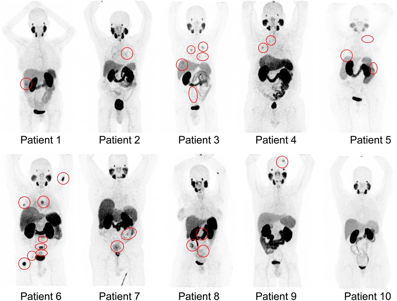
68Ga-PSMA-11 PET imaging of 10 patients with metastatic CRC (Supplemental Tables 2 and 3) resulted in no adverse events. Maximum-intensity projections of participants are shown in Figure 1, and SUVmax is in Figure 2, including TheraP criteria 1 and 2 (19). The metastases of all patients fell significantly short of satisfying criteria 1 and 2, except for liver lesions in patient 3 and lymph nodes in patient 8. Liver metastases in patient 3 met criterion 1 but not criterion 2. Patient 3 had synchronous lung and omental metastases that had insufficient avidity to satisfy criteria 1 and 2. Primary tumor and pelvic lymph node metastases in patient 8 exhibited avidity greater than criterion 1 but not criterion 2. This patient also had locoregional lymph node metastases that failed to satisfy criteria 1 and 2. Two of 3 patients with primary tumors (patients 6 and 7) failed to satisfy both criterion 1 and criterion 2. Bone metastases in patient 6 had the greatest avidity of all lesions and satisfied criterion 1 but fell just short of satisfying criterion 2. Locoregional and retroperitoneal lymph nodes and adrenal metastases in patient 6 failed to satisfy both criterion 1 and criterion 2.

FIGURE 1.
  • Download figure
  • Open in new tab
  • Download powerpoint
FIGURE 1.

68Ga-PSMA-11 PET maximum-intensity-projection images of patients with metastatic CRC. Red circles = avid lesions.

FIGURE 2.
  • Download figure
  • Open in new tab
  • Download powerpoint
FIGURE 2.

SUVmax of 68Ga-PSMA-11 metastatic CRCs. Green line = TheraP criterion 1, SUVmax ≥ 10, required at all sites. Red line = criterion 2, SUVmax > 20, required at most avid site.

Also of note, patient metastases lacked consistency in tumor-to-liver SUVmax ratios (Fig. 3) and no patient satisfied criterion 3 that 68Ga-PSMA-11 avidity be greater than 18F-FDG avidity (Supplemental Table 4). Supplemental Table 5 lists the lesions of each patient as detected by 18F-FDG PET, contrast-enhanced CT, and 68Ga-PSMA-11 PET, including the number missed by 68Ga-PSMA-11 PET. The time between 68Ga-PSMA-11 PET imaging and 18F-FDG PET or contrast-enhanced CT is provided in Supplemental Table 6. Eight of 10 patients (patients 2, 3, 4, 5, 6, 7, 8, and 10) had lesions detected by 18F-FDG PET or contrast-enhanced CT but missed by 68Ga-PSMA-11 PET. Liver and lymph node metastases in patients 1 and 8 had heterogeneous uptake, with only a portion of lesions avid. Although patient 6 had bone metastases with significantly higher avidity during 68Ga-PSMA-11 PET than other soft-tissue and visceral lesions, avidity was still significantly lower than 18F-FDG PET avidity. Supplemental Figure 1 provides representative images of pelvic lymph node metastases with negligible avidity during 68Ga-PSMA-11 PET, compared with high 18F-FDG avidity, for patients 7 and 8. No lesions were detected by 68Ga-PSMA-11 PET that were not also identified during 18F-FDG PET. Patients 2, 3, 4, 9, and 10 had previously received neoadjuvant, adjuvant, or palliative chemotherapy, with patient 3 receiving palliative chemotherapy 8 weeks before 68Ga-PSMA-11 PET, which likely had minimal effect on avidity because tumor response was poor. For the remaining 4 patients, at least 7 months had elapsed since chemotherapy.

FIGURE 3.
  • Download figure
  • Open in new tab
  • Download powerpoint
FIGURE 3.

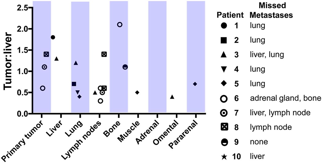
CRC tumor-to-liver (background) SUVmax.

Because resected tumors from the 10 patients were unavailable to explore the reason for the lack of tumor avidity, we performed immunohistochemistry for PSMA in matched primary tumors and metastases from an independent cohort of 37 patients (Supplemental Tables 7 and 8). PSMA was exclusive to endothelial cells of the tumor vasculature, which consistently comprised about 5% of the cells in tumors. Representative images of tumor regions displaying moderate (≥2.5–≤ 4.5% positive cells) and strong (≥4.5% positive cells) PSMA expression (Supplemental Fig. 2A) demonstrate that tumor expression was consistently very low. Quantitative analyses indicated that the invasive edge of 79% of primary tumors and 87% of central regions of primary tumors had nil or weak PSMA expression (Supplemental Figure 2B), with levels consistent between tumor regions (Supplemental Fig. 2C). In metastases, the invasive edge of tumors and the central region of 95% of tumors displayed nil or weak PSMA expression (Supplemental Fig. 2D), and expression was also consistent between these regions of metastases (Supplemental Fig. 2E). These data suggest that the low observed PSMA ligand avidity was due to consistently low PSMA expression in CRC tumors.

DISCUSSION

Responding to the recent call for prospective trials to assess the utility of PSMA-targeted theranostic agents for cancers beyond PC (12), this study indicated that 68Ga-PSMA-11 PET has low avidity in metastatic CRC, with heterogeneous or nonexistent uptake in lesions. A range of factors may contribute to low tumor avidity, the most likely of which is low PSMA expression on tumor vasculature. Although PSMA expression has been reported on colorectal neovasculature (5,6), PSMA messenger RNA is 10–20 times lower in CRC than in PC (19), with our immunohistochemistry confirming low PSMA protein levels in CRC vasculature.

Although it is also possible that low avidity was due to a lack of homing of 68Ga-PSMA-11 to CRC tumors, this is unlikely because we used a protocol that identifies metastatic PC and allows sufficient time for radioligand circulation, antigen binding, and internalization by PSMA-expressing cells (8). Other potential contributing factors include heterogeneous neovascularization and microvessel density in CRC lesions (20), tumor coopting of normal vessels lacking PSMA expression (21), and vascular mimicry with tumor blood-conducting channels lined by malignant cells (22).

We estimated that 68Ga-PSMA-11 PET for metastatic CRC can be beneficial if tumor avidity is sufficient to progress at least 30% of patients to PRRT. However, none of the patients had sufficient avidity to progress to PRRT. Because our sample size was small, we cannot be definitive that 68Ga-PSMA-11 PET is not justified for CRC. However, we note that, using binomial probability, there was only a very small chance (3%) that none of the 10 patients would have sufficient tumor avidity to warrant PRRT, justifying our decision not to continue recruitment beyond 10 patients.

CONCLUSION

68Ga-PSMA-11 PET/CT is not sufficiently sensitive to detect metastatic CRC. Further research is required to identify cell-surface receptors as theranostic targets for imaging and treatment of CRC metastasis.

DISCLOSURE

The study was supported by the Redcliffe Private Practice Fund, the Royal Brisbane and Women’s Hospital Foundation, and the Mater Foundation. No other potential conflict of interest relevant to this article was reported.

KEY POINTS

  • QUESTION: Can PSMA expression on CRC neovasculature be targeted using 68Ga-PSMA-11 with high sensitivity and avidity to qualify patients for 177Lu-PSMA-617 therapy?

  • PERTINENT FINDINGS: This prospective pilot study assessed the tumor avidity of 10 patients with metastatic CRC using 68Ga-PSMA-11. Overall, 68Ga-PSMA-11 was insensitive in detecting CRC metastases. Identified lesions had avidity that was insufficient to warrant PSMA-targeted therapy.

  • IMPLICATIONS FOR PATIENT CARE: Theranostic ligands targeting specific receptors on metastatic CRC cells should be sought in place of targeting PSMA expressed by tumor neovasculature.

Acknowledgments

We acknowledge the generous support of the Royal Brisbane and Women’s Hospital radiochemistry staff in performing radiolabeling, the Herston Imaging Research Facility in performing PET/CT imaging, and the Royal Brisbane and Women’s Hospital and the Redcliffe Hospital in recruiting patients.

Footnotes

  • Published online May 1, 2020.

  • © 2020 by the Society of Nuclear Medicine and Molecular Imaging.

REFERENCES

  1. 1.↵
    1. Arnold M,
    2. Sierra MS,
    3. Laversanne M,
    4. Soerjomataram I,
    5. Jemal A,
    6. Bray F
    . Global patterns and trends in colorectal cancer incidence and mortality. Gut. 2017;66:683–691.
    OpenUrlAbstract/FREE Full Text
  2. 2.↵
    1. Turner JH
    . An introduction to the clinical practice of theranostics in oncology. Br J Radiol. 2018;91:20180440.
    OpenUrl
  3. 3.↵
    1. Tse BW,
    2. Cowin GJ,
    3. Soekmadji C,
    4. et al
    . PSMA-targeting iron oxide magnetic nanoparticles enhance MRI of preclinical prostate cancer. Nanomedicine (Lond). 2015;10:375–386.
    OpenUrl
  4. 4.↵
    1. Kiess AP,
    2. Banerjee SR,
    3. Mease RC,
    4. et al
    . Prostate-specific membrane antigen as a target for cancer imaging and therapy. Q J Nucl Med Mol Imaging. 2015;59:241–268.
    OpenUrlPubMed
  5. 5.↵
    1. Abdel-Hadi M,
    2. Ismail Y,
    3. Younis L
    . Prostate-specific membrane antigen (PSMA) immunoexpression in the neovasculature of colorectal carcinoma in Egyptian patients. Pathol Res Pract. 2014;210:759–763.
    OpenUrl
  6. 6.↵
    1. Haffner MC,
    2. Kronberger IE,
    3. Ross JS,
    4. et al
    . Prostate-specific membrane antigen expression in the neovasculature of gastric and colorectal cancers. Hum Pathol. 2009;40:1754–1761.
    OpenUrlCrossRefPubMed
  7. 7.↵
    1. Benešová M,
    2. Schafer M,
    3. Bauder-Wust U,
    4. et al
    . Preclinical evaluation of a tailor-made DOTA-conjugated PSMA inhibitor with optimized linker moiety for imaging and endoradiotherapy of prostate cancer. J Nucl Med. 2015;56:914–920.
    OpenUrlAbstract/FREE Full Text
  8. 8.↵
    1. Hofman MS,
    2. Murphy DG,
    3. Williams SG,
    4. et al
    . A prospective randomized multicentre study of the impact of gallium-68 prostate-specific membrane antigen (PSMA) PET/CT imaging for staging high-risk prostate cancer prior to curative-intent surgery or radiotherapy (proPSMA study): clinical trial protocol. BJU Int. 2018;122:783–793.
    OpenUrl
  9. 9.↵
    1. Hofman MS,
    2. Violet J,
    3. Hicks RJ,
    4. et al
    . 177Lu-PSMA-617 radionuclide treatment in patients with metastatic castration-resistant prostate cancer (LuPSMA trial): a single-centre, single-arm, phase 2 study. Lancet Oncol. 2018;19:825–833.
    OpenUrl
  10. 10.↵
    1. Huang YT,
    2. Fong W,
    3. Thomas P
    . Rectal carcinoma on 68Ga-PSMA PET/CT. Clin Nucl Med. 2016;41:e167–e168.
    OpenUrlCrossRefPubMed
  11. 11.↵
    1. Hangaard L,
    2. Jochumsen MR,
    3. Vendelbo MH,
    4. Bouchelouche K
    . Metastases from colorectal cancer avid on 68Ga-PSMA PET/CT. Clin Nucl Med. 2017;42:532–533.
    OpenUrl
  12. 12.↵
    1. Salas Fragomeni RA,
    2. Amir T,
    3. Sheikhbahaei S,
    4. et al
    . Imaging of nonprostate cancers using PSMA-targeted radiotracers: rationale, current state of the field, and a call to arms. J Nucl Med. 2018;59:871–877.
    OpenUrlAbstract/FREE Full Text
  13. 13.↵
    1. Minamimoto R,
    2. Hancock S,
    3. Schneider B,
    4. et al
    . Pilot comparison of 68Ga-RM2 PET and 68Ga-PSMA-11 PET in patients with biochemically recurrent prostate cancer. J Nucl Med. 2016;57:557–562.
    OpenUrlAbstract/FREE Full Text
  14. 14.↵
    1. Sawicki LM,
    2. Buchbender C,
    3. Boos J,
    4. et al
    . Diagnostic potential of PET/CT using a 68Ga-labelled prostate-specific membrane antigen ligand in whole-body staging of renal cell carcinoma: initial experience. Eur J Nucl Med Mol Imaging. 2017;44:102–107.
    OpenUrl
  15. 15.↵
    1. Eder M,
    2. Lohr T,
    3. Bauder-Wust U,
    4. et al
    . Pharmacokinetic properties of peptidic radiopharmaceuticals: reduced uptake of (EH)3-conjugates in important organs. J Nucl Med. 2013;54:1327–1330.
    OpenUrlAbstract/FREE Full Text
  16. 16.↵
    1. Rhee H,
    2. Blazak J,
    3. Tham CM,
    4. et al
    . Pilot study: use of gallium-68 PSMA PET for detection of metastatic lesions in patients with renal tumor. EJNMMI Res. 2016;6:76.
    OpenUrl
  17. 17.↵
    1. Hofman MS,
    2. Emmett L,
    3. Violet J,
    4. et al
    . TheraP: a randomized phase 2 trial of 177Lu-PSMA-617 theranostic treatment vs cabazitaxel in progressive metastatic castration-resistant prostate cancer (Clinical Trial Protocol ANZUP 1603). BJU Int. 2019;124(suppl 1):5–13.
    OpenUrl
  18. 18.↵
    1. Cohen JF,
    2. Korevaar DA,
    3. Altman DG,
    4. et al
    . STARD 2015 guidelines for reporting diagnostic accuracy studies: explanation and elaboration. BMJ Open. 2016;6:e012799.
    OpenUrlAbstract/FREE Full Text
  19. 19.↵
    1. Weinstein JN,
    2. Collisson EA,
    3. Mills GB,
    4. et al
    . The Cancer Genome Atlas Pan-Cancer analysis project. Nat Genet. 2013;45:1113–1120.
    OpenUrlCrossRefPubMed
  20. 20.↵
    1. Kwak Y,
    2. Lee HE,
    3. Kim WH,
    4. Kim DW,
    5. Kang SB,
    6. Lee HS
    . The clinical implication of cancer-associated microvasculature and fibroblast in advanced colorectal cancer patients with synchronous or metachronous metastases. PLoS One. 2014;9:e91811.
    OpenUrlPubMed
  21. 21.↵
    1. Qian CN,
    2. Tan MH,
    3. Yang JP,
    4. Cao Y
    . Revisiting tumor angiogenesis: vessel co-option, vessel remodeling, and cancer cell-derived vasculature formation. Chin J Cancer. 2016;35:10.
    OpenUrl
  22. 22.↵
    1. Baeten CI,
    2. Hillen F,
    3. Pauwels P,
    4. de Bruine AP,
    5. Baeten CG
    . Prognostic role of vasculogenic mimicry in colorectal cancer. Dis Colon Rectum. 2009;52:2028–2035.
    OpenUrlCrossRefPubMed
  • Received for publication July 20, 2019.
  • Accepted for publication March 9, 2020.
PreviousNext
Back to top

In this issue

Journal of Nuclear Medicine: 61 (11)
Journal of Nuclear Medicine
Vol. 61, Issue 11
November 1, 2020
  • Table of Contents
  • Table of Contents (PDF)
  • About the Cover
  • Index by author
Print
Download PDF
Article Alerts
Sign In to Email Alerts with your Email Address
Email Article

Thank you for your interest in spreading the word on Journal of Nuclear Medicine.

NOTE: We only request your email address so that the person you are recommending the page to knows that you wanted them to see it, and that it is not junk mail. We do not capture any email address.

Enter multiple addresses on separate lines or separate them with commas.
PET Imaging Quantifying 68Ga-PSMA-11 Uptake in Metastatic Colorectal Cancer
(Your Name) has sent you a message from Journal of Nuclear Medicine
(Your Name) thought you would like to see the Journal of Nuclear Medicine web site.
Citation Tools
PET Imaging Quantifying 68Ga-PSMA-11 Uptake in Metastatic Colorectal Cancer
Tahleesa J. Cuda, Andrew D. Riddell, Cheng Liu, Vicki L. Whitehall, Jennifer Borowsky, David K. Wyld, Matthew E. Burge, Elizabeth Ahern, Alison Griffin, Nicholas J.R. Lyons, Stephen E. Rose, David A. Clark, Andrew R.L. Stevenson, John D. Hooper, Simon Puttick, Paul A. Thomas
Journal of Nuclear Medicine Nov 2020, 61 (11) 1576-1579; DOI: 10.2967/jnumed.119.233312

Citation Manager Formats

  • BibTeX
  • Bookends
  • EasyBib
  • EndNote (tagged)
  • EndNote 8 (xml)
  • Medlars
  • Mendeley
  • Papers
  • RefWorks Tagged
  • Ref Manager
  • RIS
  • Zotero
Share
PET Imaging Quantifying 68Ga-PSMA-11 Uptake in Metastatic Colorectal Cancer
Tahleesa J. Cuda, Andrew D. Riddell, Cheng Liu, Vicki L. Whitehall, Jennifer Borowsky, David K. Wyld, Matthew E. Burge, Elizabeth Ahern, Alison Griffin, Nicholas J.R. Lyons, Stephen E. Rose, David A. Clark, Andrew R.L. Stevenson, John D. Hooper, Simon Puttick, Paul A. Thomas
Journal of Nuclear Medicine Nov 2020, 61 (11) 1576-1579; DOI: 10.2967/jnumed.119.233312
Twitter logo Facebook logo LinkedIn logo Mendeley logo
  • Tweet Widget
  • Facebook Like
  • Google Plus One
Bookmark this article

Jump to section

  • Article
    • Abstract
    • MATERIALS AND METHODS
    • RESULTS
    • DISCUSSION
    • CONCLUSION
    • DISCLOSURE
    • Acknowledgments
    • Footnotes
    • REFERENCES
  • Figures & Data
  • Supplemental
  • Info & Metrics
  • PDF

Related Articles

  • This Month in JNM
  • PubMed
  • Google Scholar

Cited By...

  • No citing articles found.
  • Google Scholar

More in this TOC Section

Oncology

  • Chimeric antigen receptor (CAR) T cells Imaging: Clinical Needs and Strategies for Success
  • Metastatic NUT Midline Carcinoma
  • Radionuclides used in Nuclear Therapeutic Medicine: a brief history, properties and main relevant studies of radionuclides with mass number less than 100
Show more Oncology

Clinical

  • TauIQ: A Canonical Image Based Algorithm to Quantify Tau PET Scans
  • Dual PET Imaging in Bronchial Neuroendocrine Neoplasms: The NETPET Score as a Prognostic Biomarker
  • Addition of 131I-MIBG to PRRT (90Y-DOTATOC) for Personalized Treatment of Selected Patients with Neuroendocrine Tumors
Show more Clinical

Similar Articles

Keywords

  • metastatic colorectal cancer
  • peptide receptor radionuclide therapy
  • PET
  • theranostics
SNMMI

© 2025 SNMMI

Powered by HighWire