


Article Figures & Data
Figures
- FIGURE 1.
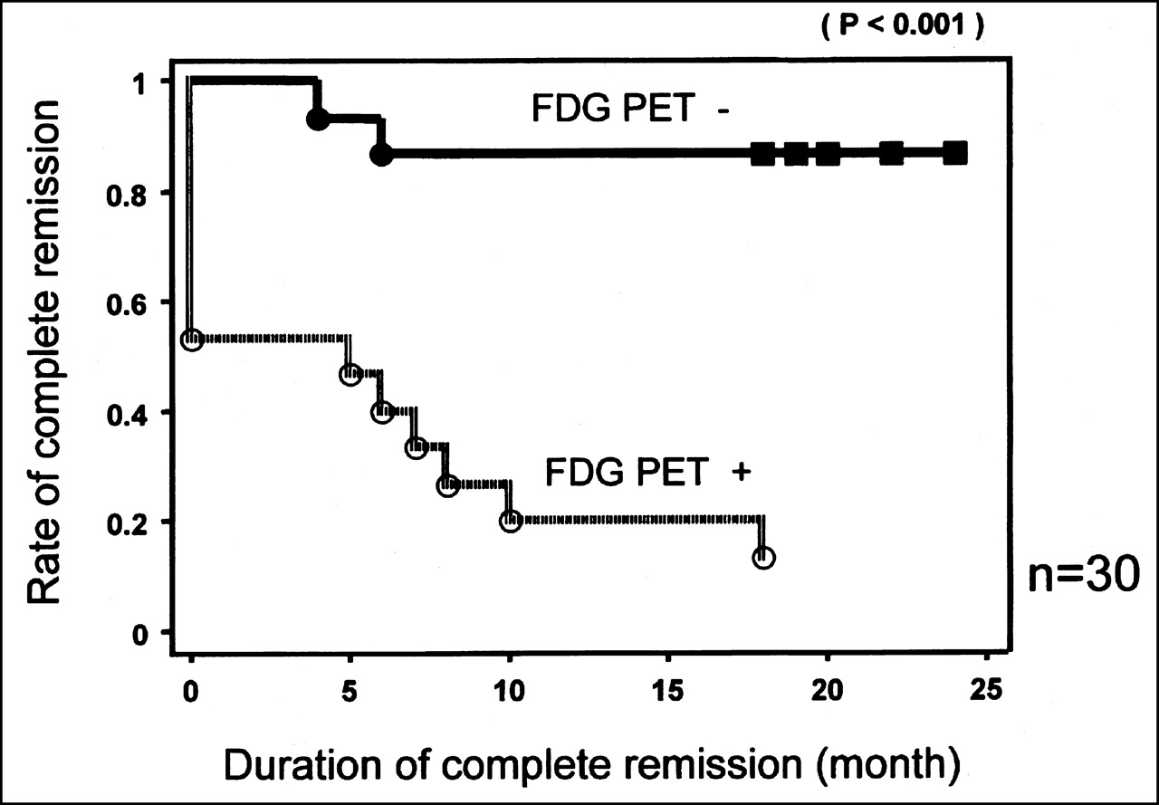
In entire group of patients who underwent 18F-FDG PET after first cycle of chemotherapy (30 patients), Kaplan-Meier estimate of PFS for 15 patients with positive 18F-FDG PET results is compared with that for 15 patients with negative 18F-FDG PET results after first cycle of chemotherapy. Statistically significant difference in PFS was found between positive and negative 18F-FDG PET results (P < 0.001).
- FIGURE 2.
In group of patients who underwent both early and late 18F-FDG PET (23 patients), Kaplan-Meier estimate of PFS for 6 patients with positive 18F-FDG PET results is compared with that for 17 patients with negative 18F-FDG PET results at completion of chemotherapy. Statistically significant difference in PFS was found between positive and negative 18F-FDG PET results (P = 0.001).
- FIGURE 3.
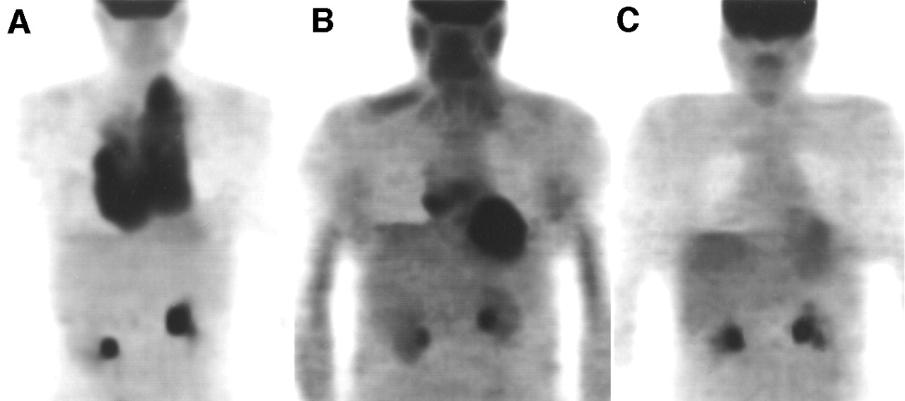
A 35-y-old man with bulky HD underwent 18F-FDG PET before (A), after first cycle of (B), and at completion of (C) chemotherapy. Pretherapy 18F-FDG PET images reveal radiotracer uptake in anterior mediastinum involving both hilar regions and extending into left supraclavicular region. Patient underwent chemotherapy with doxorubicin, bleomycin, vinblastine, and dacarbazine. 18F-FDG PET after first cycle of chemotherapy reveals residual disease in right anterior mediastinum, whereas 18F-FDG PET at completion of chemotherapy shows no evidence of residual lymphoma in corresponding regions. Disease relapsed in mediastinum after PFS of 6 mo. Images obtained after first cycle show physiologic uptake in salivary glands, oral mucosa, right shoulder (trapezius muscle), and heart.
- FIGURE 4.
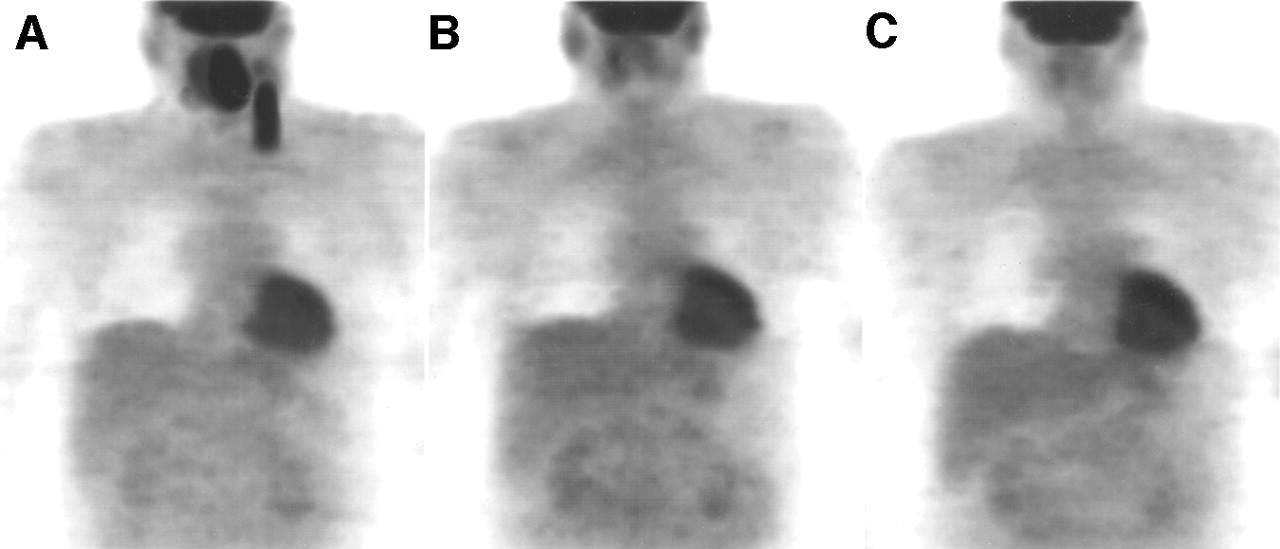
A 45-y-old man with NHL underwent 18F-FDG PET before (A), after first cycle of (B), and at completion of (C) chemotherapy. Pretherapy 18F-FDG PET images reveal radiotracer uptake in nasopharynx and left cervical lymph nodes. Patient underwent chemotherapy with cyclophosphamide, doxorubicin, vincristine, and prednisone. Both after first cycle and at completion of chemotherapy, 18F-FDG PET reveals no evidence of residual disease. Disease was still in remission after PFS of 18 mo. All images show physiologic uptake in heart.
- FIGURE 5.
In group of patients who underwent both early and late 18F-FDG PET (23 patients), Kaplan-Meier estimate of PFS for 10 patients with positive 18F-FDG PET results is compared with that for 13 patients with negative 18F-FDG PET results after first cycle of chemotherapy. Statistically significant difference in PFS was found between positive and negative 18F-FDG PET results (P < 0.001).


Tables
Characteristic All patients (n = 30) Patients examined twice* (n = 23) Age (y) Mean ± SD 52.3 ± 16.0 50.1 ± 14.0 Range 26–77 26–77 Sex Male 16 12 Female 14 11 Histologic diagnosis NHL (n = 17) Diffuse large B-cell lymphoma 13 10 Follicular large cell lymphoma 2 2 Lymphoblastic lymphoma 2 1 HD (n = 13) 13 10 No. of patients examined At initial staging 17 13 At relapse 13 10 ↵* These patients had 18F-FDG PET studies both after first cycle and at completion of therapy.
- TABLE 2
Characteristics of Patients with Relapse and with Poor Prognostic Features vs. Comparative 18F-FDG PET Results
Patient no. Age (y) Histology Ann Arbor stage Tumor size After 1 cycle After completion Outcome PFS (mo) 1 >60 DLCL IIIB Bulky + − (FN) Relapse 18 2 >60 DLCL IIA Nonbulky + − (FN) Relapse 10 3 <60 DLCL IIB Bulky + − (FN) Relapse 6 4 >60 FLC IIIA Bulky − − Remission 24 5 <60 HD IIB Bulky + (FP) + (FP) Remission 18 6 <60 HD IIA Bulky − − Remission 20 7 >60 DLCL IIA Bulky − − Remission 19 8 >60 FLC IV Nonbulky − − Remission 20 9 DLCL + − (FN) Relapse 7 10 DLCL + + NFOD 0 11 HD − (FN) + Relapse 4 12 LL + + NFOD 0 13 HD + − (FN) Relapse 5 14 DLCL + + NFOD 0 15 DLCL − (FN) − (FN) Relapse 6 16 DLCL − − Remission 18 17 HD + + NFOD 0 18 DLCL − − Remission 18 After 1 cycle = 18F-FDG PET after first cycle of chemotherapy; After completion = 18F-FDG PET after completion of chemotherapy; DLCL = diffuse large cell lymphoma; FN = false-negative; FLC = follicular large cell lymphoma; FP = false-positive; NFOD = never free of disease; LL = lymphoblastic lymphoma.
Patients 1–8 had poor prognostic features at initial staging; patients 9–18 were included in study at relapse.
Category 18F-FDG PET + 18F-FDG PET − Relapse 13 2 Remission 2 13 Total 15 15 Median PFS* (mo) 0 Not reached ↵* Statistically significant difference between negative and positive 18F-FDG PET results (P < 0.0001).
Sensitivity = 87%; specificity = 87%; negative predictive value = 87%; positive predictive value = 87%; accuracy = 87%.
Category After 1 cycle After completion 18F-FDG PET + 18F-FDG PET − 18F-FDG PET + 18F-FDG PET − Relapse 9 2 5 6 Remission 1 11 1 11 Total 10 13 6 17 Median PFS (mo) 5 Not reached 0 Not reached
In this issue


Jump to section
Related Articles
Cited By...
- In Vivo Treatment Sensitivity Testing With Positron Emission Tomography/Computed Tomography After One Cycle of Chemotherapy for Hodgkin Lymphoma
- Validation of Several SUV-Based Parameters Derived from 18F-FDG PET for Prediction of Survival After SIRT of Hepatic Metastases from Colorectal Cancer
- Interim FDG-PET in Hodgkin lymphoma: a compass for a safe navigation in clinical trials?
- 18F-FDG PET/CT Predicts Survival After Radioembolization of Hepatic Metastases from Breast Cancer
- Interim [18F]Fluorodeoxyglucose Positron Emission Tomography Scan in Diffuse Large B-Cell Lymphoma Treated With Anthracycline-Based Chemotherapy Plus Rituximab
- Specific biomarkers of receptors, pathways of inhibition and targeted therapies: clinical applications
- PET/CT with 18F-FLT: Does It Improve the Therapeutic Management of Metastatic Germ Cell Tumors?
- Mixed Response in Interim Positron Emission Tomography/Computed Tomography Leads to Detection of a Gastrointestinal Stromal Tumor in a Patient With Mediastinal Diffuse Large B-Cell Lymphoma
- Early and Late Therapy Response Assessment With [18F]Fluorodeoxyglucose Positron Emission Tomography in Pediatric Hodgkin's Lymphoma: Analysis of a Prospective Multicenter Trial
- Tumor Metabolic Phenotypes on 18F FDG PET
- PET/CT for Therapy Response Assessment in Lymphoma
- Fluorine-18-Fluorodeoxyglucose Positron Emission Tomography for Interim Response Assessment of Advanced-Stage Hodgkin's Lymphoma and Diffuse Large B-Cell Lymphoma: A Systematic Review
- Role of [18F]Fluorodeoxyglucose Positron Emission Tomography Scan in the Follow-Up of Lymphoma
- Prognostic Value of Interim 18F-FDG PET in Patients with Diffuse Large B-Cell Lymphoma: SUV-Based Assessment at 4 Cycles of Chemotherapy
- Reproducibility of Standardized Uptake Value Measurements Determined by 18F-FDG PET in Malignant Tumors
- Evaluation of response to fractionated radioimmunotherapy with 90Y-epratuzumab in non-Hodgkin's lymphoma by 18F-fluorodeoxyglucose positron emission tomography
- Early 18F-FDG PET for Prediction of Prognosis in Patients with Diffuse Large B-Cell Lymphoma: SUV-Based Assessment Versus Visual Analysis
- Early Interim 2-[18F]Fluoro-2-Deoxy-D-Glucose Positron Emission Tomography Is Prognostically Superior to International Prognostic Score in Advanced-Stage Hodgkin's Lymphoma: A Report From a Joint Italian-Danish Study
- A Pilot Study of [18F]Fluorodeoxyglucose Positron Emission Tomography Scans During and After Radiation-Based Therapy in Patients With Non Small-Cell Lung Cancer
- Histological verification of positive positron emission tomography findings in the follow-up of patients with mediastinal lymphoma
- Respective prognostic values of germinal center phenotype and early 18fluorodeoxyglucose-positron emission tomography scanning in previously untreated patients with diffuse large B-cell lymphoma
- Time Course of Early Response to Chemotherapy in Non-Small Cell Lung Cancer Patients with 18F-FDG PET/CT
- Effect of Corticosteroids on 18F-FDG Uptake in Tumor Lesions After Chemotherapy
- Use of Positron Emission Tomography for Response Assessment of Lymphoma: Consensus of the Imaging Subcommittee of International Harmonization Project in Lymphoma
- Revised Response Criteria for Malignant Lymphoma
- Risk-adapted BEACOPP regimen can reduce the cumulative dose of chemotherapy for standard and high-risk Hodgkin lymphoma with no impairment of outcome
- Integrating PET and PET/CT into the Risk-Adapted Therapy of Lymphoma
- Cost-Effectiveness Analysis of Computerized Tomography in the Routine Follow-Up of Patients After Primary Treatment for Hodgkin's Disease
- The Role of PET in Lymphoma
- Positron Emission Tomography As an Imaging Biomarker
- Optimal Use of Prognostic Factors in Non-Hodgkin Lymphoma
- FDG-PET after two cycles of chemotherapy predicts treatment failure and progression-free survival in Hodgkin lymphoma
- Utility of Positron Emission Tomography (PET) Scanning in Managing Patients with Hodgkin Lymphoma
- PET for response assessment in oncology: radiotherapy and chemotherapy
- [18F]fluoro-2-deoxy-D-glucose positron emission tomography (FDG-PET) in aggressive lymphoma: an early prognostic tool for predicting patient outcome
- Role of Positron Emission Tomography in Lymphoma
- Use of PET for Monitoring Cancer Therapy and for Predicting Outcome
- Progress and Promise of FDG-PET Imaging for Cancer Patient Management and Oncologic Drug Development
- Imaging DNA Synthesis In Vivo with 18F-FMAU and PET
- Diffuse Large B-Cell Lymphoma: Risk Stratification and Management of Relapsed Disease
- Decreased 18F-FDG Uptake 1 Day After Initiation of Chemotherapy for Malignant Lymphomas
- Early Identification of Therapeutic Failure in Nonseminomatous Germ Cell Tumors by Assessing Serum Tumor Marker Decline During Chemotherapy: Still Not Ready for Routine Clinical Use
- Chemotherapy With or Without Radiotherapy in Limited-Stage Diffuse Aggressive Non-Hodgkin's Lymphoma: Eastern Cooperative Oncology Group Study 1484
- PET/CT in Oncology: Integration into Clinical Management of Lymphoma, Melanoma, and Gastrointestinal Malignancies
- Hodgkin's Lymphoma: Evolving Concepts with Implications for Practice
- PET Scans in the Staging of Lymphoma: Current Status
- Prognostic Value of PET Using 18F-FDG in Hodgkin's Disease for Posttreatment Evaluation
- Early Response to Chemotherapy in Hypopharyngeal Cancer: Assessment with 11C-Methionine PET, Correlation with Morphologic Response, and Clinical Outcome
- 18F-FDG PET Evaluation of the Response to Therapy for Lymphoma and for Breast, Lung, and Colorectal Carcinoma
- Assessment of Lymphoma Therapy Using 18F-FDG PET