


Abstract
Neurofibrillary tangles in Alzheimer disease (AD) brains are composed of the microtubule-associated protein tau. Noninvasive monitoring of tau protein aggregates in the living brain will provide useful information regarding tau pathophysiology in AD. However, no PET probes are currently available for selective detection of tau pathology in AD. We have previously reported 18F-labeled THK-523 (18F-6-(2-fluoroethoxy)-2-(4-aminophenyl)quinoline) as a tau imaging radiotracer candidate for PET. After compound optimization, we developed novel 18F-labeled arylquinoline derivatives, 18F-THK-5105 and 18F-THK-5117, for use as tau imaging PET tracers. Methods: 18F-labeled compounds were prepared from the corresponding tosylated precursors. The binding affinity of compounds to synthetic tau aggregates and tau-rich AD brain homogenates was determined by saturation and competition binding assays. The binding selectivity of compounds to tau pathology was evaluated by autoradiography of AD brain sections. The pharmacokinetics of compounds were assessed in biodistribution studies in normal mice. A 14-d toxicity study with intravenous administration of compounds was performed using rats and mice. Results: In vitro binding assays demonstrated higher binding affinity of THK-5105 and THK-5117 than THK-523 to tau protein aggregates and tau-rich AD brain homogenates. Autoradiographic analyses of AD brain sections showed that these radiotracers preferentially bound to neurofibrillary tangles and neuropil threads, which colocalized with Gallyas-positive and immunoreactive tau protein deposits. The distribution of this radiotracer binding in AD brain sections was completely different from that of 11C-Pittsburgh compound B, showing preferential binding to amyloid plaques. Furthermore, these derivatives demonstrated abundant initial brain uptake and faster clearance in normal mice than 18F-THK-523 and other reported 18F-labeled radiotracers. THK-5105 and THK-5117 showed no toxic effects related to the administration of these compounds in mice and rats and no significant binding for various neuroreceptors, ion channels, and transporters at 1-μM concentrations. Conclusion: 18F-labeled THK-5105 and THK-5117 are promising candidates as PET tau imaging radiotracers.
Alzheimer disease (AD) is the most common cause of dementia in the elderly. At present, approximately 18 million people worldwide have AD, and this number is estimated to double by 2025 (1). The major pathologic hallmarks of AD are senile plaques (SPs) and neurofibrillary tangles (NFTs). SPs are composed of amyloid-β protein (Aβ), a 39–43 amino acid protein product derived from the proteolytic cleavage of the amyloid precursor protein. Abnormalities in the production or clearance of Aβ are considered to be the initiating events in AD pathogenesis (2). Excessive Aβ concentrations lead to its aggregation and formation of SPs, followed by NFT formations, synaptic dysfunction, and neuronal death. NFTs are composed of hyperphosphorylated tau, a microtubule-associated protein that stabilizes microtubule assembly in axons (3). Tau accumulation is also recognized as neuropil threads and dystrophic neurites in the AD brain (4). Phosphorylation of tau decreases its ability to bind to microtubules, which are destabilized, leading to neuronal death. NFT lesions follow a stereotypical pattern, initially appearing in the transentorhinal cortex, followed by the entorhinal cortex and the hippocampus, and subsequently the neocortex (5). In AD patients, the severity of tau pathology is closely related to neuronal loss (6,7) and cognitive impairment (8,9). The deposition of NFTs is thought to begin before extensive neuronal loss and cognitive decline occur. Thus, noninvasive detection of tau pathology would be useful to predict future cognitive decline in the preclinical stages of AD and to track disease progression before extensive neuronal loss occurs.
Several researchers have focused on developing radiotracers for imaging tau pathology in the human brain (10–17). Tau imaging radiotracers need to cross the blood–brain barrier and to have a high binding affinity to NFTs with minimal nonspecific binding (18). 2-(1-(6-[(2-18F-fluoroethyl)(methyl)amino]-2-naphthyl)ethylidene)malononitrile (18F-FDDNP) is claimed as the only PET tracer that allows measurement of the amount of tau protein deposits in the human brain (19). However, 18F-FDDNP was found to have lower binding affinity for protein fibrils than 11C-Pittsburgh compound B (11C-PiB) (20,21). In addition, this tracer has been claimed to bind to both SPs and NFTs equally (22). In the neocortex of the AD brain, SPs and NFTs colocalize with each other, where Aβ concentrations are 5–20 times higher than that of tau (23,24). In such cases, the signal from the SPs would be so overwhelming that it would obscure the signal from the NFTs. Therefore, the development of selective tau imaging tracers is necessary for accurate and quantitative evaluation of tau pathology in AD.
In the past few years, we also have screened more than 2,000 compounds to develop novel radiotracers with high affinity and selectivity for tau aggregates. Consequently, we identified a series of novel quinoline and benzimidazole derivatives that bind NFTs and, to a lesser extent, Aβ plaques (10). Serial analyses of these compounds led to the design and synthesis of the novel tau imaging agent 18F-6-(2-fluoroethoxy)-2-(4-aminophenyl)quinoline (18F-THK-523) (15,17). Preclinical analyses of 18F-THK-523 indicated that this tracer selectively labels tau pathology in the AD brain. However, the preclinical data suggest that the pharmacokinetics and binding characteristics of 18F-THK-523 might not reach the necessary optimal levels required for PET tracers. Through our optimization process, we developed novel 18F-labeled 2-arylquinoline derivatives that are promising candidates for in vivo tau imaging probes. In this study, we performed the preclinical evaluation of the binding and pharmacokinetic properties of these compounds.
MATERIALS AND METHODS
Synthesis and Radiosynthesis of 2-Arylquinoline Derivatives
The chemical structures of 6-[(3-18F-fluoro-2-hydroxy)propoxy]-2-(4-dimethylaminophenyl)quinoline (18F-THK-5105), 6-[(3-18F-fluoro-2-hydroxy)propoxy]-2-(4-methylaminophenyl)quinoline (18F-THK-5117), 6-[(3-18F-fluoro-2-hydroxy)propoxy]-2-(4-aminophenyl)quinoline (18F-THK-5116), and 18F-THK-523 are shown in Figure 1. 18F-THK-5105, 18F-THK-5116, and 18F-THK-5117 were prepared from the corresponding tosylate precursors according to the scheme as indicated in the Figure 2. Details on their syntheses will be described elsewhere (S. Furumoto et al., unpublished data, 2013). Briefly, the aqueous 18F− contained in the K2CO3 solution (1.59–3.69 GBq) and Kryptofix222 (15 mg) were placed in a brown vial. Water was removed by azeotropic evaporation with acetonitrile. After being dried, the activated 18F-KF/Kryptofix222 was reacted with the precursor (3 mg) in dimethylsulfoxide (0.7 mL) at 110°C for 10 min. Then, 2 M HCl was added to the solution, followed by an additional 3-min reaction for deprotection of the hydroxyl group. After neutralization with 4 M AcOK, the product was purified by semipreparative high-performance liquid chromatography (HPLC) (column: Inertsil ODS-4 [GL Sciences, Inc.]; mobile phase: 20 mM NaH2PO4/acetonitrile [55/45 for THK-5105 and THK-5117, 65/35 for THK-5116]; flow rate: 5.0 mL/min). The radiolabeled product was dissolved in ethanol, dimethylsulfoxide, or saline with polysobate-80 (<0.1%) for biologic evaluation.
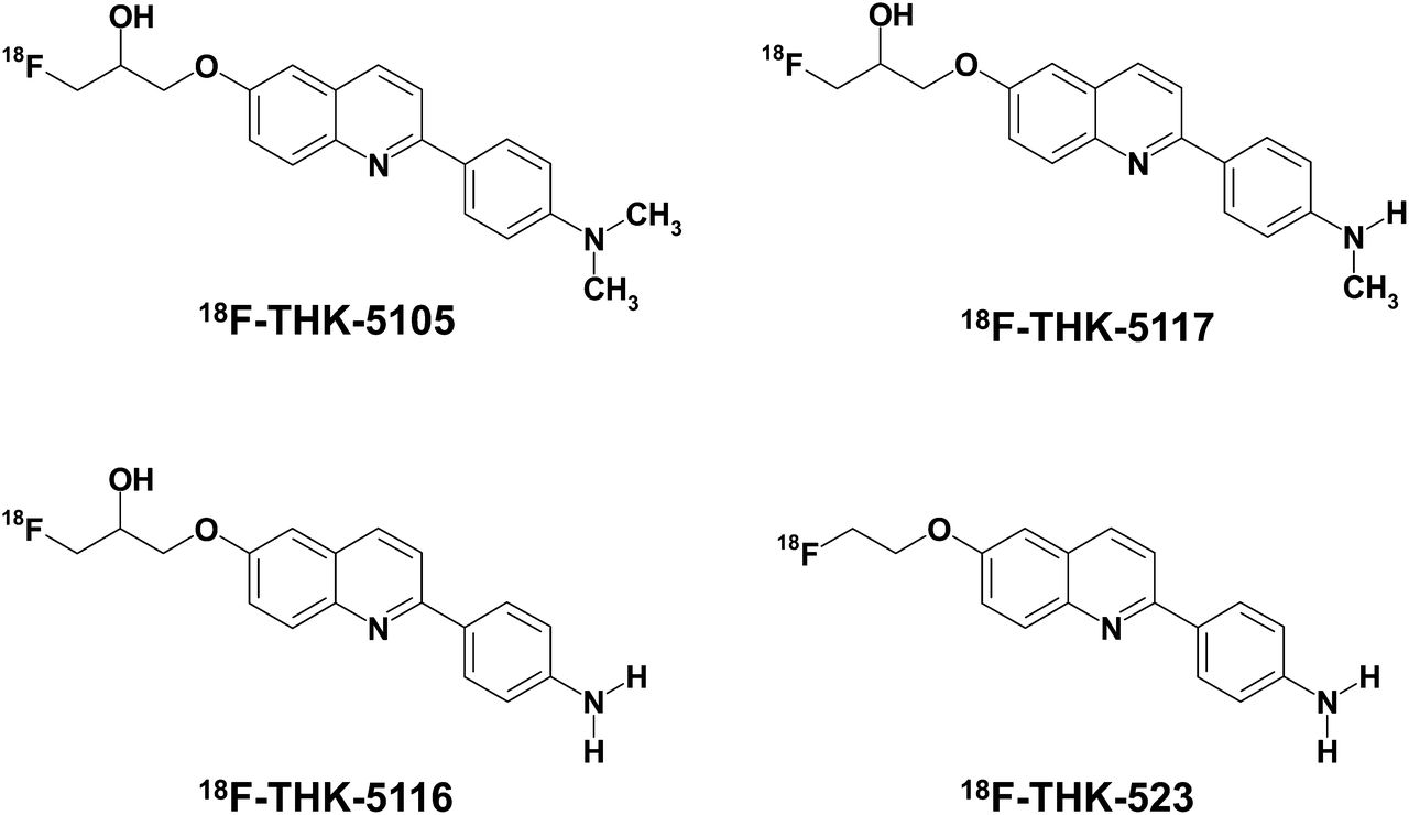
Chemical structures of 18F-THK-5105, 18F-THK-5116, 18F-THK-5117, and 18F-THK-523.

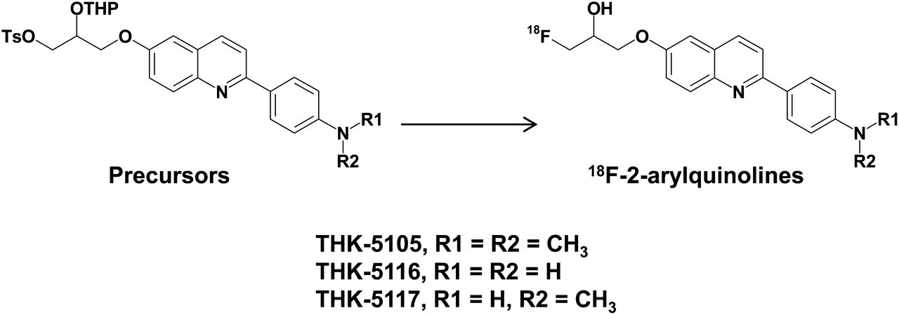
Radiosynthesis scheme of 18F-2-arylquinolines.
18F-THK-523 and 18F-FDDNP were also prepared in a manner similar to the one described above using the corresponding tosylate precursors reported previously (15,25,26). 11C-PiB was radiolabeled using its precursor (2-(4-aminophenyl)-6-methoxymethoxybenzothiazole) and 11C-methyl triflate, as previously described (27).
Determination of Log P Values
Log P values were determined by the HPLC method according to the guideline of the Organisation for Economic Co-operation and Development (OECD Guideline for Testing of Chemicals: Partition Coefficient (n-octanol/water), High Performance Liquid Chromatography [HPLC] Method), with slight modification. Briefly, 12 reference compounds whose log P values ranged between 0.5 and 4.0 were analyzed by HPLC under the following conditions: HPLC, a JASCO LC-2000 Plus series (JASCO); column, Inertsil ODS-4 (4.6 × 150 mm, 5 μm; GL Sciences, Inc.); mobile phase, 20 mM NaH2PO4 (pH 7.4)/acetonitrile (55/45); flow rate, 1.5 mL/min; ultraviolet absorbance, 245 nm; and column temperature, 40°C. Then, a calibration curve of log (tR − t0) (tR, retention time; t0, dead time) versus log P of each reference compound was created (R2 = 0.9469). Test compounds listed in Table 1 were also analyzed by the same HPLC method to measure log (tR − t0) values that were used for determination of log P values from the calibration curve.
Log P and Brain Uptake After Intravenous Administration of 18F-Labeled Compounds in Mice
In Vitro Binding Assays
Synthetic human Aβ1–42 was purchased from Peptide Institute Inc. Recombinant K18ΔK280-tau protein was obtained from Life Technologies Japan Ltd. Fibrils of Aβ1–42 and K18ΔK280-tau were prepared as described previously (15). Briefly, synthetic Aβ1–42 (200 μM) and K18Δ280K-tau (20 μM) solutions in phosphate-buffered saline (PBS) were incubated at 37°C with agitation for 3–4 d. We additionally prepared AD brain homogenates for binding assay, because the structural conformation of synthetic protein fibrils does not fully correlate with the structure of native protein deposits in the human brain. Human brain tissue was isolated from a mesial temporal frozen sample of an AD patient and homogenized in PBS. Brain tissue homogenate aliquots were taken and frozen at −80°C until used. Insoluble Aβ and tau levels were determined using a human β-amyloid enzyme-linked immunosorbent assay (ELISA) kit (Wako) and a human tau ELISA kit (Life Technologies Japan Ltd.), respectively. Next, brain homogenates and the solutions of synthetic Aβ1–42 or K18Δ280K-tau fibrils were incubated with increasing concentrations of 18F-THK-5105 (0.1–250 nM). To account for nonspecific binding of 18F-THK-5105, the reactions were performed in triplicate in the presence of 2 μM unlabeled THK-5105. The binding reactions were incubated for 1 h at room temperature in assay buffer (Dulbecco PBS; 0.1% bovine serum albumin). Bound radioactive compounds were separated from free radioactive compounds by filtration under reduced pressure (MultiScreen HTS Vacuum Manifold; Millipore). Filters were washed three times with assay buffer, and the radioactivity contained within the filters was counted in a γ-counter (AccuFLEX γ7000, Aloka, Tokyo, Japan). Binding data were analyzed using curve-fitting software that calculates the Kd and Bmax (Kd is dissociation constant and Bmax is maximum number of binding sites, respectively) using nonlinear regression (GraphPad Prism; GraphPad Software).
For inhibition studies, the assay buffer containing each compound (0.1–1,000 nM), 18F-THK-5105 (1.76 nM, ∼37 kBq), K18Δ280K-tau (200 nM), and 0.1% bovine serum albumin was incubated at room temperature for 1 h. Nonspecific binding was determined in the presence of 10 μM THK-5105. The mixture was filtered through Multiscreen HTS 96-well filtration plates, followed by washing three times with PBS (0.1% bovine serum albumin), and the filters containing bound 18F ligand were counted in a γ-counter. The percentage of bound radioligand at each concentration was measured in triplicate and then plotted against the inhibitor concentration. Half-maximal inhibitory concentration values were determined from the displacement curves using the GraphPad Prism software. Inhibition constant (Ki) values were calculated from the half-maximal inhibitory concentration values using the Cheng–Prusoff equation (28).
Tissue Staining
Experiments were performed under the regulations of the ethics committee of Tohoku University School of Medicine. Paraffin-embedded hippocampal brain sections from an autopsy-confirmed AD case (78-y-old woman) were used for tissue staining with THK-5105. Brain sections were obtained from Fukushimura Hospital. After deparaffinization, autofluorescence quenching was performed as previously described (29). Quenched tissue sections were immersed for 10 min in a 100-μM THK-5105 solution containing 50% ethanol. Sections were then dipped briefly into water, rinsed in PBS, coverslipped with FluorSave Reagent (Calbiochem), and examined using an Eclipse microscope (Nikon) equipped with a blue-violet filter (excitation, 400–440 nm; dichroic mirror, 455 nm; barrier filter, 470 nm). Sections stained with THK-5105 were subsequently immunostained with the AT8 anti-tau antibody (diluted 1:20; Innogenetics). After incubation at 4°C in the primary antibody for 16 h, sections were processed by the avidin-biotin method using a Pathostain ABC-POD(M) Kit (Wako) and diaminobenzidine as a chromogen. Sections were additionally stained using a modified Gallyas–Braak method (pretreatment with 0.3% potassium permanganate for 10 min, followed by 0.1% oxalic acid for 3 min) (30).
Autoradiography of Human Brain Sections
For the autoradiographic study, 8-μm-thick paraffin-embedded brain sections from a healthy control (62-y-old man) and 2 AD patients (69-y-old man and 92-y-old woman) were used. After deparaffinization, sections were incubated for 10 min at room temperature with radiolabeled compounds (0.5 MBq/mL) and washed briefly with water and 50% ethanol. After being dried, the labeled sections were exposed overnight to a BAS-III imaging plate (Fuji Film). The autoradiographic images were obtained using a BAS-5000 phosphoimaging instrument (Fuji Film). The neighboring sections were stained using a modified Gallyas–Braak method or immunostained using the AT8 anti-tau monoclonal antibody (diluted 1:20; Innogenetics), the 4G8 Aβ antibody (diluted 1:100; Signet), or the 6F/3D Aβ antibody (diluted 1:50; Dako). For correlational analysis of the autoradiographic and immunohistochemical images, 36 circular regions of interest (the area of each region of interest was ˜7 mm2) were placed on the gray matter of the hippocampus, parahippocampal gyrus, fusiform gyrus, temporal gyri (superior, middle, and inferior), insula, pre- and postcentral gyri, superior frontal gyrus, paracentral lobule, and cingulate gyrus. The percentage area of positive signals in each region of interest was calculated using ImageJ software (National Institutes of Health). A correlational analysis between percentage areas of tracer binding and positive immunostaining was performed using Pearson simple correlation.
Biodistribution in Mice
The experimental protocol of animal study was approved by the Ethics Committee of Tohoku University School of Medicine. 18F-labeled tracers (1.1–6.3 MBq) were injected into the tail vein of male ICR mice (n = 20; mean weight, 28–32 g). Mice were then sacrificed by decapitation at 2, 10, 30, 60, and 120 min after injection. The brain, blood, liver, kidney, and femur were removed and weighed, and radioactivity was counted with an automatic γ-counter. The percentage injected dose per gram of tissue (%ID/g) was calculated by comparing tissue counts to tissue weight. Each %ID/g value is expressed as a mean ± SD of 4 separate experiments.
Animal Toxicity Studies
A 14-d toxicity study with intravenous administration of a single dose of THK-5105 and THK-5117 was performed using Sprague–Dawley rats and ICR mice. Briefly, the study included 3 groups of male and female rats and mice that were administered 0 (group 1), 0.1 (group 2), and 1 (group 3) mg/kg of test article (10% dimethylsulfoxide/90% distilled water) per rat or mouse by intravenous injection on study day 1. The study included clinical observations plus body weight measurements for a 14-d observation period. Hematology and pathologic examinations were conducted on study days 2 and 15. Detailed necropsies with external examinations were also performed.
Receptor Binding Assays
Receptor binding screens were conducted by Sekisui Medical Inc. Binding inhibition effects of 1 μM THK-5105 and THK-5117 were evaluated in competitive radioligand assays against 60 common neurotransmitter receptors, ion channels, and transporters. Percentage inhibition ratios were calculated by the following equation: inhibition ratio (%) = [1 – (B − N)/(B0 − N)] × 100, where N is the nonspecific bound radioactivity, and B and B0 are the bound radioactivity in the presence and absence of tested compounds, respectively. Data are expressed as the mean values of duplicate samples.
RESULTS
Radiosynthesis
All radiolabeled compounds were obtained in greater than 97% radiochemical purities after HPLC purification. The decay-corrected average radiochemical yields of 18F-THK-523, 18F-THK-5105, 18F-THK-5116, 18F-THK-5117, and 18F-FDDNP were 58%, 48%, 41%, 48%, and 22%, respectively. The specific activities of 18F-labeded compounds ranged from 37 to 110 GBq/μmol, corrected at the end of synthesis. The mean specific activity of 11C-PiB was 35 GBq/μmol.
In Vitro Binding Assays
The binding properties of phenylquinoline derivatives to tau fibrils was investigated and compared with Aβ1–42 fibrils. Although only a single class of 18F-THK-5105 binding sites was identified on Aβ1–42 fibrils, 2 classes of 18F-THK-5105 binding sites were identified on K18Δ280-tau fibrils. As shown in Table 2, the Kd for the first class of K18Δ280-tau binding sites was 1.45 nM, indicating higher binding affinity to tau fibrils than to Aβ1–42 fibrils (Kd = 35.9 nM). Further, competitive binding assays with 18F-THK-5105 displayed high binding affinity of phenylquinoline derivatives to tau fibrils (Fig. 3). The Ki for THK-5117 was 10.5 nM, indicating that THK-5117 has higher binding affinity for tau fibrils than THK-523 (Ki = 59.3 nM). In contrast, the Ki for FDDNP was 263 nM. In binding assays using mesial temporal brain homogenates containing a high density of tau (1,075 pmol/g) and moderate density of Aβ (434 pmol/g), both 18F-THK-5105 (Kd = 2.63 nM; Bmax = 358 pmol/g of tissue) and 18F-THK-5117 (Kd = 5.19 nM; Bmax = 338 pmol/g of tissue) showed higher affinity for mesial temporal brain homogenates than 18F-THK-523 (Kd = 86.5 nM; Bmax = 647.1 pmol/g of tissue) (Supplemental Fig. 1; supplemental materials are available online only at http://jnm.snmjournals.org).
Kd and Bmax Values of 18F-THK-5105 for Synthetic Tau and Aβ1–42 Fibrils

Competitive inhibition of 18F-THK-5105 binding by 2-arylquinolines and FDDNP to tau protein fibrils. Ki values for inhibition of 18F-THK-5105 binding to tau are shown.
Tissue Staining and Autoradiography
The selective binding ability of the compounds was further examined using AD brain sections. The fluorescent compound THK-5105 clearly stained NFTs and neuropil threads in the hippocampal section of an AD patient (Fig. 4A). Selective binding of this compound with tau pathology was confirmed by comparing with the results of tau immunohistochemistry for the same sections (Fig. 4B). In contrast, SPs were faintly stained with THK-5105. Further, we compared findings of THK-5105 staining with those of Gallyas–Braak silver staining, a conventional technique used to visualize tau pathology in the AD brain (Figs. 4C and 4D), and the binding of THK-5105 to NFTs and neuropil threads was confirmed. The images of staining with THK-5116 and THK-5117 were similar to those with THK-5105 (data not shown).
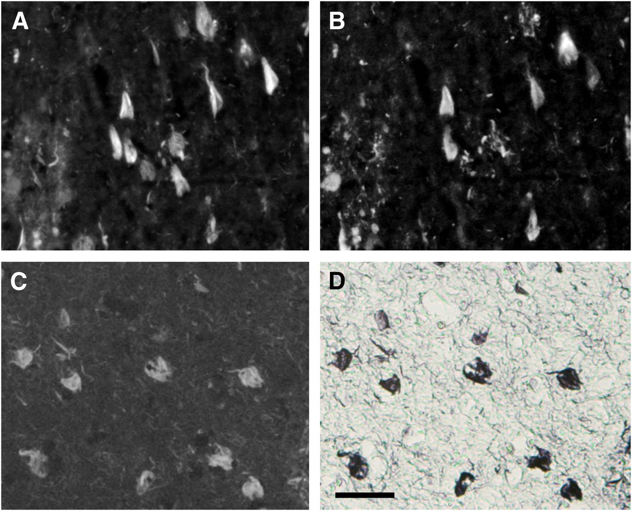
Neuropathologic staining of brain sections from AD patients. Neurofibrillary tangles and neuropil threads were clearly stained with THK-5105 (A and C). These stainings were consistent with tau immunostaining (B) and Gallyas–Braak staining (D) in same sections. Bar = 50 μm.
To investigate the binding ability of 18F-THK-5105 and 18F-THK-5117 to NFTs at tracer doses, in vitro autoradiography was performed in postmortem AD brain sections, and the findings were compared with Gallyas–Braak staining and immunohistochemistry. In the mesial temporal sections, laminar distributions of 18F-THK-5105 and 18F-THK-5117 were observed in the deep layer of gray matter (Fig. 5A). A high density of tracer accumulation was observed in the CA1 area of the hippocampus, which is reported as the most frequent site for NFTs in AD (31). These tracer distributions coincided with Gallyas–Braak staining and tau immunostaining (Fig. 5B) but not with the distribution of 11C-PiB (Fig. 5A) and Aβ immunostaining (Fig. 5B). In contrast, no significant accumulation of 18F-THK-5105 and 18F-THK-5117 was observed in the hippocampus of the healthy control subject (Supplemental Fig. 2). 18F-THK-5116 failed to give a specific signal in the AD brain sections (data not shown).

(A) Autoradiographic images of 18F-THK-5105, 18F-THK-5117, and 11C-PiB binding in mesial temporal section from AD patient. (B) Gallyas–Braak silver staining (left) and immunostaining with anti-tau (center) and anti-Aβ (right) antibodies in adjacent brain sections. Arrowheads = CA1 area of hippocampus; longer arrows = entorhinal cortex.
To further assess the regional differences of tracer binding in the AD brain, 18F-THK-5105 autoradiography was conducted using AD hemibrain sections and compared with the Aβ PET tracer 11C-PiB (32). 18F-THK-5105 densely accumulated in the gray matter of the hippocampus, parahippocampal gyrus, fusiform gyrus, inferior and middle temporal gyri, insula, and cingulate gyrus (Fig. 6A), regions known for the abundance of tau pathology in AD (33). In contrast, tracer binding in the parietal areas was modest. The pattern of tracer distribution correlated with the known distribution of tau pathology (Fig. 6A) but not with the known distribution of Aβ or the binding of 11C-PiB (data not shown). In addition, quantitative analyses of these images demonstrated a significant correlation of 18F-THK-5105 binding with tau immunostained areas but not with the areas of Aβ immunostaining (Fig. 6B; Supplemental Fig. 3). In contrast, 11C-PiB bindings showed a good correlation with Aβ deposition but not with tau deposition (Supplemental Fig. 3).

(A) Autoradiography of hemibrain sections from AD patient with 18F-THK-5105 and tau immunostaining in neighboring section. (B) Region-of-interest analysis indicated that percentage areas of 18F-THK-5105 binding (line plots) were significantly correlated with percentage areas of tau immunostaining (gray bars) but not with that of Aβ immunostaining (white bars). CG = cingulate gyrus; HIP = hippocampus; FUG = fusiform gyrus; IHC = immunohistochemistry; INS = insula; ITG = inferior temporal gyrus; MTG = middle temporal gyrus; PCL = paracentral lobule; PHG = parahippocampal gyrus; POG = postcentral gyrus; PRG = precentral gyrus; SFG = superior frontal gyrus; STG = superior temporal gyrus.
Pharmacokinetics in Mice
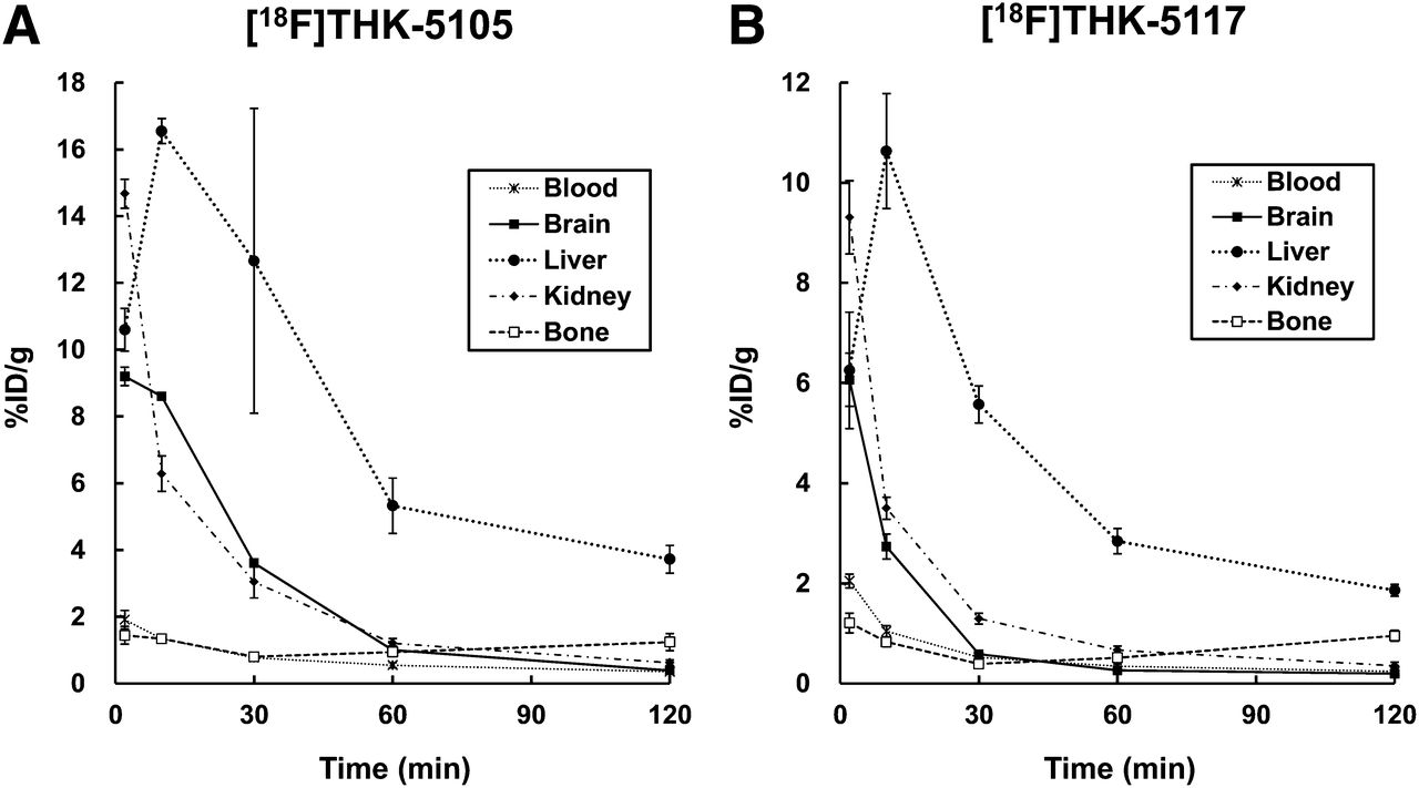
All tested compounds exhibited sufficient amounts of tracer uptake in the mouse brain immediately after intravenous administration. Compared with 18F-THK-523, new compounds showed significantly higher brain uptake at 2 min after injection (Table 1). 18F-THK-5105 showed the highest brain uptake. In addition, clearance of these derivatives from normal brain tissue was faster than that of 18F-THK-523 and 18F-FDDNP (Table 1). The brain uptake ratio at 2 versus 60 min was highest for 18F-THK-5117, followed by 18F-THK-5105, 18F-THK-5116, 18F-FDDNP, and 18F-THK-523. After injection of 18F-THK-5105 and 18F-THK-5117, the regional tracer uptake in the liver was highest at 10 min after injection, and the tracer was then slowly washed out from the body (Fig. 7). Compared with 18F-THK-5105, 18F-THK-5117 tended to have faster clearance from the brain, blood, liver, and kidney. No remarkable accumulation of 18F-THK-5105 and 18F-THK-5117 was observed in the bone.

Time–activity curves after intravenous administration of 18F-THK-5105 (A) and 18F-THK-5117 (B) in mice.
Animal Toxicity Studies
A single intravenous administration of THK-5105 and THK-5117 at 1 mg/kg, equivalent to 100,000-fold the intended clinical dose for humans, caused no systemic toxicity in rats or mice. There were no unscheduled deaths or morbidity detected in this study. During the experimental period, the body weight of all animals increased normally, and no treatment-related changes were noted in any animals. There were no major clinical, biochemical, or histopathologic findings associated with the administration of THK-5105 and THK-5117.
Receptor Binding Assays
Binding inhibition of THK-5105 and THK-5117 was assessed in competitive radioligand binding assays against 60 common neurotransmitter receptors, ion channels, and transporters. As a result, no remarkable inhibition (<50%) was observed for various receptors, ion channels, and transporters at 1-μM concentrations of THK-5105 and THK-5117.
DISCUSSION
These findings suggest that 18F-THK-5105 and 18F-THK-5117 are promising candidates as tau imaging PET probes. Although previous saturation analysis showed the high binding affinity of 18F-THK-523 for tau fibrils (Kd = 1.67 nM), the current competition assay demonstrated relatively lower binding affinity of THK-523 for tau fibrils (Ki = 59.3 nM) than THK-5105 (Ki = 7.8 nM) and THK-5117 (Ki = 10.5 nM). 18F-THK-5105 showed higher affinity for tau pathology than for Aβ pathology in AD brain sections. Most amyloid imaging agents potentially bind to both tau and Aβ fibrils, because both protein fibrils share a common β-sheet secondary structure. To ensure the binding specificity of these compounds as tau-selective PET probes, the binding affinity to Aβ fibrils should be below the in vivo detection threshold. In vitro binding assays indicated that the binding affinity of 18F-THK-5105 for Aβ fibrils (Kd = 35.9 nM) was 25 times lower than for tau fibrils (Kd = 1.45 nM). This Kd would allow selective detection of tau pathology, because the usual required Kd values for imaging Aβ are below 20 nM (34). However, the required Kd value for imaging tau deposits is still unknown. Considering that the concentrations of tau are about an order of magnitude lower than those of Aβ, the Kd value for tau should be well below 20 nM, in the low nanomolar range, to allow sensitive detection of tau pathology. In that respect, the binding affinities of both 18F-THK-5105 and 18F-THK-5117 to tau fibrils may be sufficient for in vivo detection of tau pathology in the brain. However, in vitro binding assay data should be carefully interpreted, because the structural conformation of synthetic tau fibrils does not fully correlate with the structure of NFTs and neuropil threads in the human brain. Actually, 18F-THK-523 showed substantially lower affinity for AD brain homogenates (Kd = 86.5 nM) than for synthetic tau protein fibrils (Kd = 1.67 nM) (15). In the future, in vitro binding data should be compared with in vivo PET data to determine the required Kd value for in vivo tau detection.
In vitro assays using human brain samples are considered more reliable for evaluating the binding selectivity of radiotracers to tau and Aβ pathology at tracer doses. Autoradiography studies using human brain sections demonstrated the preferential binding of 18F-THK-5105 and 18F-THK-5117 to tau protein deposits in the AD brain. We observed a high density of 18F-THK-5105 and 18F-THK-5117 binding in the CA1 region of AD hippocampus, which contained substantial amounts of NFTs and neuropil threads. In addition, these tracers clearly visualized the laminar distribution of tau in the pri-α layer of the transentorhinal and temporal cortices, which is typically observed in the AD brain (5). The distribution pattern of THK tracer binding in AD brains was different from that of the Aβ imaging probe PiB and BF-227, which showed diffuse punctate distribution in broad neocortical gray matter and less tracer distribution in the mesial temporal region. These findings strongly suggest that binding properties of 18F-THK-5105 and 18F-THK-5117 are different from those of currently available Aβ PET probes. Compared with 18F-THK-523 (17), both 18F-THK-5105 and 18F-THK-5117 showed higher contrast of tau pathology in autoradiographic images. These findings most likely reflect the increased binding affinity to tau by methylation of the amino group, as indicated by in vitro binding assays. Similar findings were previously reported in an arylbenzothiazole derivative (35). Compared with 18F-THK-5105, 18F-THK-5117 showed lesser tracer binding in the gray matter containing high density of Aβ plaques, suggesting low binding affinity to Aβ and high selectivity to tau. 18F-THK-5105 tends to show higher signals in the gray matter, and some of the images of 18F-THK-5105 binding showed the patchy pattern as observed for 11C-PiB binding. One possible reason for this is the binding of 18F-THK-5105 to tau protein in dystrophic neurites. Another possible reason is binding of 18F-THK-5105 to Aβ fibrils. However, the latter explanation seems unlikely given that 18F-THK-5105 binding, as clearly shown in Figure 6, was correlated with tau, and not Aβ, deposits.
In vitro binding assays using AD brain homogenates are generally used to measure the binding affinity of Aβ imaging radiotracers to SPs or NFTs and the number of binding sites in real AD pathology (36). For most of the useful Aβ imaging radiotracers, the reported Kd or Ki values for neocortical brain samples are below 10 nM (36,37). In this study, the Kd values for high-affinity sites of AD mesial temporal homogenates were 2.63 nM for 18F-THK-5105 and 5.19 nM for 18F-THK-5117. These binding affinities were higher than that for 18F-THK-523 and appear to be sufficient for the in vivo detection of AD pathology in the mesial temporal region at tracer doses. Furthermore, the Bmax/Kd ratios of 18F-THK-5105 and 18F-THK-5117 for AD brain homogenates were 136.1 and 65.1, respectively, which fulfills the criteria (Bmax/Kd ratio > 10) for a good neuroimaging agent (35).
The optimization of pharmacokinetics is an important aspect in the development of a PET tracer (38). 18F-THK-5105, 18F-THK-5116, and 18F-THK-5117 fulfilled the criteria of appropriate log P value (log P = 1–3) for brain entry (39). In mice, these tracers showed sufficient brain uptake and rapid washout from normal brain tissue. 18F-THK-5105 and 18F-THK-5117 exhibited high initial brain uptake in normal mice (>6 %ID/g at 2 min). These values, which are equivalent to over 100% injected dose index in a 25-g mouse, meet the prerequisites for useful PET imaging agents (34). The 2- to 60-min ratio of radioactivity concentrations for 18F-THK-5117 was 23.1, indicating faster washout from normal brain for these compounds than for other currently available 18F-labeled tracers such as 18F-FDDNP (2.91), 18F-florbetaben (4.83) (40), and 18F-florbetapir (3.90) (37). Compared with 18F-THK-523, 18F-THK-5116 washed out faster from normal brain tissue of mice, indicating that the hydroxylation of the fluoroalkoxy group improves pharmacokinetics in mice. However 18F-THK-5116 is not a suitable compound for clinical application, because of its lower initial brain uptake and binding affinity than the other 2 compounds.
CONCLUSION
18F-THK-5105 and 18F-THK-5117 should be considered as promising candidates for PET tau imaging radiotracers. Future clinical studies will clarify the usefulness of these radiotracers for the early detection of AD tau pathology.
DISCLOSURE
The costs of publication of this article were defrayed in part by the payment of page charges. Therefore, and solely to indicate this fact, this article is hereby marked “advertisement” in accordance with 18 USC section 1734. This study was supported by the research fund from GE Healthcare; the Industrial Technology Research Grant Program of the NEDO in Japan (09E51025a); Health and Labor Sciences Research grants from the Ministry of Health, Labor, and Welfare of Japan; and a Grant-in-Aid for Scientific Research (B) (23390297) and “Japan Advanced Molecular Imaging Program (J-AMP)” of the Ministry of Education, Culture, Sports, Science and Technology (MEXT), Japan. No other potential conflict of interest relevant to this article was reported.
Footnotes
Published online Jul. 15, 2013.
- © 2013 by the Society of Nuclear Medicine and Molecular Imaging, Inc.
REFERENCES
- Received for publication November 19, 2012.
- Accepted for publication February 19, 2013.


Jump to section
Related Articles
Cited By...
- Preclinical Characterization of the Tau PET Tracer [18F]SNFT-1: Comparison of Tau PET Tracers
- Preclinical Characterization of the Tau PET Tracer [18F]SNFT-1: Comparison of Tau PET Tracers
- 18F-SMBT-1: A Selective and Reversible PET Tracer for Monoamine Oxidase-B Imaging
- Test-Retest Reproducibility for the Tau PET Imaging Agent Flortaucipir F 18
- Comparative In Vitro and In Vivo Quantifications of Pathologic Tau Deposits and Their Association with Neurodegeneration in Tauopathy Mouse Models
- Cerebrospinal Fluid Clearance in Alzheimer Disease Measured with Dynamic PET
- Biodistribution and Radiation Dosimetry for the Tau Tracer 18F-THK-5351 in Healthy Human Subjects
- Interactions between Microtubule-Associated Protein Tau (MAPT) and Small Molecules
- Kinetic Modeling of the Tau PET Tracer 18F-AV-1451 in Human Healthy Volunteers and Alzheimer Disease Subjects
- In Vivo Comparison of Tau Radioligands 18F-THK-5351 and 18F-THK-5317
- Tau Positron Emission Tomography Imaging
- Reference Tissue-Based Kinetic Evaluation of 18F-AV-1451 for Tau Imaging
- In vivo visualization of tau deposits in corticobasal syndrome by 18F-THK5351 PET
- Kinetics of the Tau PET Tracer 18F-AV-1451 (T807) in Subjects with Normal Cognitive Function, Mild Cognitive Impairment, and Alzheimer Disease
- Temporal T807 binding correlates with CSF tau and phospho-tau in normal elderly
- In Vivo Tau, Amyloid, and Gray Matter Profiles in the Aging Brain
- Small-Animal PET Imaging of Tau Pathology with 18F-THK5117 in 2 Transgenic Mouse Models
- Tracer Kinetic Analysis of (S)-18F-THK5117 as a PET Tracer for Assessing Tau Pathology
- Structure-Activity Relationship of 2-Arylquinolines as PET Imaging Tracers for Tau Pathology in Alzheimer Disease
- 18F-THK5351: A Novel PET Radiotracer for Imaging Neurofibrillary Pathology in Alzheimer Disease
- Cortical Laminar Binding of PET Amyloid and Tau Tracers in Alzheimer Disease
- New Targets for the Development of PET Tracers for Imaging Neurodegeneration in Alzheimer Disease
- Molecular Imaging of Alzheimer Disease Pathology
- Molecular Imaging Insights into Neurodegeneration: Focus on Tau PET Radiotracers