


Article Figures & Data
Figures
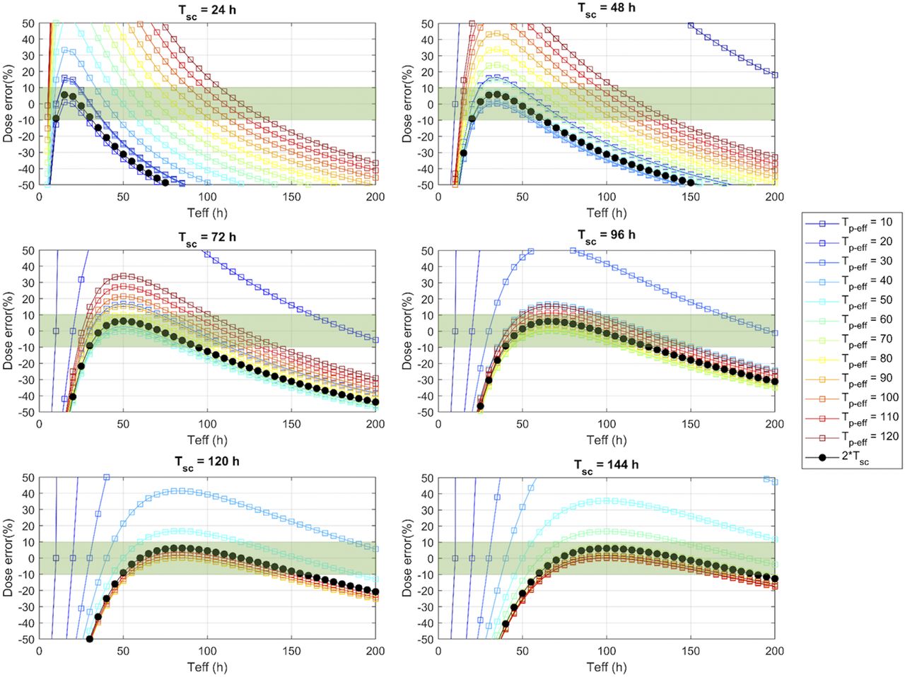
- FIGURE 1.
DEs (%) resulting from method 1 (black line) and method 2 (with Tp-eff ranging from 10 to 120 h; colored lines) relative to true Teff of radiopharmaceutical washout. DEs within ±10% are highlighted in green.
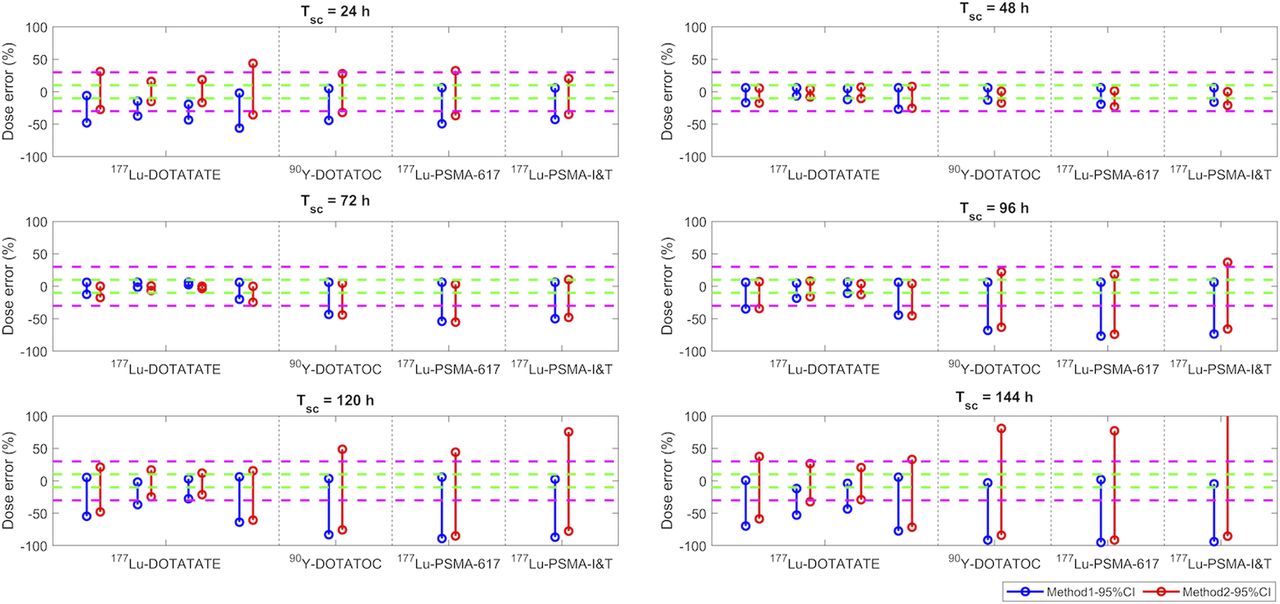
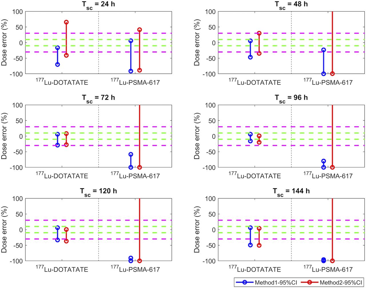
- FIGURE 2.
DEs (%) of kidney doses estimated using method 1 (blue) and method 2 (red) when patient Teff is within simulated 95% CI range listed in Table 2. Green and magenta dashed lines indicate ±10% and ±30% of DEs, respectively. Four sets of results shown in 177Lu-DOTATATE column correspond to Teff data from studies 1–4.
- FIGURE 3.
DEs (%) of bone marrow doses estimated using method 1 (blue) and method 2 (red) when patient Teff is within simulated 95% CI range from Table 2. Green and magenta dashed lines indicate ±10% and ±30% of DEs, respectively. Two sets of results correspond to data from studies 4 and 7.
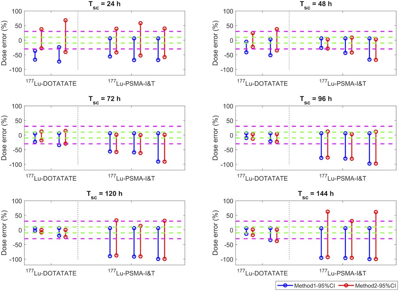
- FIGURE 4.
DEs (%) of tumor doses estimated using method 1 (blue) and method 2 (red) when patient Teff is within 95% CI range from Table 2. Green and magenta dashed lines indicate ±10% and ±30% of DEs, respectively. Data in 177Lu-DOTATATE column correspond to studies 3 and 4, whereas data in 177Lu-PSMA-I&T column correspond to bone and lymph node metastasis data from studies 8 and 9.


Tables
Method Approximation Conclusion 1 (3) 
Error < 10% if 2 (4,7) Smallest errors can be observed if []- TABLE 2
Mean Teff and SD, and Computed 95% CI, of Organs and Lesions for Commonly Applied Radiopharmaceuticals That Were Used in This Investigation
Agent Study Reference Patients (n) Organ or target Median mean (h) SD (h)95% CI (h) 177Lu-DOTATATE 1 Hou et al. 2019 (12) 30 (87) Kidney* 46 (30–82) 47.0 11.6 28.5–73.6 2 Heikkonen et al. 2016 (13) 24 (24) Kidney NA (36–59) 45.3 5.9 34.8–57.4 3 Hänscheid et al. 2018 (3)† 27 (54) Kidney‡ 51 (40–68) 51.0 7.0 38.8–66.0 25 (25) Liver‡ 67 (55–117) 76.5 15.5 51.4–110.7 27 (27) Spleen‡ 68 (52–99) 68.0 11.8 49.5–91.6 22 (22) Tumor‡ 77 (56–130) 85.4 18.5 56.0–125.8 4 Del Prete et al. 2018 (11) 158 (1,117) Kidney* ‡ 47 (23–159) 50.8 16.9 25.6–91.0 Desy et al. 2020 (14) 158 (474) Bone marrow* ‡ 70 (29–160) 76.4 27.6 36.2–142.8 158 (2,166) Tumor* ‡ 84 (16–161) 87.8 30.5 42.8–160.6 90Y-DOTATOC 5 Menda et al. 2018 (15) 25 (69) Kidney NA (25–92) 37.5 12.5 19.4–67.2 177Lu-PSMA-617 6 Kurth et al. 2018 (16) 25 (25) Whole body NA (22–86) 40.5 15.8 18.8–79.1 7 Sarnelli et al. 2019 (17) 9 (9) Parotid gland 33 (26–61) 35.4 10.6 22.2–53.2 9 (9) Kidney 31 (12–81) 39.2 20.9 17.2–76.2 9 (9) Red marrow 8 (3–15) 8.0 4.7 3.2–16.3 9 (9) Liver 25 (13–63) 33.5 20.0 13.4–69.1 9 (9) Whole body 40 (32–80) 52.4 22.2 27.2–90.5 177Lu-PSMA-I&T 8 Written communications 15 (290) Bone metastases* 38 (13–191) 42.6 19.1 16.9–90.0 9 Baum et al. 2016 (18) 30 (NA) Bone metastases‡ 52 (14–149) 52.0 30.0 16.2–132.6 30 (NA) Lymph nodemetastases‡ 43 (25–160) 43.0 32.0 9.9–132.7 30 (NA) Kidney‡ 33 (19–83) 33.0 14.0 14.8–64.9 30 (NA) Parotid gland‡ 25 (20–43) 25.0 5.0 16.7–36.1 ↵* Teff of each individual ROI (organ or lesion) was available (i.e., complete listing of Teff for all patients).
↵† Overall dataset (29 patients) was primarily 177Lu-DOTATATE (22 patients) but also included 177Lu-DOTATOC (7 patients).
↵‡ Teff was published as median and range. For studies 3 and 9, corresponding mean and SD were calculated using method of Hozo et al. 2005 (19). For study 4, we had access to complete listing of Teff.
NA = not applicable.
Data in parentheses are range (for median) or total number of ROIs (for number of patients).


Supplemental Data
Files in this Data Supplement:


Jump to section
Related Articles
Cited By...
- Can 177Lu-DOTATATE Kidney Absorbed Doses be Predicted from Pretherapy SSTR PET? Findings from Multicenter Data
- Characterization of Effective Half-Life for Instant Single-Time-Point Dosimetry Using Machine Learning
- Impact of Posttreatment SPECT/CT on Patient Management During 177Lu-PSMA-617 Radiopharmaceutical Therapy
- Impact of the Reference Multiple-Time-Point Dosimetry Protocol on the Validity of Single-Time-Point Dosimetry for [177Lu]Lu-PSMA-I&T Therapy
- Assessing Response to PSMA Radiopharmaceutical Therapies with Single SPECT Imaging at 24 Hours After Injection
- ISIT-QA: In Silico Imaging Trial to Evaluate a Low-Count Quantitative SPECT Method Across Multiple Scanner-Collimator Configurations for 223Ra-Based Radiopharmaceutical Therapies
- Single-Time-Point Renal Dosimetry Using Nonlinear Mixed-Effects Modeling and Population-Based Model Selection in [177Lu]Lu-PSMA-617 Therapy
- Single-Time-Point Imaging for Dosimetry After [177Lu]Lu-DOTATATE: Accuracy of Existing Methods and Novel Data-Driven Models for Reducing Sensitivity to Time-Point Selection
- Toward Single-Time-Point Image-Based Dosimetry of 177Lu-PSMA-617 Therapy
- Prostate-Specific Membrane Antigen Radioligand Therapy Using 177Lu-PSMA I&T and 177Lu-PSMA-617 in Patients with Metastatic Castration-Resistant Prostate Cancer: Comparison of Safety, Biodistribution, and Dosimetry
- Reply: Single-Time-Point Tumor Dosimetry Assuming Normal Distribution of Tumor Kinetics
- Reimbursement Approaches for Radiopharmaceutical Dosimetry: Current Status and Future Opportunities