
Main menu

  • Home
  • Content
    • Current
    • Ahead of print
    • Past Issues
    • JNM Supplement
    • SNMMI Annual Meeting Abstracts
    • Continuing Education
    • JNM Podcasts
  • Subscriptions
    • Subscribers
    • Institutional and Non-member
    • Rates
    • Journal Claims
    • Corporate & Special Sales
  • Authors
    • Submit to JNM
    • Information for Authors
    • Assignment of Copyright
    • AQARA requirements
  • Info
    • Reviewers
    • Permissions
    • Advertisers
  • About
    • About Us
    • Editorial Board
    • Contact Information
  • More
    • Alerts
    • Feedback
    • Help
    • SNMMI Journals
  • SNMMI
    • JNM
    • JNMT
    • SNMMI Journals
    • SNMMI

User menu

  • Subscribe
  • My alerts
  • Log in
  • My Cart

Search

  • Advanced search
Journal of Nuclear Medicine
  • SNMMI
    • JNM
    • JNMT
    • SNMMI Journals
    • SNMMI
  • Subscribe
  • My alerts
  • Log in
  • My Cart
Journal of Nuclear Medicine

Advanced Search

  • Home
  • Content
    • Current
    • Ahead of print
    • Past Issues
    • JNM Supplement
    • SNMMI Annual Meeting Abstracts
    • Continuing Education
    • JNM Podcasts
  • Subscriptions
    • Subscribers
    • Institutional and Non-member
    • Rates
    • Journal Claims
    • Corporate & Special Sales
  • Authors
    • Submit to JNM
    • Information for Authors
    • Assignment of Copyright
    • AQARA requirements
  • Info
    • Reviewers
    • Permissions
    • Advertisers
  • About
    • About Us
    • Editorial Board
    • Contact Information
  • More
    • Alerts
    • Feedback
    • Help
    • SNMMI Journals
  • View or Listen to JNM Podcast
  • Visit JNM on Facebook
  • Join JNM on LinkedIn
  • Follow JNM on Twitter
  • Subscribe to our RSS feeds
Research ArticleRadiochemistry

Preclinical Evaluation of Benzazepine-Based PET Radioligands (R)- and (S)-11C-Me-NB1 Reveals Distinct Enantiomeric Binding Patterns and a Tightrope Walk Between GluN2B- and σ1-Receptor–Targeted PET Imaging

Achi Haider, Adrienne Müller Herde, Stefanie D. Krämer, Jasmine Varisco, Claudia Keller, Katrin Frauenknecht, Yves P. Auberson, Louisa Temme, Dina Robaa, Wolfgang Sippl, Roger Schibli, Bernhard Wünsch, Linjing Mu and Simon M. Ametamey
Journal of Nuclear Medicine August 2019, 60 (8) 1167-1173; DOI: https://doi.org/10.2967/jnumed.118.221051
Achi Haider
1Institute of Pharmaceutical Sciences, ETH Zurich, Zurich, Switzerland
  • Find this author on Google Scholar
  • Find this author on PubMed
  • Search for this author on this site
Adrienne Müller Herde
1Institute of Pharmaceutical Sciences, ETH Zurich, Zurich, Switzerland
  • Find this author on Google Scholar
  • Find this author on PubMed
  • Search for this author on this site
Stefanie D. Krämer
1Institute of Pharmaceutical Sciences, ETH Zurich, Zurich, Switzerland
  • Find this author on Google Scholar
  • Find this author on PubMed
  • Search for this author on this site
Jasmine Varisco
1Institute of Pharmaceutical Sciences, ETH Zurich, Zurich, Switzerland
  • Find this author on Google Scholar
  • Find this author on PubMed
  • Search for this author on this site
Claudia Keller
1Institute of Pharmaceutical Sciences, ETH Zurich, Zurich, Switzerland
  • Find this author on Google Scholar
  • Find this author on PubMed
  • Search for this author on this site
Katrin Frauenknecht
2Institute of Neuropathology, University of Zurich/University Hospital Zurich, Zurich, Switzerland
  • Find this author on Google Scholar
  • Find this author on PubMed
  • Search for this author on this site
Yves P. Auberson
3Novartis Institutes for BioMedical Research, Basel, Switzerland
  • Find this author on Google Scholar
  • Find this author on PubMed
  • Search for this author on this site
Louisa Temme
4Institute of Pharmaceutical and Medicinal Chemistry, University of Münster, Münster, Germany
  • Find this author on Google Scholar
  • Find this author on PubMed
  • Search for this author on this site
Dina Robaa
5Institute of Pharmacy, Martin-Luther University of Halle-Wittenberg, Halle/Saale, Germany; and
  • Find this author on Google Scholar
  • Find this author on PubMed
  • Search for this author on this site
Wolfgang Sippl
5Institute of Pharmacy, Martin-Luther University of Halle-Wittenberg, Halle/Saale, Germany; and
  • Find this author on Google Scholar
  • Find this author on PubMed
  • Search for this author on this site
Roger Schibli
1Institute of Pharmaceutical Sciences, ETH Zurich, Zurich, Switzerland
6Department of Nuclear Medicine, University Hospital Zurich, Zurich, Switzerland
  • Find this author on Google Scholar
  • Find this author on PubMed
  • Search for this author on this site
Bernhard Wünsch
4Institute of Pharmaceutical and Medicinal Chemistry, University of Münster, Münster, Germany
  • Find this author on Google Scholar
  • Find this author on PubMed
  • Search for this author on this site
Linjing Mu
1Institute of Pharmaceutical Sciences, ETH Zurich, Zurich, Switzerland
6Department of Nuclear Medicine, University Hospital Zurich, Zurich, Switzerland
  • Find this author on Google Scholar
  • Find this author on PubMed
  • Search for this author on this site
Simon M. Ametamey
1Institute of Pharmaceutical Sciences, ETH Zurich, Zurich, Switzerland
  • Find this author on Google Scholar
  • Find this author on PubMed
  • Search for this author on this site
  • Article
  • Figures & Data
  • Supplemental
  • Info & Metrics
  • PDF
Loading

Article Figures & Data

Figures

  • Additional Files
  • FIGURE 1.
    • Download figure
    • Open in new tab
    • Download powerpoint
    FIGURE 1.

    Absolute configuration and binding behavior of (R)- and (S)-Me-NB1. (A) ORTEP view of (R)- and (S)-Me-NB1 in solid state. Me-NB1 exhibits one chiral center depicted at position C13. Thermal ellipsoids are set to 30% probability. For all nonhydrogen atoms, anisotropic displacement parameters were used. Hydrogen atoms were refined in idealized positions using riding model. (B) Representative in vitro autoradiograms of (R)/(S)-11C-Me-NB1 incubated with Wistar rat brain sections. Although (S)-enantiomer binds to virtually all brain regions, (R)-enantiomer exhibits selectivity for GluN2B-rich forebrain. Cx = cortex; CPu = corpus striatum; Hp = hippocampus; Th = thalamus; Cb = cerebellum.

  • FIGURE 2.
    • Download figure
    • Open in new tab
    • Download powerpoint
    FIGURE 2.

    Autoradiographic screening of clinically tested GluN2B ligands CERC-301, EVT-101, and CP101,606, as well as σ1R ligands SA4503, fluspidine, and (+)-pentazocine, as blocking agents for (R)-11C-Me-NB1 and (S)-11C-Me-NB1. (R)-11C-Me-NB1 showed heterogeneous binding behavior that was GluN2B-specific whereas (S)-11C-Me-NB1 exhibited considerable σ1R binding.

  • FIGURE 3.
    • Download figure
    • Open in new tab
    • Download powerpoint
    FIGURE 3.

    Whole-brain time–activity curves of racemic and enantiomerically pure 11C-Me-NB1 in Wistar rat brain are depicted as SUVs. GluN2B-specific (R)-form exhibited higher SUVs than (S)-form, whereas racemic mixture displayed time–activity curve that lies between the two enantiomers. For blockade experiment, 2 mg/kg dose of GluN2B antagonist eliprodil was used.
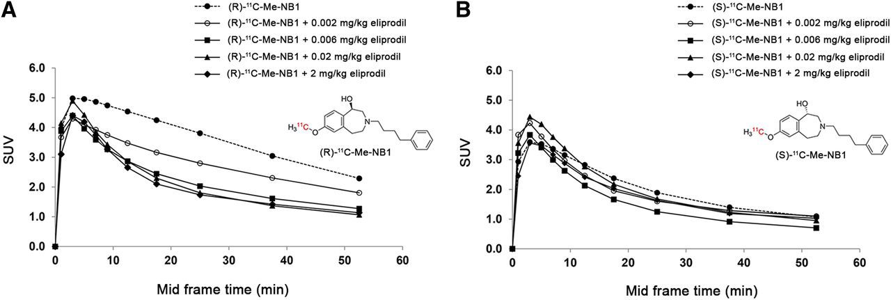
  • FIGURE 4.
    • Download figure
    • Open in new tab
    • Download powerpoint
    FIGURE 4.

    Dose–response curves of (R)-11C-Me-NB1 and (S)-11C-Me-NB1 with eliprodil in Wistar rat brain. Results are reported as SUVs calculated from PET experiments (0–60 min). (A) (R)-11C-Me-NB1 revealed consistent dose–response profile on stepwise dose escalation of eliprodil. (B) (S)-11C-Me-NB1 did not show dose-dependent blockade.

  • FIGURE 5.
    • Download figure
    • Open in new tab
    • Download powerpoint
    FIGURE 5.

    Representative ex vivo autoradiogram of Wistar rat brain at 15 min after injection of (R)-11C-Me-NB1. Selectivity for cortex, striatum, thalamus, and hippocampus over cerebellum was observed. Cb = cerebellum; CPu = corpus striatum; Cx = cortex; Hp = hippocampus; Th = thalamus.

  • FIGURE 6.
    • Download figure
    • Open in new tab
    • Download powerpoint
    FIGURE 6.

    Receptor occupancy by clinically tested GluN2B antagonist CP101,606 and GluN2B PET radioligand (R)-11C-Me-NB in Wistar rats (B), calculated from experimental SUV0–60 min (A), revealed plasma concentration of 158 nmol/L, or 52 ng/mL, required for 50% receptor occupancy. Clinically applied target plasma concentration of 200 ng/mL occupies 80% of GluN2B binding sites in our rat model. Rat brain PET images were superimposed on PMOD MRI template (C). Bs = brain stem; Cb = cerebellum; CPu = corpus striatum; Cx = cortex; Hp = hippocampus; Th = thalamus.

  • FIGURE 7.
    • Download figure
    • Open in new tab
    • Download powerpoint
    FIGURE 7.

    Representative autoradiograms on rodent and human brain tissue. (A) Ex vivo autoradiography at 15 min after injection of (R)-11C-Me-NB1 into Wistar rat and subsequent in vitro displacer screening with GluN2B ligands CERC-301, EVT-101, CP101,606, and σ1R ligands fluspidine, (+)-pentazocine, and (S)-Me-NB1. Displacement of (R)-11C-Me-NB1 binding is observed only for GluN2B antagonists. (B) Representative autoradiograms of (R)-11C-Me-NB1 incubated with human postmortem brain sections. GluN2B-specific binding was observed in temporal cortex (indicated by blockade experiment with GluN2B antagonist CERC-301), whereas cerebellum did not reveal any specific binding of radioligand.

Additional Files

  • Figures
  • Supplemental Data

    Files in this Data Supplement:

    • Supplemental Data
PreviousNext
Back to top

In this issue

Journal of Nuclear Medicine: 60 (8)
Journal of Nuclear Medicine
Vol. 60, Issue 8
August 1, 2019
  • Table of Contents
  • Table of Contents (PDF)
  • About the Cover
  • Index by author
Print
Download PDF
Article Alerts
Sign In to Email Alerts with your Email Address
Email Article

Thank you for your interest in spreading the word on Journal of Nuclear Medicine.

NOTE: We only request your email address so that the person you are recommending the page to knows that you wanted them to see it, and that it is not junk mail. We do not capture any email address.

Enter multiple addresses on separate lines or separate them with commas.
Preclinical Evaluation of Benzazepine-Based PET Radioligands (R)- and (S)-11C-Me-NB1 Reveals Distinct Enantiomeric Binding Patterns and a Tightrope Walk Between GluN2B- and σ1-Receptor–Targeted PET Imaging
(Your Name) has sent you a message from Journal of Nuclear Medicine
(Your Name) thought you would like to see the Journal of Nuclear Medicine web site.
Citation Tools
Preclinical Evaluation of Benzazepine-Based PET Radioligands (R)- and (S)-11C-Me-NB1 Reveals Distinct Enantiomeric Binding Patterns and a Tightrope Walk Between GluN2B- and σ1-Receptor–Targeted PET Imaging
Achi Haider, Adrienne Müller Herde, Stefanie D. Krämer, Jasmine Varisco, Claudia Keller, Katrin Frauenknecht, Yves P. Auberson, Louisa Temme, Dina Robaa, Wolfgang Sippl, Roger Schibli, Bernhard Wünsch, Linjing Mu, Simon M. Ametamey
Journal of Nuclear Medicine Aug 2019, 60 (8) 1167-1173; DOI: 10.2967/jnumed.118.221051

Citation Manager Formats

  • BibTeX
  • Bookends
  • EasyBib
  • EndNote (tagged)
  • EndNote 8 (xml)
  • Medlars
  • Mendeley
  • Papers
  • RefWorks Tagged
  • Ref Manager
  • RIS
  • Zotero
Share
Preclinical Evaluation of Benzazepine-Based PET Radioligands (R)- and (S)-11C-Me-NB1 Reveals Distinct Enantiomeric Binding Patterns and a Tightrope Walk Between GluN2B- and σ1-Receptor–Targeted PET Imaging
Achi Haider, Adrienne Müller Herde, Stefanie D. Krämer, Jasmine Varisco, Claudia Keller, Katrin Frauenknecht, Yves P. Auberson, Louisa Temme, Dina Robaa, Wolfgang Sippl, Roger Schibli, Bernhard Wünsch, Linjing Mu, Simon M. Ametamey
Journal of Nuclear Medicine Aug 2019, 60 (8) 1167-1173; DOI: 10.2967/jnumed.118.221051
Twitter logo Facebook logo LinkedIn logo Mendeley logo
  • Tweet Widget
  • Facebook Like
  • Google Plus One
Bookmark this article

Jump to section

  • Article
    • Abstract
    • MATERIALS AND METHODS
    • RESULTS
    • DISCUSSION
    • CONCLUSION
    • DISCLOSURE
    • Acknowledgments
    • Footnotes
    • REFERENCES
  • Figures & Data
  • Supplemental
  • Info & Metrics
  • PDF

Related Articles

  • This Month in JNM
  • PubMed
  • Google Scholar

Cited By...

  • Evaluation of (rac)-, (R)-, and (S)-18F-OF-NB1 for Imaging GluN2B Subunit-Containing N-Methyl-D-Aspartate Receptors in Nonhuman Primates
  • First-in-Humans Brain PET Imaging of the GluN2B-Containing N-methyl-D-aspartate Receptor with (R)-11C-Me-NB1
  • Preclinical Development of 18F-OF-NB1 for Imaging GluN2B-Containing N-Methyl-D-Aspartate Receptors and Its Utility as a Biomarker for Amyotrophic Lateral Sclerosis
  • Evaluation of 11C-NR2B-SMe and Its Enantiomers as PET Radioligands for Imaging the NR2B Subunit Within the NMDA Receptor Complex in Rats
  • Google Scholar

More in this TOC Section

Radiochemistry

  • Cyclotron-Produced 132La as a PET Imaging Surrogate for Therapeutic 225Ac
  • Cyclotron Production and Separation of Scandium Radionuclides from Natural Titanium Metal and Titanium Dioxide Targets
  • Synthesis of the PET Tracer 124I-Trametinib for MAPK/ERK Kinase Distribution and Resistance Monitoring
Show more Radiochemistry

Basic

  • Cyclotron-Produced 132La as a PET Imaging Surrogate for Therapeutic 225Ac
  • Cyclotron Production and Separation of Scandium Radionuclides from Natural Titanium Metal and Titanium Dioxide Targets
  • Synthesis of the PET Tracer 124I-Trametinib for MAPK/ERK Kinase Distribution and Resistance Monitoring
Show more Basic

Similar Articles

Keywords

  • PET imaging
  • GluN2B
  • σ1-receptor
  • receptor occupancy
  • neurodegeneration
SNMMI

© 2025 SNMMI

Powered by HighWire