


Article Figures & Data
Figures
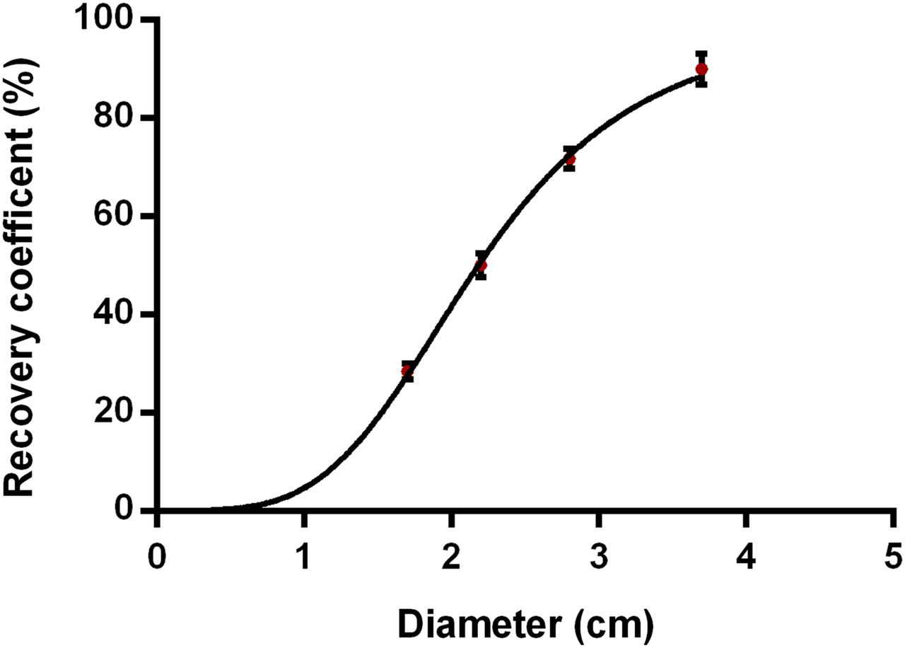
- FIGURE 1.
RC vs. sphere diameter (3.7, 2.8, 2.2, and 1.7). For each sphere in National Electrical Manufacturers Association image quality phantom, 6 RCs were determined, and the mean and SD of these 6 values are shown. Solid line represents 2-parameter sigmoid fit.
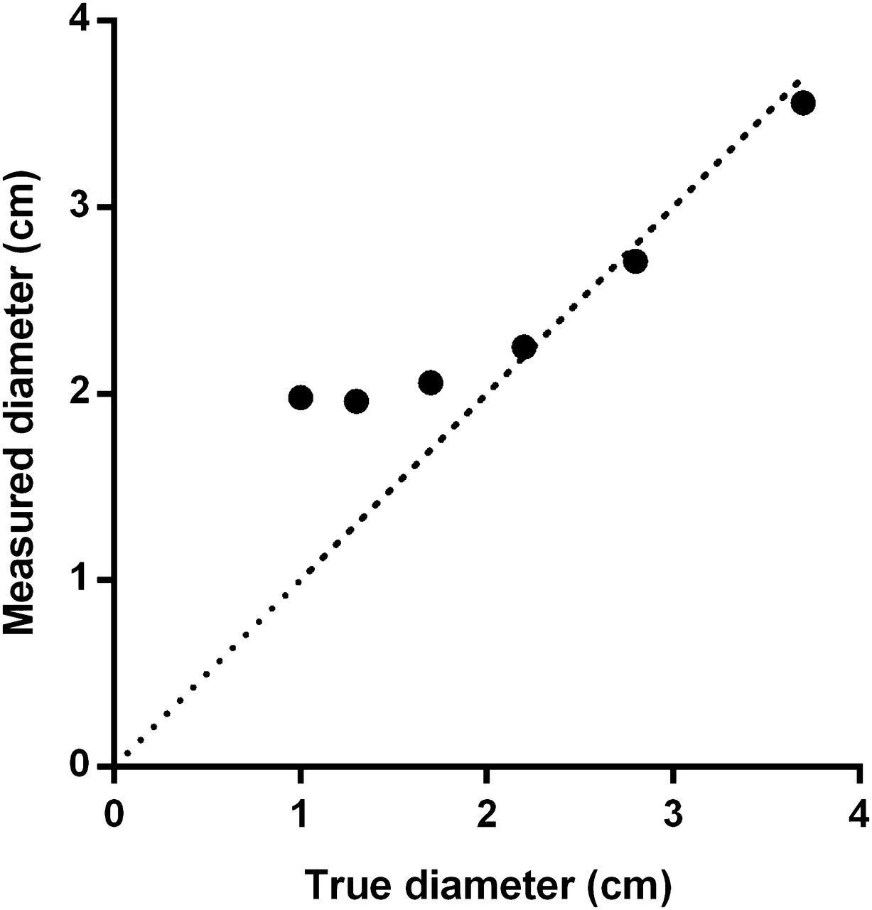
- FIGURE 2.
Measured sphere diameter using a 42% isocontour VOI vs. true sphere diameter in National Electrical Manufacturers Association image quality phantom. Dashed line represents line of identity.
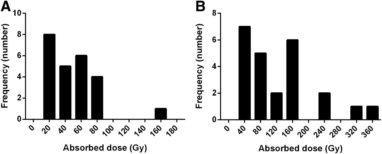
- FIGURE 3.
Frequency distribution of absorbed doses in 24 tumors. (A) Absorbed dose distribution during first treatment cycle. (B) Distribution of tumor-absorbed dose until best response. Numbers on x axes are centers of absorbed dose intervals used for each bar (20 Gy = 20 ± 10 Gy [A], 40 Gy = 40 ± 20 Gy [B]).
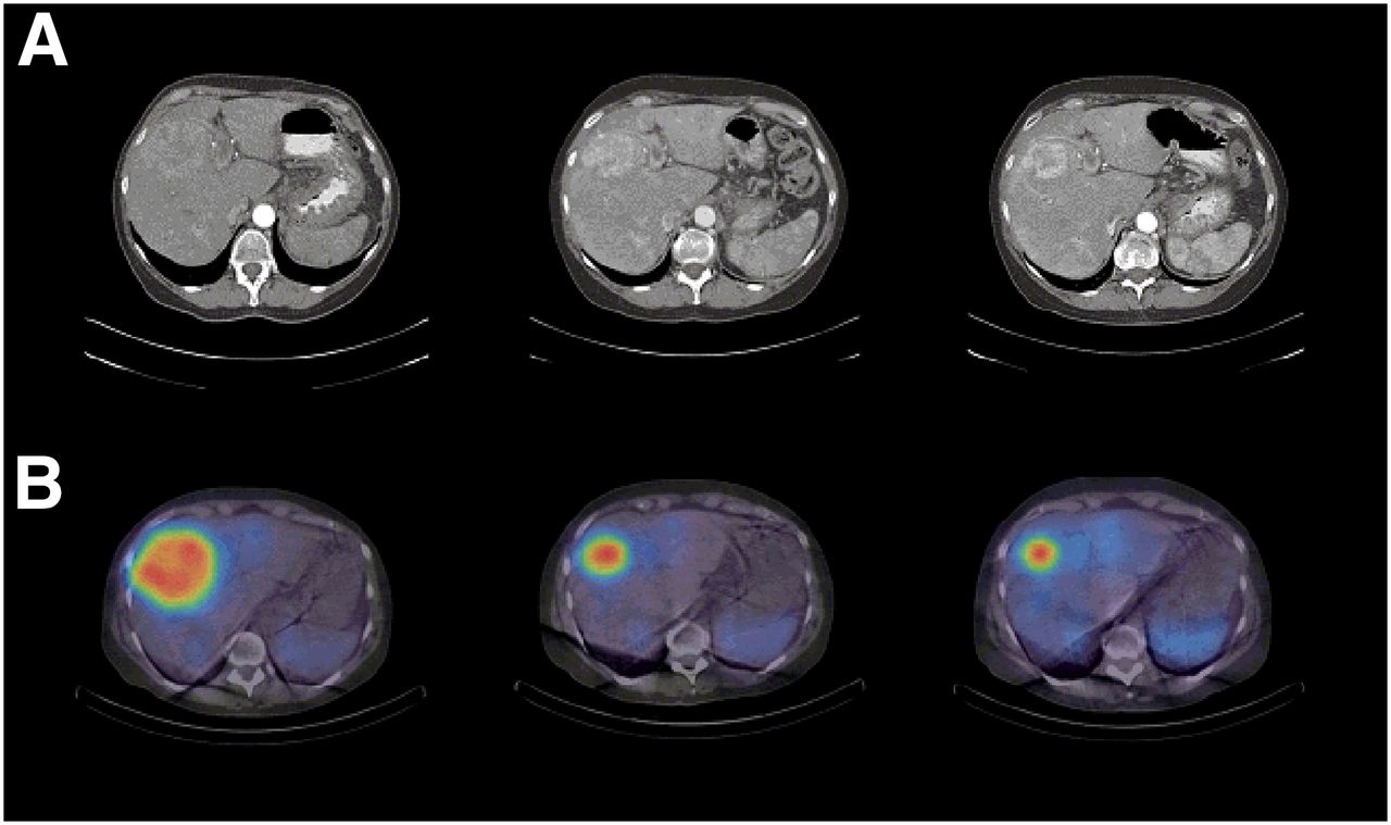
- FIGURE 4.
Example of dose response in patient with inoperable metastasized PNET in liver (patient 19). (A) Transversal contrast-enhanced CT images in late arterial phase. (B) Fused SPECT/CT images. Shown left to right are examinations at baseline and after 2 and 3 cycles of PRRT.
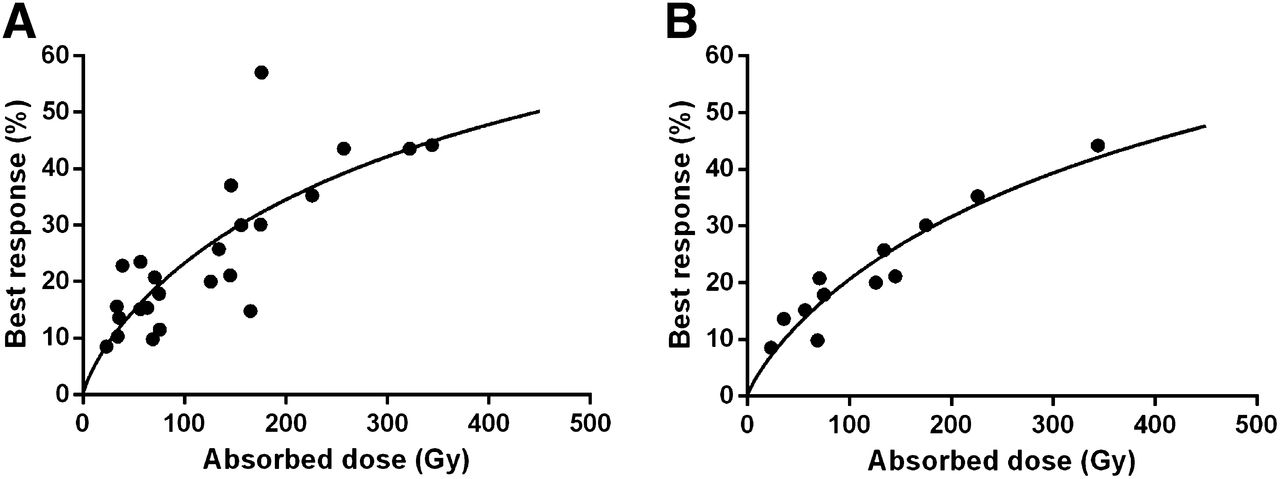
- FIGURE 5.
Tumor dose–response relationship for patients with PNETs treated with PRRT using 177Lu-DOTATATE, including tumors larger than 2.2 cm (A) and only tumors larger than 4 cm (B). Solid lines represent 2-parameter sigmoid fits (y = 100/(1+ (α/x)β)), where α and β are fitting parameters. Parameters α and β were 445 and 0.79, with SEs of 104 and 0.14, respectively, for tumors larger than 2.2 cm and 504 and 0.84, with SEs of 83 and 0.1, respectively, for tumors larger than 4 cm. Pearson correlation coefficients (R2) were 0.64 (A) and 0.91 (B).


Tables
Patient no. Sex No. of tumors A1 (GBq) A2 (GBq) A3 (GBq) A4 (GBq) A5 (GBq) A6 (GBq) 1 Female 2 7.4* 7.4 7.4 7.4* 7.4 7.4 2 Female 1 7.4* 7.4 7.4 7.4 — — 3 Female 1 7.4* 7.4 7.4 7.4* 7.4 — 4 Male 3 7.4* 7.4 6.0 — — — 5 Male 1 7.4* 7.4 7.4 — — — 6 Male 1 7.4* 7.4 7.4 7.4* 7 Female 1 7.4* 7.4 7.4 7.4* 7.4 — 8 Female 1 7.4* 7.4 7.4 7.4* 7.4 7.4 9 Male 2 7.4* 7.4 7.4* 7.4 7.4 — 10 Male 1 7.4* 7.4 7.4 7.4* — — 11 Male 3 7.4* 7.4 — — — — 12 Male 1 7.4* 7.4 6.0* 7.4 7.4* — 13 Male 2 7.4* 7.4* 7.4 7.4 — — 14 Female 2 7.4* 7.4 6.0* 7.4 — — 15 Female 1 7.4* 5.0 7.4* 4.0 — — 16 Male 1 7.4* 7.4 7.4 7.4* — — 17 Male 4 7.4* 7.4 7.4* 7.4 — — 18 Male 1 7.4* 7.4 7.4* 7.4 — — 19 Female 1 7.4* 7.4 7.4* — — — 20 Female 3 7.4* 7.4 7.4* 7.4 7.4 — 21 Male 1 7.4* 7.4 7.4* — — — 22 Female 4 7.4* 7.4 7.4 7.4* — — 23 Female 1 7.4* 7.4 5.0* — — — 24 Male 3 7.4* 5.0 5.0* — — — ↵* Complete dosimetric evaluation.
A1–6 is amount of administered activity at each treatment up to 6 cycles.


Jump to section
Related Articles
Cited By...
- Assessment of [177Lu]Lu-DOTATATE Dosimetry from High-Speed Whole-Body Recordings Provided by a 360{degrees} Cadmium-Zinc-Telluride Camera Compared with Results from a Conventional Anger-Camera Protocol
- Dosimetry of [177Lu]Lu-DOTATATE in Patients with Advanced Midgut Neuroendocrine Tumors: Results from a Substudy of the Phase III NETTER-1 Trial
- Impact of the Reference Multiple-Time-Point Dosimetry Protocol on the Validity of Single-Time-Point Dosimetry for [177Lu]Lu-PSMA-I&T Therapy
- Relationship Between Absorbed Dose and Response in Neuroendocrine Tumors Treated with [177Lu]Lu-DOTATATE
- Assessing Response to PSMA Radiopharmaceutical Therapies with Single SPECT Imaging at 24 Hours After Injection
- Absorbed Dose-Response Relationship in Patients with Gastroenteropancreatic Neuroendocrine Tumors Treated with [177Lu]Lu-DOTATATE: One Step Closer to Personalized Medicine
- The Impact of Posttreatment Imaging in Peptide Receptor Radionuclide Therapy
- Prediction of 177Lu-DOTATATE PRRT Outcome Using Multimodality Imaging in Patients with Gastroenteropancreatic Neuroendocrine Tumors: Results from a Prospective Phase II LUMEN Study
- A Pipeline for Automated Voxel Dosimetry: Application in Patients with Multi-SPECT/CT Imaging After 177Lu-Peptide Receptor Radionuclide Therapy
- Dosimetric Quantities in Neuroendocrine Tumors over Treatment Cycles with 177Lu-DOTATATE
- A Randomized, Factorial Phase II Study to Determine the Optimal Dosing Regimen for 68Ga-Satoreotide Trizoxetan as an Imaging Agent in Patients with Gastroenteropancreatic Neuroendocrine Tumors
- Tumor Response to Radiopharmaceutical Therapies: The Knowns and the Unknowns
- Reimbursement Approaches for Radiopharmaceutical Dosimetry: Current Status and Future Opportunities
- Succinylated Gelatin Improves the Theranostic Potential of Radiolabeled Exendin-4 in Insulinoma Patients
- Variability and Repeatability of Quantitative Uptake Metrics in 18F-FDG PET/CT of Non-Small Cell Lung Cancer: Impact of Segmentation Method, Uptake Interval, and Reconstruction Protocol
- Twelve-Year Follow-up After Peptide Receptor Radionuclide Therapy
- Molecular radiotheranostics for neuroendocrine tumours
- Individualized Dosimetry for Theranostics: Necessary, Nice to Have, or Counterproductive?
- Tumor-Absorbed Dose for Non-Hodgkin Lymphoma Patients Treated with the Anti-CD37 Antibody Radionuclide Conjugate 177Lu-Lilotomab Satetraxetan
- Dose Deposits from 90Y, 177Lu, 111In, and 161Tb in Micrometastases of Various Sizes: Implications for Radiopharmaceutical Therapy
- Feasibility of Affibody-Based Bioorthogonal Chemistry-Mediated Radionuclide Pretargeting
- MIRD Pamphlet No. 26: Joint EANM/MIRD Guidelines for Quantitative 177Lu SPECT Applied for Dosimetry of Radiopharmaceutical Therapy