


Article Figures & Data
Figures
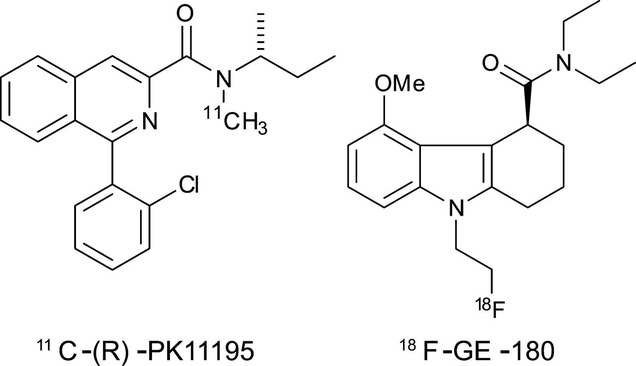
- FIGURE 1.
Chemical structures of the 2 radiotracers, 11C-(R)-PK11195 and 18F-GE-180.
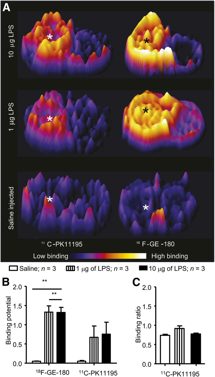
- FIGURE 2.
LPS causes unilateral upregulation of TSPO binding of 11C-(R)-PK11195 and 18F-GE-180. (A) Representative coronal striatal images obtained when animals were imaged with 11C-(R)-PK11195 or 18F-GE-180. * = injection site. (B) Autoradiography analysis reveals significant (**P < 0.01) increase in 18F-GE-180 binding after intracerebral injection of LPS (10 or 1 μg); similar significant increase was not observed with 11C-(R)-PK11195. Unprocessed images are shown in Supplemental Fig. 3C. There is no significant increase (P = 0.106) in signal in contralateral hemisphere, using cerebellum as reference region. Bars = SEM.
- FIGURE 3.
Selectivity of 11C-(R)-PK11195 and 18F-GE-180. (A) Intracerebral injection of LPS causes significant increase in OX-42–positive cells in striatum and cortex in injected hemisphere (P < 0.0001) when compared with control group. There was no significant difference between animals imaged with 18F-GE-180, compared with 11C-(R)-PK11195, or animals injected with 10 μg of LPS, compared with 1 μg. ***P < 0.0001. 11C-(R)-PK11195 and 18F-GE-180 bind specifically to same target in vitro. (B) Representative in vitro autoradiography images from coronal striatal sections demonstrating total binding of 11C-(R)-PK11195 and 18F-GE-180 and reduction in signal when 11C-(R)-PK11195 signal was blocked by GE-180 or vice versa. (C) GE-180 significantly reduces (P < 0.0001) specific signal observed when using 11C-(R)-PK11195 to image the increase in TSPO expression. Correspondingly, (R)-PK11195 significantly (P < 0.0001) reduces specific signal observed when using 18F-GE-180 as radiotracer. Bars = SEM.
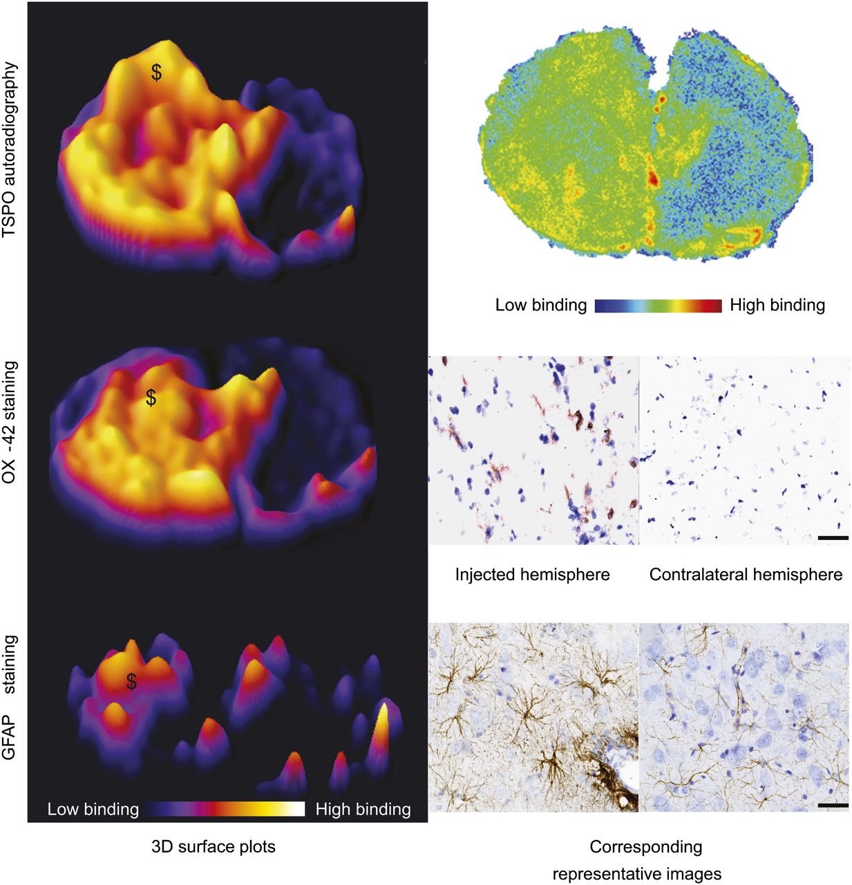
- FIGURE 4.
Specificity studies; histologic comparison to autoradiography results. (Left) Three-dimensional surface plots demonstrate area of increased signal observed in autoradiography from animals imaged with 18F-GE-180 and increase in OX-42 and GFAP immunoreactivity in sequential sections. (Right) Shown are corresponding images or high powered photomicrographs from injection site obtained from 18F-GE-180 autoradiography, OX-42 (activated microglia), and GFAP (astrocytes) staining. $ marks increase in autoradiography signal where there is corresponding area of OX-42–positive cells and increased GFAP immunoreactivity. Scale bar = 50 μm.
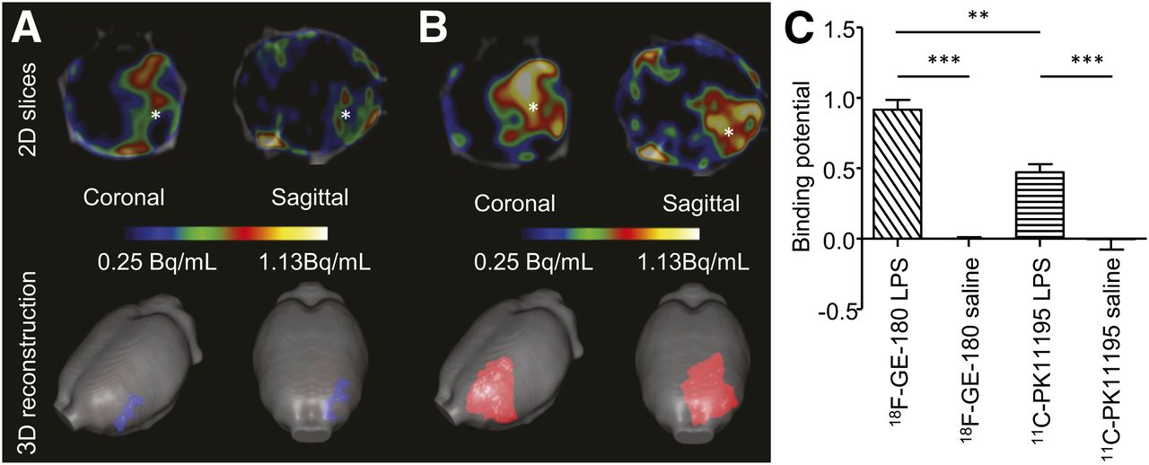
- FIGURE 5.
Radiotracer 18F-GE-180 is superior to 11C-(R)-PK11195 in detection of TSPO upregulation after brain injury. (A) Averaged (n = 3) normalized (bound-to-free pixelwise modeling) images from animals imaged in vivo with 11C-(R)-PK11195. (B) Corresponding images using 18F-GE-180. PET/CT 2-dimensional slices at level of striatum show increase in signal at site of injury (shown by *). 3-dimensional reconstruction demonstrates greater area, which can be segmented when imaging is performed with 18F-GE-180. (C) Quantification of binding demonstrates that there is significant increase in binding of both 11C-(R)-PK11195 (P < 0.0001) and 18F-GE180 (P < 0.0001) after injection of LPS (10 μg) into left striatum, compared with animals injected with saline. There is significantly (P < 0.01) higher binding of 18F-GE-180, compared with 11C-(R)-PK11195 in animals injected with LPS (10 μg). Bars = SEM.


Additional Files
Supplemental Data
Files in this Data Supplement:


Jump to section
Related Articles
Cited By...
- Pre-therapeutic Microglia Activation and Sex Determine Therapy Effects of Chronic Immunomodulation
- Nondisplaceable Binding Is a Potential Confounding Factor in 11C-PBR28 Translocator Protein PET Studies
- Sexually dimorphic responses to MPTP found in microglia, inflammation and gut microbiota in a progressive monkey model of Parkinsons disease
- PET imaging of microglia by targeting macrophage colony-stimulating factor 1 receptor (CSF1R)
- Imaging microglial activation in tacrolimus-associated CNS vasculitis with translocator protein PET
- Head-to-Head Comparison of 11C-PBR28 and 18F-GE180 for Quantification of the Translocator Protein in the Human Brain
- Neuroinflammation Appears Early on PET Imaging and Then Plateaus in a Mouse Model of Alzheimer Disease
- The FTD-like syndrome causing TREM2 T66M mutation impairs microglia function, brain perfusion, and glucose metabolism
- Flutriciclamide (18F-GE180) PET: First-in-Human PET Study of Novel Third-Generation In Vivo Marker of Human Translocator Protein
- Serial Quantitative TSPO-Targeted PET Reveals Peak Microglial Activation up to 2 Weeks After an Epileptogenic Brain Insult
- Glial Activation and Glucose Metabolism in a Transgenic Amyloid Mouse Model: A Triple-Tracer PET Study
- In Vivo Detection of Age- and Disease-Related Increases in Neuroinflammation by 18F-GE180 TSPO MicroPET Imaging in Wild-Type and Alzheimer's Transgenic Mice
- In Vivo PET Imaging Demonstrates Diminished Microglial Activation After Fingolimod Treatment in an Animal Model of Multiple Sclerosis