


Article Figures & Data
Figures
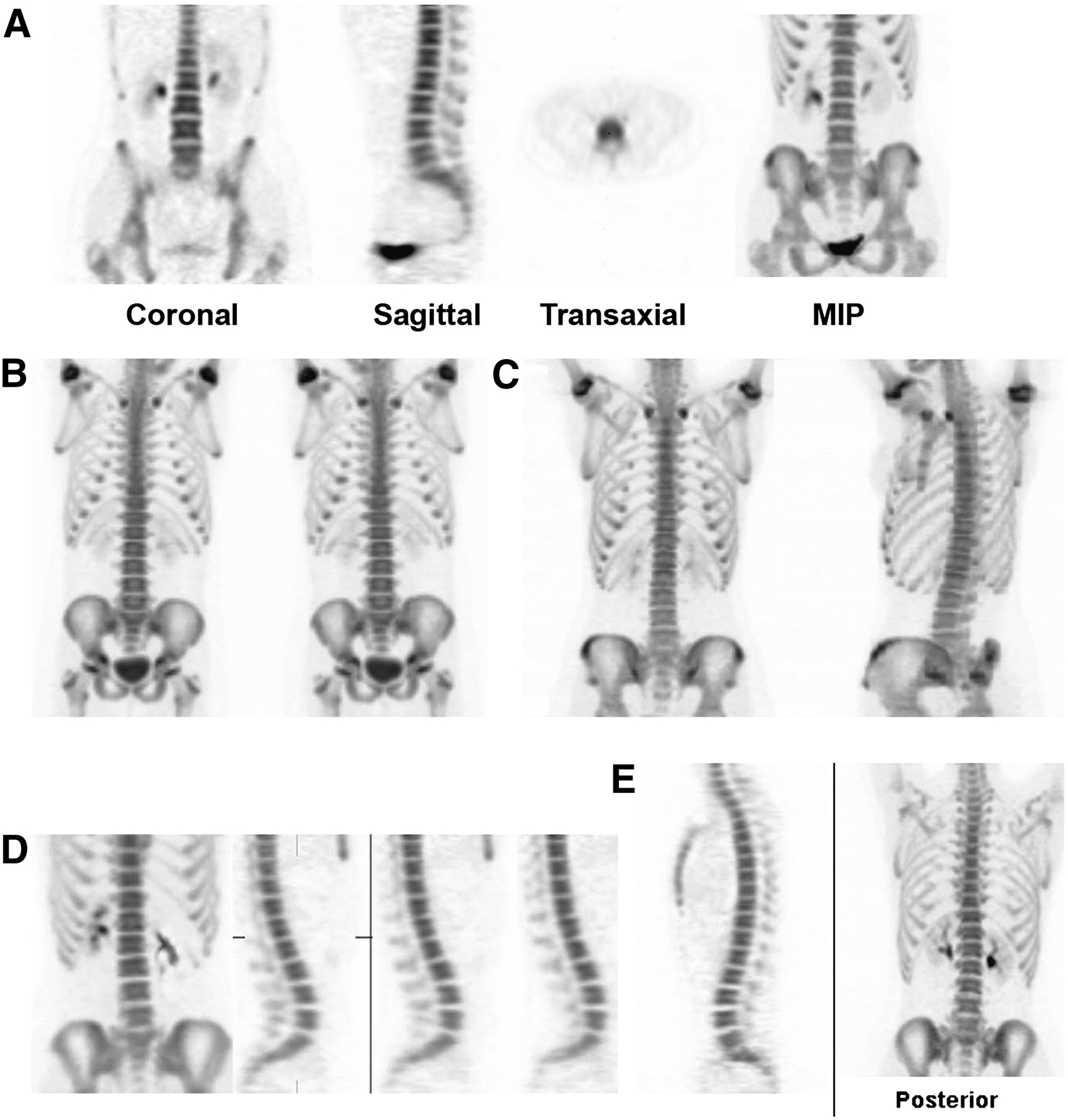
- FIGURE 1.
Normal 18F-fluoride PET skeletal findings for patients aged 5 y (A), 11 y (B), 15 y (C), 19 y (D), and 30 y (E). Pattern of 18F− uptake in skeleton is similar to pattern seen with more familiar 99mTc-labeled bisphosphonate bone scans and illustrates changes that occur with maturation of skeleton. Compared with 99mTc-MDP SPECT, 18F-fluoride bone PET provides higher-quality images, better ratios of bone uptake to soft-tissue uptake, and shorter studies. MIP = maximum-intensity projection.
- FIGURE 2.
In 8-y-old patient with Ewing's sarcoma of right distal fibula, 18F-fluoride PET projection image shows extent of primary tumor and absence of skeletal metastases.
- FIGURE 3.
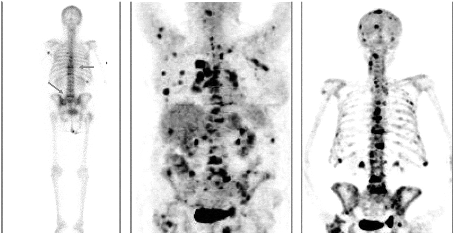
Compared with 99mTc-MDP scintigraphy (left, arrows indicate sites of thoracic and lumbar spine disease), both 18F-FDG PET (middle) and 18F-fluoride PET (right) show higher sensitivity for detecting skeletal lesions. Compared with 18F-FDG PET, 18F-fluoride PET demonstrates minimal soft-tissue uptake, which increases sensitivity for detecting bone lesions that are adjacent to sites of physiologic 18F-FDG uptake or sites of 18F-FDG–avid soft-tissue disease (Reprinted with permission of (23).).
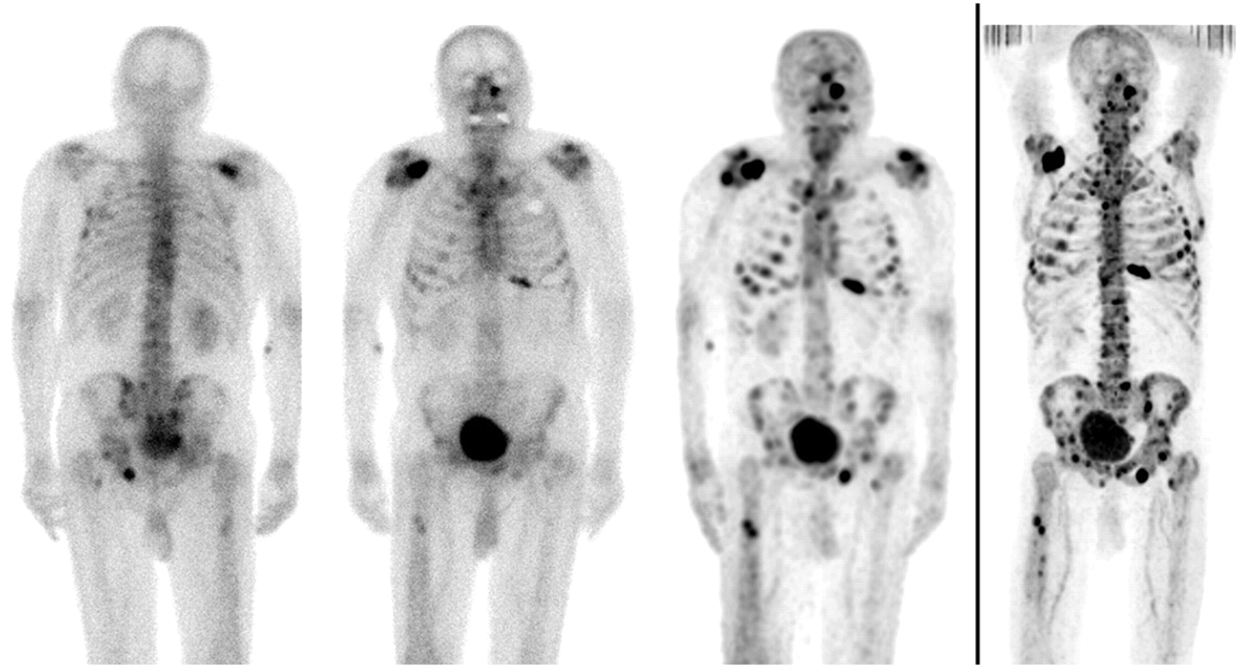
- FIGURE 4.
From left to right: posterior and anterior 99mTc-MDP planar scintigraphy, 99mTc-MDP multiple-field-of-view SPECT, and 18F-fluoride PET of 82-y-old patient with numerous bone metastases. As in this patient, more lesions are typically detected by SPECT than by planar imaging, and 18F-fluoride PET detects more lesions than does SPECT.
- FIGURE 5.
In 2-y-old girl with stage IV neuroblastoma, uptake in multiple soft-tissue tumors on 18F-fluoride PET bone scan demonstrates 18F-fluoride avidity for sites of soft-tissue calcification. A = anterior; R = right.
- FIGURE 6.
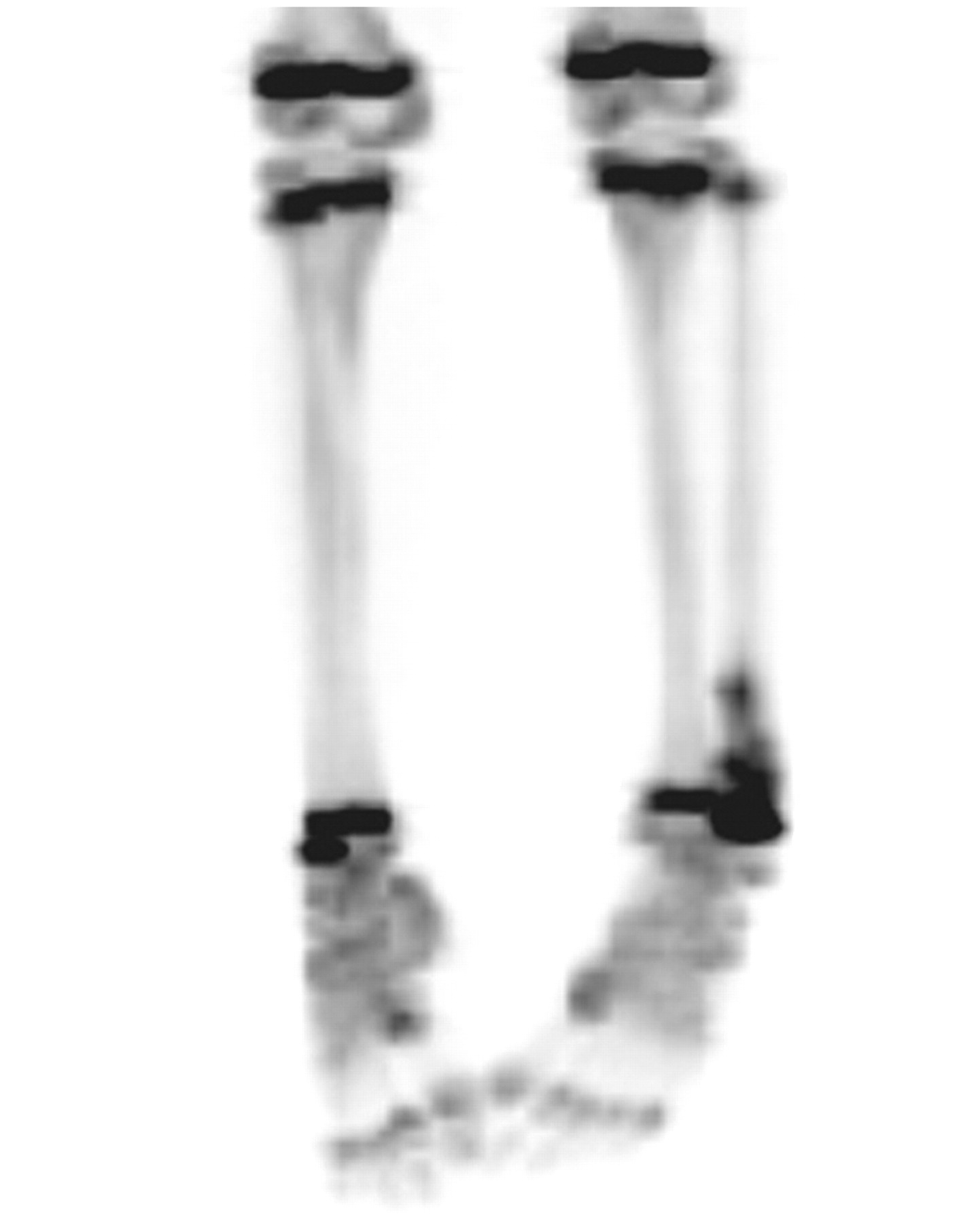
In 18-y-old female marathon runner with severe lower leg and foot pain, projection PET image shows increased 18F-fluoride uptake in tibiae and both feet, indicating multiple sites of stress injury.
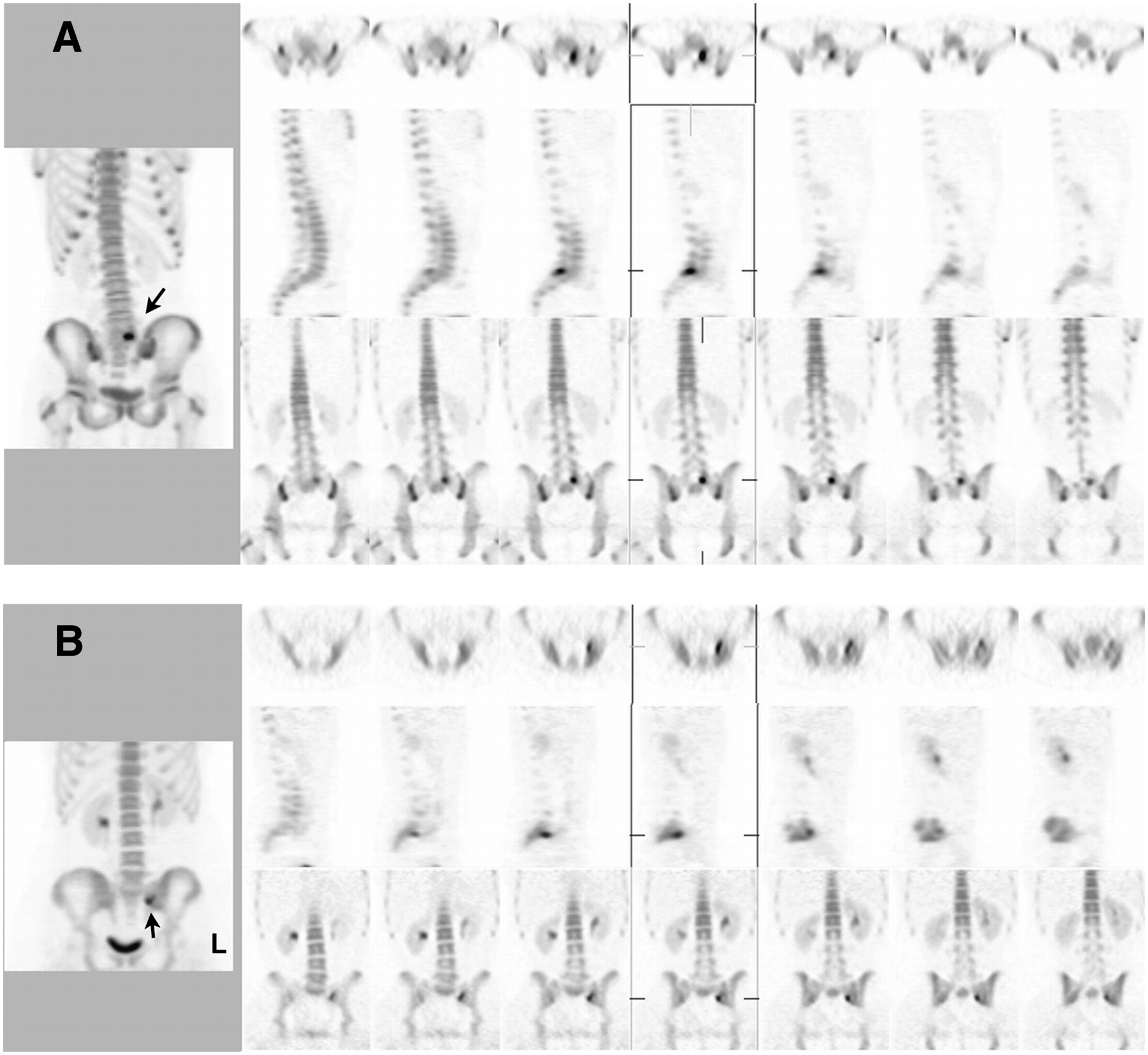
- FIGURE 7.
(A) In 12-y-old female gymnast with lower back pain, 18F-fluoride PET image shows increased focal uptake indicating stress changes in posterior elements of L5 vertebra (arrow). (B) In 21-y-old runner, 18F-fluoride PET image shows stress changes in left sacroiliac region (arrow).
- FIGURE 8.
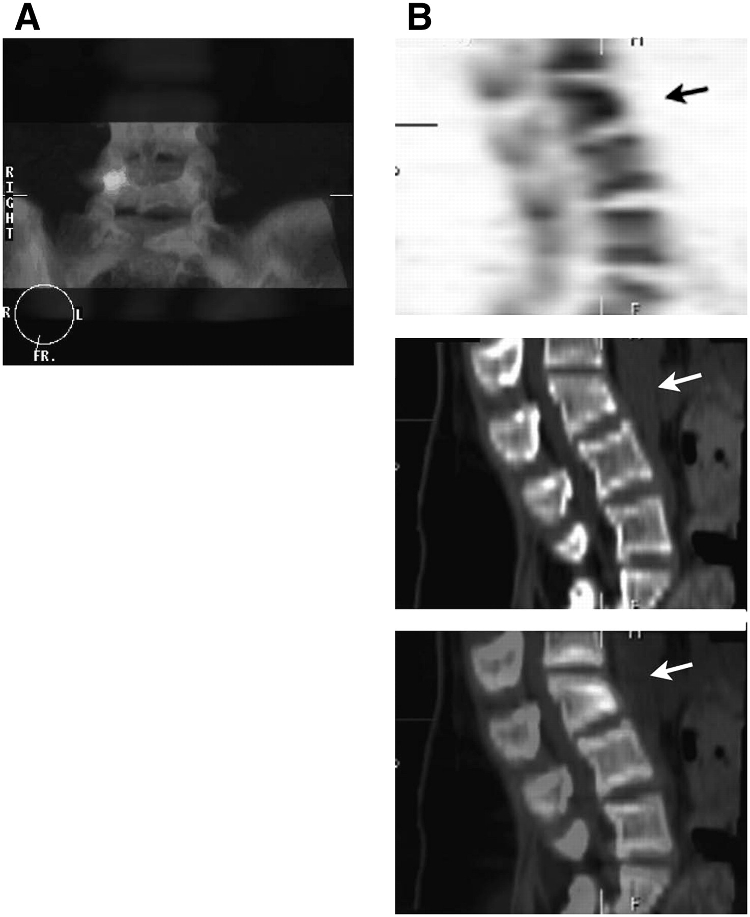
(A) Fusion of 18F-fluoride PET and CT images of 17-y-old male athlete with back pain that worsened on hyperextension. 18F-Fluoride PET had identified focally increased tracer uptake in region of right pars interarticularis of L5 vertebra. Fusion image demonstrates correlation of site of 18F− uptake with nondisplaced fracture of pars that was identified by CT. (B) From top to bottom, sagittal 18F-fluoride PET, CT, and fusion images of 15-y-old female athlete with back pain after landing from high jump. Increased 18F-fluoride uptake and deformity on PET correlates to wedge compression fracture of L3 vertebra (arrows) identified on CT.
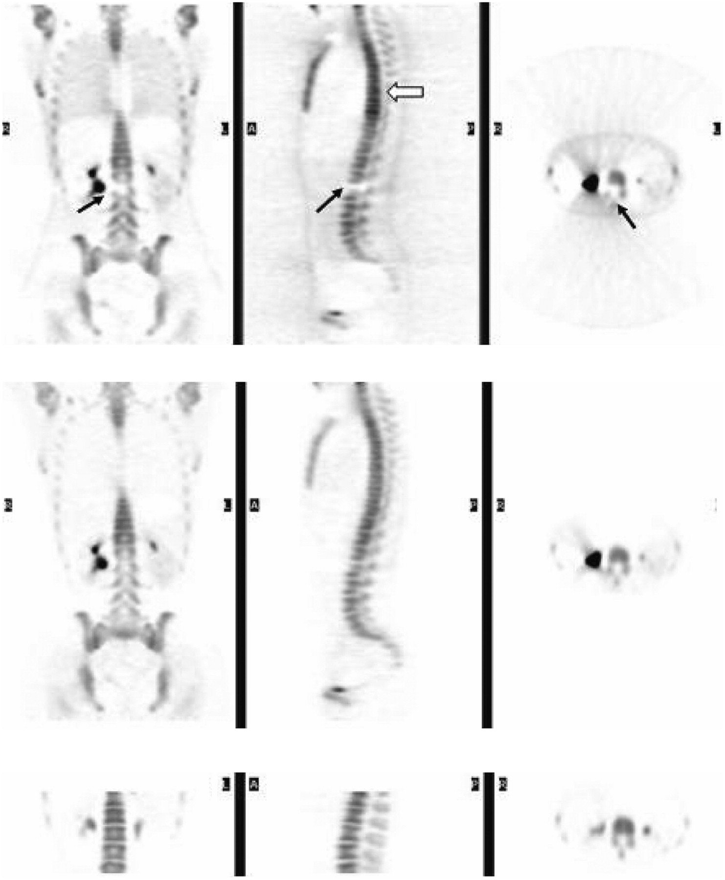
- FIGURE 9.
In 18F-fluoride PET bone scan without attenuation correction (top row, with coronal, sagittal, and transverse slices appearing from left to right), streak artifact caused by activity in renal collecting system leads to apparent loss of signal in right lumbar spine (solid arrows). Apparent increased signal in thoracic spine results from reduction of attenuation in lung region (open arrow). Both artifacts are greatly reduced when attenuation correction is applied using rotating rod sources of 68Ge/68Ga (middle row). After patient voided, repeated image (bottom row) acquired in single bed position shows resolution of artifact caused by activity in renal collecting system.


Tables
Type of imaging Adult (70 kg) 15-y-old child (55 kg) 10-y-old child (32 kg) 5-y-old child (19 kg) 1-y-old child (9.8 kg) 99mTc-MDP* Administered activity (MBq) 518 407 237 141 73 Effective dose in mSv/MBq (mSv) 0.0057 (3.0) 0.0070 (2.8) 0.0110 (2.6) 0.0140 (2.0) 0.0270 (2.0) Bladder wall in mGy/MBq (mGy) 0.048 (24.9) 0.060 (24.4) 0.088 (20.9) 0.073 (10.3) 0.130 (9.5) Bone surfaces (mGy) 0.063 (32.6) 0.082 (33.4) 0.130 (30.8) 0.220 (31.0) 0.53 (38.7) Red marrow (mGy) 0.0092 (4.8) 0.010 (4.1) 0.017 (4.0) 0.033 (4.7) 0.067 (4.9) 18F-labeled NaF† Administered activity (MBq) 148 116 68 40 21 Effective dose in mSv/MBq (mSv) 0.027 (4.0) 0.034 (3.9) 0.052 (3.5) 0.086 (3.4) 0.170 (3.6) Bladder wall in mGy/MBq (mGy) 0.22 (32.6) 0.27 (31.3) 0.40 (27.2) 0.61 (24.4) 1.10 (23.1) Bone surfaces in mGy/MBq (mGy) 0.040 (5.9) 0.050 (5.8) 0.079 (5.4) 0.130 (5.2) 0.300 (6.3) Red marrow in mGy/MBq (mGy) 0.040 (5.9) 0.053 (6.1) 0.088 (6.0) 0.180 (7.2) 0.380 (8.0) Type of disease Type of assessment Specific goal Oncologic Metastatic disease in skeleton (e.g. prostate, lung, breast, neuroblastoma) Perform initial evaluation (staging) Assess response of skeletal metastases to therapy Detect skeletal metastases during follow-up Bone pain in patients with known cancer Primary bone tumors Identify sites of disease (initial staging) Assess response to therapy Differentiate postoperative changes from residual/recurrent disease Detect recurrent/metastatic disease during follow-up Benign bone Pediatric/young adult back pain Assess vertebral spondylolysis Assess other stress injuries Bone viability Assess femoral head avascular necrosis Assess bone graft viability (long bones, mandible) Paget's disease Assess extent of disease Monitor response to therapy


Jump to section
Related Articles
Cited By...
- [99mTc]Tc-MY6349 Probe for Trop2-Targeted SPECT Imaging: From Preclinical to Pilot Clinical Study
- 18F-NaF PET/CT of Obese Patients on a Lutetium-Yttrium Oxyorthosilicate PET/CT System: Patient Dosimetry, Optimization of Injected Activity, and Acquisition Time
- 18F-Sodium Fluoride PET: History, Technical Feasibility, Mechanism of Action, Normal Biodistribution, and Diagnostic Performance in Bone Metastasis Detection Compared with Other Imaging Modalities
- Characterization of Bone Lesions in Myeloma Before and During Anticancer Therapy Using 18F-FDG-PET/CT and 18F-NaF-PET/CT
- Three-Hour Delayed Imaging Improves Assessment of Coronary 18F-Sodium Fluoride PET
- Comparison of the Variability of SUV Normalized by Skeletal Volume with the Variability of SUV Normalized by Body Weight in 18F-Fluoride PET/CT
- Assessment of Physiologic Intracranial Calcification in Healthy Adults Using 18F-NaF PET/CT
- Hospice Admission and Survival After 18F-Fluoride PET Performed for Evaluation of Osseous Metastatic Disease in the National Oncologic PET Registry
- The Role of 18F-Sodium Fluoride PET/CT Bone Scans in the Diagnosis of Metastatic Bone Disease from Breast and Prostate Cancer
- Bone-Targeted Imaging and Radionuclide Therapy in Prostate Cancer
- Evaluation of Prostate Cancer Bone Metastases with 18F-NaF and 18F-Fluorocholine PET/CT
- Prospective Study Evaluating Na18F PET/CT in Predicting Clinical Outcomes and Survival in Advanced Prostate Cancer
- 18F-Fluoride PET in the Assessment of Malignant Bone Disease
- Prognostic Factors in Patients Treated with 223Ra: The Role of Skeletal Tumor Burden on Baseline 18F-Fluoride PET/CT in Predicting Overall Survival
- Semiquantitative Analysis of the Biodistribution of the Combined 18F-NaF and 18F-FDG Administration for PET/CT Imaging
- Evaluation of 18F-Fluoride PET/MR and PET/CT in Patients with Foot Pain of Unclear Cause
- 18F-Fluoride PET Used for Treatment Monitoring of Systemic Cancer Therapy: Results from the National Oncologic PET Registry
- AEG-1 Promoter-Mediated Imaging of Prostate Cancer
- Impact of 18F-Fluoride PET on Intended Management of Patients with Cancers Other Than Prostate Cancer: Results from the National Oncologic PET Registry
- Impact of 18F-Fluoride PET in Patients with Known Prostate Cancer: Initial Results from the National Oncologic PET Registry
- Reply: Regarding Dynamic Bone Imaging with 99mTc-Labeled Diphosphonates and 18F-NaF: Mechanisms and Applications
- Bisphosphonate-Induced Osteonecrosis of the Jaw: Comparison of Disease Extent on Contrast-Enhanced MR Imaging, [18F] Fluoride PET/CT, and Conebeam CT imaging
- Dynamic Bone Imaging with 99mTc-Labeled Diphosphonates and 18F-NaF: Mechanisms and Applications
- Clinical utility of fluoride-18 positron emission tomography/CT in temporomandibular disorder with osteoarthritis: comparisons with 99mTc-MDP bone scan
- Combined 18F-Fluoride and 18F-FDG PET/CT Scanning for Evaluation of Malignancy: Results of an International Multicenter Trial
- Biodistribution, Tumor Detection, and Radiation Dosimetry of 18F-DCFBC, a Low-Molecular-Weight Inhibitor of Prostate-Specific Membrane Antigen, in Patients with Metastatic Prostate Cancer
- Validation of a Paper Chromatographic Methodology as an Alternative for Determination of the Radiochemical Purity of Na18F
- Utility of 18F-Fluoride PET/CT and 18F-FDG PET/CT in the Detection of Bony Metastases in Heightened-Risk Head and Neck Cancer Patients
- Radiation Exposure Should Not Limit Bone Scintigraphy with 18F-NaF
- The Kinetics and Reproducibility of 18F-Sodium Fluoride for Oncology Using Current PET Camera Technology
- Coronary Arterial 18F-Sodium Fluoride Uptake: A Novel Marker of Plaque Biology
- Assessment of Valvular Calcification and Inflammation by Positron Emission Tomography in Patients With Aortic Stenosis
- Nuclear Medicine in the First Year of Life
- Molecular Mechanisms of Bone 18F-NaF Deposition
- SNM Practice Guideline for Sodium 18F-Fluoride PET/CT Bone Scans 1.0
- In Vivo Molecular Imaging Analysis of a Nasal Vaccine That Induces Protective Immunity against Botulism in Nonhuman Primates
- Imaging of Back Pain in Children
- Potential Iatrogenic Alteration to 18F-Fluoride Biodistribution
- Dosimetry of Pediatric PET/CT
- Bone Metastases in Patients with Neuroendocrine Tumor: 68Ga-DOTA-Tyr3-Octreotide PET in Comparison to CT and Bone Scintigraphy
- The Role of Radiotracer Imaging in the Diagnosis and Management of Patients with Breast Cancer: Part 1--Overview, Detection, and Staging
- Radiodefluorination of 3-Fluoro-5-(2-(2-[18F](fluoromethyl)-thiazol-4-yl)ethynyl)benzonitrile ([18F]SP203), a Radioligand for Imaging Brain Metabotropic Glutamate Subtype-5 Receptors with Positron Emission Tomography, Occurs by Glutathionylation in Rat Brain