


Abstract
There is renewed interest in 18F-NaF bone imaging with PET or PET/CT. The current brief discussion focuses on the molecular mechanisms of 18F-NaF deposition in bone and presents model-based approaches to quantifying bone perfusion and metabolism in the context of preclinical and clinical applications of bone imaging with PET.
There is renewed interest in imaging malignant, metabolic, degenerative, traumatic, and inflammatory bone diseases using 18F-NaF, a bone imaging probe that was initially introduced by Blau et al. in 1962 (1). 18F-NaF became the standard method for bone scintigraphy in the 1960s but was largely replaced by 99Tc-labeled diphosphonate compounds in the 1970s (2) because of their better physical characteristics for imaging with conventional γ-cameras.
The reemergence of 18F-NaF bone imaging was ignited by the introduction of PET (3) and PET/CT (4) (Fig. 1), which permits assessment of bone diseases with a higher accuracy than planar imaging or SPECT; the availability of electronic generators allowing for the widespread use of 18F-NaF (5); and the well-documented shortage of 99Mo/99Tc generators leading to limited availability of 99Tc.
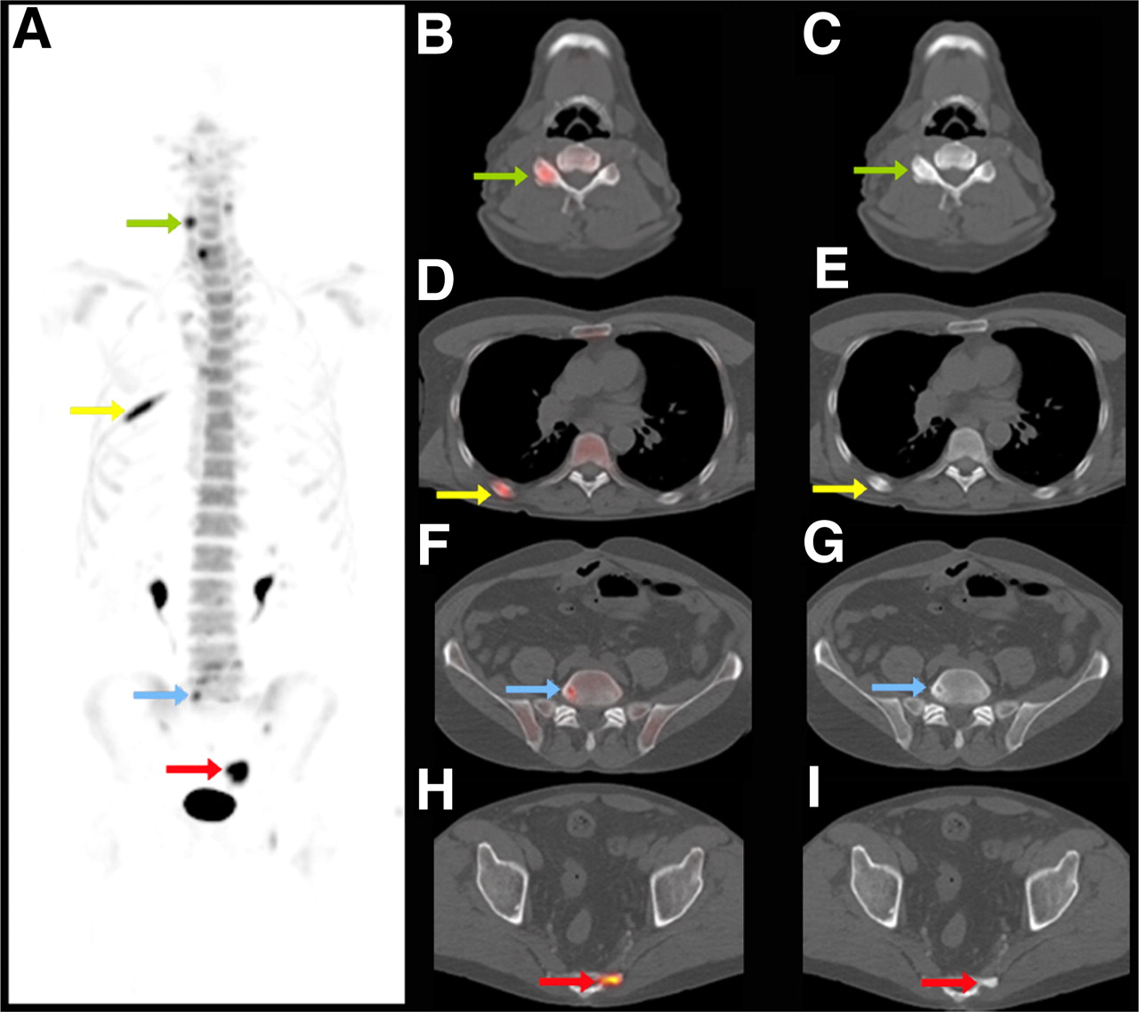
Maximum-intensity-projection PET image (A) and selected axial PET/CT (B, D, F, H) and CT (C, E, G, I) images of 18F-NaF PET/CT scan of 63-y-old man with prostate cancer. Increased 18F-NaF uptake can be seen in benign changes of right cervical facet joint (green arrow in A, B, and C), healing rib fracture (yellow arrow in A, D, and E), benign lumbar vertebral bone cyst (blue arrow in A, F, and G), and blastic metastasis in sacrum (red arrow in A, H, and I). Additional value of CT to characterize focally increased tracer uptake is evident.
Because clinical studies using PET and PET/CT bone imaging approaches have been reviewed extensively (6–9), the current discussion focuses on the molecular mechanisms of 18F-NaF deposition in bone. After a brief description of the radiosynthesis, the kinetics and biodistribution of 18F-NaF in vivo are described. Furthermore, model-based approaches to quantifying bone perfusion and metabolism are presented in the context of preclinical and clinical applications of bone imaging with PET.
PREPARATION OF 18F-NaF
18F-fluoride is produced by 11-MeV proton irradiation of 18O-water in a tantalum target body (10) using a cyclotron. The irradiated aqueous solution containing 18F-fluoride is diluted with sterile water (5 mL) and passed through a cation exchange (H+ form) cartridge. The eluent from the cation exchange cartridge is passed through an anion exchange (HCO3− form) cartridge to trap the 18F-fluoride. The anion exchange cartridge is then flushed with 10 mL of sterile water, and the 18F fluoride is then eluted with 10 mL of sterile normal saline and is passed through a sterile filter into a sterile multidose vial. The quality control tests for the 18F-NaF are conducted according to the method of the U.S. Pharmacopeia (11).
KINETICS OF 18F-NaF
The rate-limiting step of 18F-NaF bone uptake is blood flow (12), and almost all 18F-NaF delivered is retained by bone after a single pass of blood (13). The initial 18F-NaF distribution therefore represents blood flow that varies among different bones (14). Uptake of tracer by the bone marrow is negligible (15). Around 30% of the injected 18F-NaF is present in erythrocytes, but this fact does not impede ion exchange in bone because 18F-NaF is freely diffusible across membranes (16). 18F-NaF is rapidly cleared from plasma and excreted by the kidneys. One hour after injection, only 10% of 18F-NaF remains in plasma, and the 5-h integrated plasma concentration was only 1.1%–2.6% of the dose per liter. The 5-h cumulative urine excretion was 7.4%–24.8% in 8 patients (17).
For bone deposition, 18F-ions need to “pass from plasma through the extracellular fluid space into the shell of bound water surrounding each crystal, onto the crystal surface and in the interior of the crystal,” as described by Blau et al. (12). After chemisorption onto hydroxyapatite, 18F exchanges rapidly for OH on the surface of the hydroxyapatite matrix (Ca10(PO4)6OH2) to form fluoroapatite (Ca10(PO4)6F2) (18). The incorporation of 18F into bone matrix as fluoroapatite is slow (days to weeks) (12).
Bone surface encompasses 300 m2/g of bone tissue for a total of 3 × 10⁶ m2 for the whole body (19). 18F-NaF uptake and retention in bone depends on the area of the “exposed” bone surface, which is larger in a variety of benign and malignant bone disorders. The relationship between osteoblastic and osteoclastic activity determines the incorporation of 18F-NaF into the bone matrix (20). Negligible plasma protein binding, rapid blood and renal clearance (21), and high bone uptake early after injection result in high target-to-background ratios, which in turn permit whole-body imaging as early as 45–60 min after tracer injection.
TRACER KINETIC MODELS
Quantitative bone imaging is potentially important because several metabolic bone diseases are subtle at onset, diffuse in nature, and difficult to detect with other techniques, including blood- or urine-derived biomarkers or other imaging techniques (22). For instance, measurements of bone density are of unknown prognostic value for fracture risk assessments and unproven for monitoring disease during treatment (23). Quantitative measurements of bone blood flow and metabolism can be derived from dynamic PET with 18F-NaF and appropriate tracer kinetic models. Such models can provide quantitative measurements in patients with a variety of bone diseases and might greatly aid drug response assessments.
The model that best describes fluoride incorporation into bone is a simple 3-compartment model (18) consisting of a vascular, an extravascular, and a bone compartment. The model describes regional rather than whole-body pharmacokinetics (24,25) (Fig. 2) and is similar to the standard in vivo model for 18F-FDG (26). This and other models have been described previously (27,28).
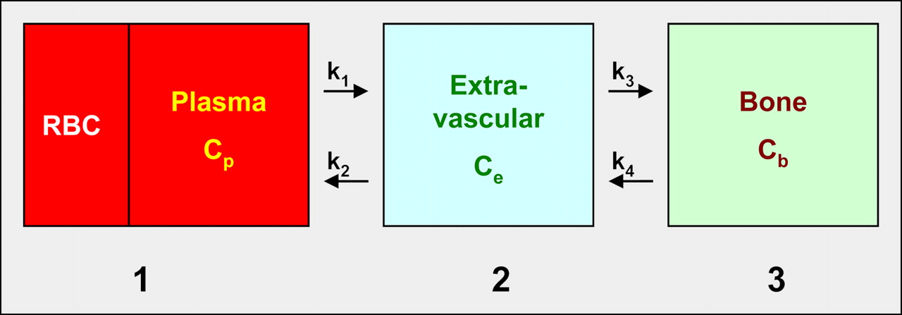
18F-NaF kinetic model with 3 compartments: vascular (1), extravascular (2), and bone (3). Plasma clearance of 18F-NaF is measured in left ventricle or aorta with PET scanner or from arterial blood draw; k1–k4 are rate constants; k1 and k2 represent forward and reverse transport from plasma, and k3 and k4 represent uptake and release from bone. If extraction fraction equals 1, then k1 represents local bone blood flow. Influx rate Ki = k1 · k3 · (k2 + k3)−1 is related to Ca2+ influx and bone apposition rate and, presumably, represents bone remodeling rate. Ki is determined by both bone blood flow and bone turnover. Therefore, measurements have to be interpreted in context of each individual study. Respective concentrations of fluoride are denoted as Cp (in plasma), Ce (in extravascular space), and Cb (in bone compartment). RBC = red blood cells
ANIMAL EXPERIMENTAL STUDIES
PET has been used to measure bone blood flow (29), first-pass bone extraction fraction (30), whole-body biodistribution (21,31), tracer kinetics (24,25), and the pharmacokinetics (17) of 18F-NaF. These and other studies were reviewed extensively by Blake et al. (8).
The normal kinetics and biodistribution of 18F-NaF have also been exploited to study physiologic processes. For instance, assessing renal function is of critical importance in preclinical drug development. Traditionally, this requires blood and urine sampling, which is difficult in small animals. Because renal fluoride clearance is related to the glomerular filtration rate, the pharmacokinetics of 18F-NaF were used to assess renal function in rats (32). The 18F-NaF–based measurements correlated well with traditional measurements of renal function derived from imaging (99Tc-mercaptoacetyltriglycine) and from serum markers, suggesting that this quantitative method could be used for measuring renal function preclinically and clinically. However, given the existence of other accurate tests, this application is less likely to be adopted clinically in the near future.
Several animal experimental studies have used quantitative approaches to describe alterations in bone blood flow and metabolism in a variety of diseases.
Trauma
Dworkin et al. (21) examined the biodistribution and pharmacokinetics of 18F-NaF in fractured and nonfractured bone in dogs at variable times after a surgically induced tibia fracture. They observed a time-dependent accumulation of 18F-NaF without increased tracer uptake at the fracture site 3 h after injury but increasing uptake from 3 wk to 3 mo, suggesting that bone healing and therapeutic interventions can be monitored in animal experimental models.
To investigate the impact of chronic stress and fatigue, the ulna of rats was exposed to cyclic compressions at various subfracture levels (33). This resulted in increased bone vascularity as early as 1 d after stress, which was followed by periosteal bone formation at day 3. 18F-NaF uptake correlated with these histopathologic changes, which in turn reflected the severity of induced stress. Tracer uptake returned to normal at 24 d after stress, suggesting that this assay could be used to monitor stress responses of bone.
Metabolic Bone Diseases
Novel agents targeting bone loss are being developed (34). The potential of PET-derived quantitative measurements of treatment-induced changes in bone flow and metabolism in drug development and clinical trials is obvious.
The equilibrium between bone formation and resorption is altered in many metabolic bone disorders, including primary and secondary hyperparathyroidism, renal osteodystrophy, osteoporosis, and others. Histomorphometric measurements can be used to evaluate bone formation invasively. PET was used to demonstrate in mini pigs that quantitative measurements of bone blood flow and metabolism can be derived from dynamic 18F-NaF imaging (20) and that these noninvasive measurements correlate well with invasive assessments of bone turnover.
Murine Tumor Models
18F-NaF PET bone imaging protocols have been optimized in mice. The coefficient of variation from 4 consecutive measurements as an index of reproducibility of 18F-NaF bone uptake was less than 15% when bone counts per pixel per minute were normalized to a region of interest encompassing the whole skeleton (35). The excellent reproducibility supports the use of 18F-NaF small-animal PET to study a variety of bone diseases.
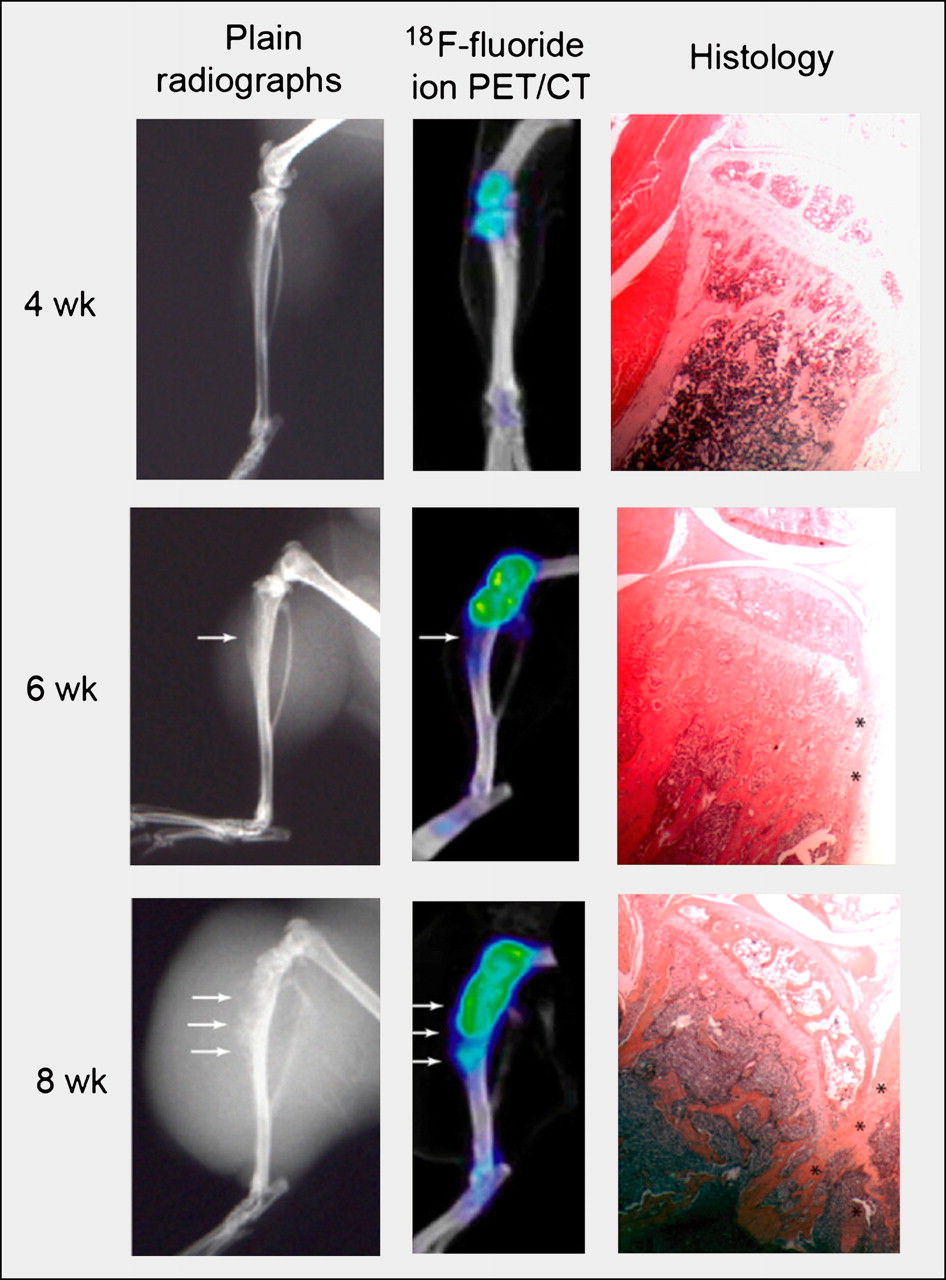
Small-animal PET or PET/CT has been used to image relevant models of metastatic prostate cancer and to monitor natural disease progression. For instance, the formation of osteolytic, osteoblastic, and mixed bone metastases from prostate cancer was studied with small-animal 18F-NaF and 18F-FDG PET/CT scans, radiographs, and histology or histomorphometry (36). 18F-NaF and 18F-FDG PET/CT accurately identified osteoblastic lesions and their progression (Fig. 3). Mixed blastic–lytic lesions were well visualized only with 18F-FDG, whereas 18F-NaF uptake was low or absent. In pure lytic lesions, 18F-FDG uptake increased over time. In contrast, 18F-NaF uptake was initially high but decreased to background levels after 6 wk, suggesting early increases in tumor vascularization resulting in flow-dependent accumulation of the probe.

Radiographs (left), 18F-NaF PET/CT scans (middle), and photomicrographs of histologic specimen (right). PET/CT images reveal osteoblastic lesion earlier (4 wk) than radiography (arrows denote bone lesions). Increasing 18F-NaF uptake over time corresponds to increased bone formation seen on histology (asterisks). (Reprinted from (36).)
Another group injected human Ewing sarcoma cells subcutaneously or intravenously into immune-deficient mice (37). Small-animal PET with 18F-FDG and 18F-NaF was then used to search for soft-tissue, lung, and bone metastases. Only intravenous tumor cell injection reproduced the human metastatic pattern (lung, bone, and soft tissue). In contrast to the study by Hsu et al. (36), Ewing sarcoma bone lesions exhibited decreased 18F-NaF uptake relative to normal bone. This difference is most likely explained by the use of a different tumor model and the different timing of PET scans after tumor cell injection.
Thus, relevant animal models of primary and metastatic bone lesions that mimic the biologic behavior of these tumors in humans were generated. These models can be used to evaluate novel bone imaging agents or to monitor the effects of therapeutic interventions on the course of primary or secondary bone malignancies.
QUANTITATIVE HUMAN STUDIES
The long-term precision and reproducibility of quantitative bone metabolism measurements with 18F-NaF PET was established by Frost et al. (38) in patients with osteopenia or osteoporosis. All underwent PET scans of the lumbar spine at baseline and after 6 mo. Measurement precision and reproducibility were compared with biomarkers of bone formation. The coefficient of variation was comparable among PET-based measurements and nonimaging biomarkers. In general, the precision of PET-based measurements ranged from 10% to 15%.
Hawkins et al. (18) measured an average influx rate of 0.036 ± 0.006 mL/min/mL in healthy bones (18). Schiepers et al. (28) reported a blood flow to lumbar vertebrae of 0.058 mL/min/mL and influx of 0.022 mL/min/mL for osteoporosis. For Paget disease, the flow rate was 0.205 mL/min/mL (or 4 times higher than flow to normal bone) and the influx was 0.114 mL/min/mL. Using the same model (Fig. 2), Cook et al. (27) also found an almost 4-fold increase in Paget-affected bones.
Messa et al. (39) measured bone flow and fluoride influx in patients with renal osteodystrophy and primary hyperparathyroidism and found significantly increased influx rates that decreased by 30%–40% after parathyroidectomy and medical therapy. Excellent correlations between influx rates and serum alkaline phosphatase and parathyroid hormone levels, as well as histomorphometric indices of bone formation rate, were reported.
The blood flow to the femoral head was estimated by Schiepers et al. (40) in 5 patients with unilateral hip trauma. A flow ratio of greater than 2 between the abnormal and normal femoral head was necessary for a successful outcome with conservative treatment. A minimum flow of 0.04 mL/min/mL was measured in 1 patient whose affected femoral head healed conservatively.
Using the model shown in Figure 2 (18,28), Frost et al. studied bone flow and turnover in women with treated and untreated metabolic bone disease (14). Bone perfusion was highly variable between the lumbar spine and the humerus, an observation that was not attainable by biomarkers of global bone turnover or by biopsy.
CONCLUSION
18F-NaF PET or PET/CT is a well-established and validated qualitative and quantitative imaging tool. Because PET is fundamentally superior to conventional planar imaging and SPECT, and because of the favorable pharmacokinetics of 18F-NaF allowing for early imaging after tracer injection, PET or PET/CT bone imaging will eventually replace conventional 99Tc-based γ-camera techniques in the clinic. Moreover, therapeutic interventions targeting metabolic, degenerative, traumatic, and neoplastic bone diseases will be assessed for their effectiveness in preclinical research and clinical practice.
Footnotes
Guest Editor: Barry Siegel, Mallinckrodt Institute of Radiology
- © 2010 by Society of Nuclear Medicine
REFERENCES
- Received for publication June 30, 2010.
- Accepted for publication August 20, 2010.


Jump to section
Related Articles
Cited By...
- Multimodality Imaging of Aortic Valve Calcification and Function in a Murine Model of Calcific Aortic Valve Disease and Bicuspid Aortic Valve
- Multimodality Imaging of Aortic Valve Calcification and Function in a Murine Model of Calcific Aortic Valve Disease and Bicuspid Aortic Valve
- 68Ga-Bisphosphonates for the Imaging of Extraosseous Calcification by Positron Emission Tomography
- Reducing Radiation Exposure from PET Patients
- 18F-NaF PET/CT of Obese Patients on a Lutetium-Yttrium Oxyorthosilicate PET/CT System: Patient Dosimetry, Optimization of Injected Activity, and Acquisition Time
- HAP-multitag, a PET and positive MRI contrast nanotracer for the longitudinal characterization of vascular calcifications in atherosclerosis
- Observer Agreement and Accuracy of 18F-Sodium Fluoride PET/CT in the Diagnosis of Bone Metastases in Prostate Cancer
- 18F-Sodium Fluoride PET: History, Technical Feasibility, Mechanism of Action, Normal Biodistribution, and Diagnostic Performance in Bone Metastasis Detection Compared with Other Imaging Modalities
- Three-Hour Delayed Imaging Improves Assessment of Coronary 18F-Sodium Fluoride PET
- Characterization of Bone Lesions in Myeloma Before and During Anticancer Therapy Using 18F-FDG-PET/CT and 18F-NaF-PET/CT
- Assessment of Physiologic Intracranial Calcification in Healthy Adults Using 18F-NaF PET/CT
- A Prospective Study Comparing 99mTc-Hydroxyethylene-Diphosphonate Planar Bone Scintigraphy and Whole-Body SPECT/CT with 18F-Fluoride PET/CT and 18F-Fluoride PET/MRI for Diagnosing Bone Metastases
- Repeatability of Quantitative 18F-NaF PET: A Multicenter Study
- The Role of 18F-Sodium Fluoride PET/CT Bone Scans in the Diagnosis of Metastatic Bone Disease from Breast and Prostate Cancer
- Bone-Targeted Imaging and Radionuclide Therapy in Prostate Cancer
- 18F-Fluoride PET in the Assessment of Malignant Bone Disease
- Impact of Personal Characteristics and Technical Factors on Quantification of Sodium 18F-Fluoride Uptake in Human Arteries: Prospective Evaluation of Healthy Subjects
- AEG-1 Promoter-Mediated Imaging of Prostate Cancer
- A Compartmental Model of Mouse Thrombopoiesis and Erythropoiesis to Predict Bone Marrow Toxicity After Internal Irradiation
- An Approach to Breast Cancer Diagnosis via PET Imaging of Microcalcifications Using 18F-NaF
- Mammary Cancer Bone Metastasis Follow-up Using Multimodal Small-Animal MR and PET Imaging
- Dynamic Bone Imaging with 99mTc-Labeled Diphosphonates and 18F-NaF: Mechanisms and Applications
- Clinical utility of fluoride-18 positron emission tomography/CT in temporomandibular disorder with osteoarthritis: comparisons with 99mTc-MDP bone scan