


Article Figures & Data
Figures
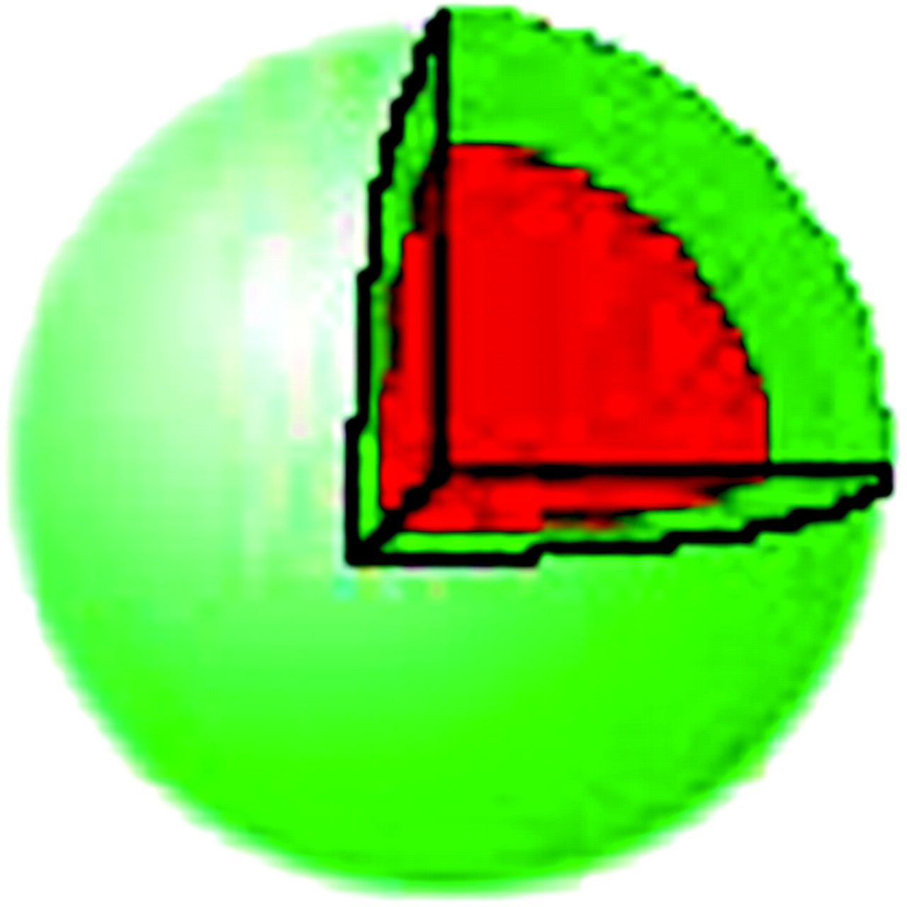
- FIGURE 1.
Uniform-density sphere with effective half-life of 2 h in outer green region and 4 h within red region. Green and red regions have equal volume in this example. Initial activity in each region is selected so that total numbers of decays are equal in both regions.
- FIGURE 2.
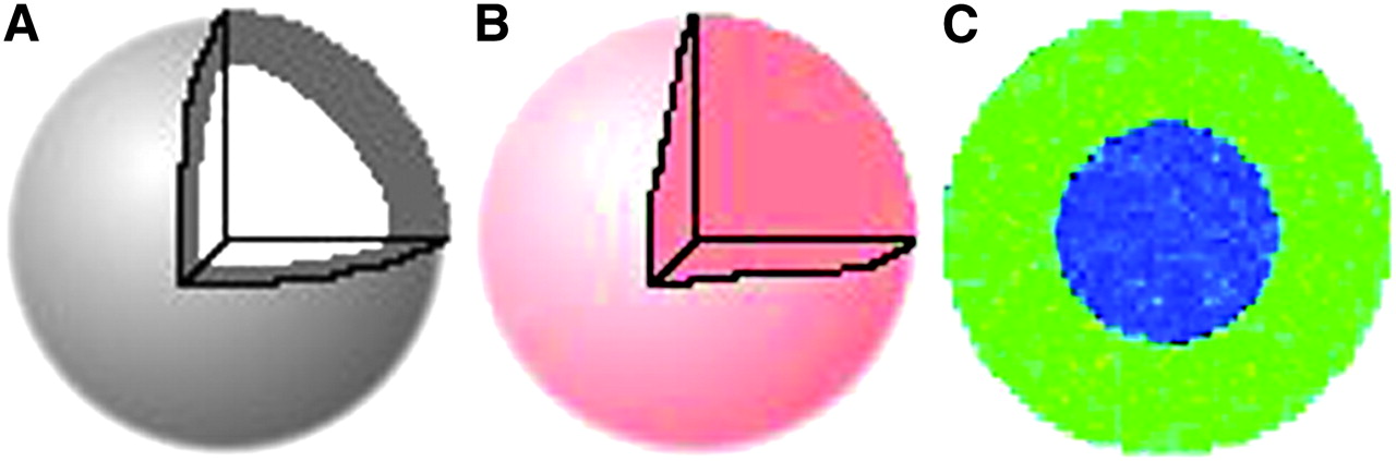
Density distribution (uniform) (A) for uniform activity distribution model (B) and nonuniform activity distribution model (C). In nonuniform distribution, same total activity as shown in Figure 3B is concentrated into half the volume (outer shell). Assuming a uniform density sphere (A), 2 activity distributions are depicted: uniform (B) and nonuniform (C). In C, the same total activity as in B is concentrated into the outer shell of the sphere.
- FIGURE 3.
(A) Spheric nonuniform density model in which inner sphere is twice unit density (2.0 g/cm3) and outer shell is at unit density (1.0 g/cm3). (B) Uniform activity distribution for density model in Figure 2A. (C) Cross-sectional slice of 3D-RD output for spheric nonuniform density model.
- FIGURE 4.
(A) Clinical CT portion of a SPECT/CT scan of patient showing nonuniform density distribution in lungs. (B) Clinical SPECT scan of patient showing nonuniform activity distribution. (C) Rate map generated from 3 longitudinally aligned SPECT images; regions with effective half-life greater than physical half-life of 131I reflect tumor uptake. (D) Cumulative activity generated from rate map and SPECT.
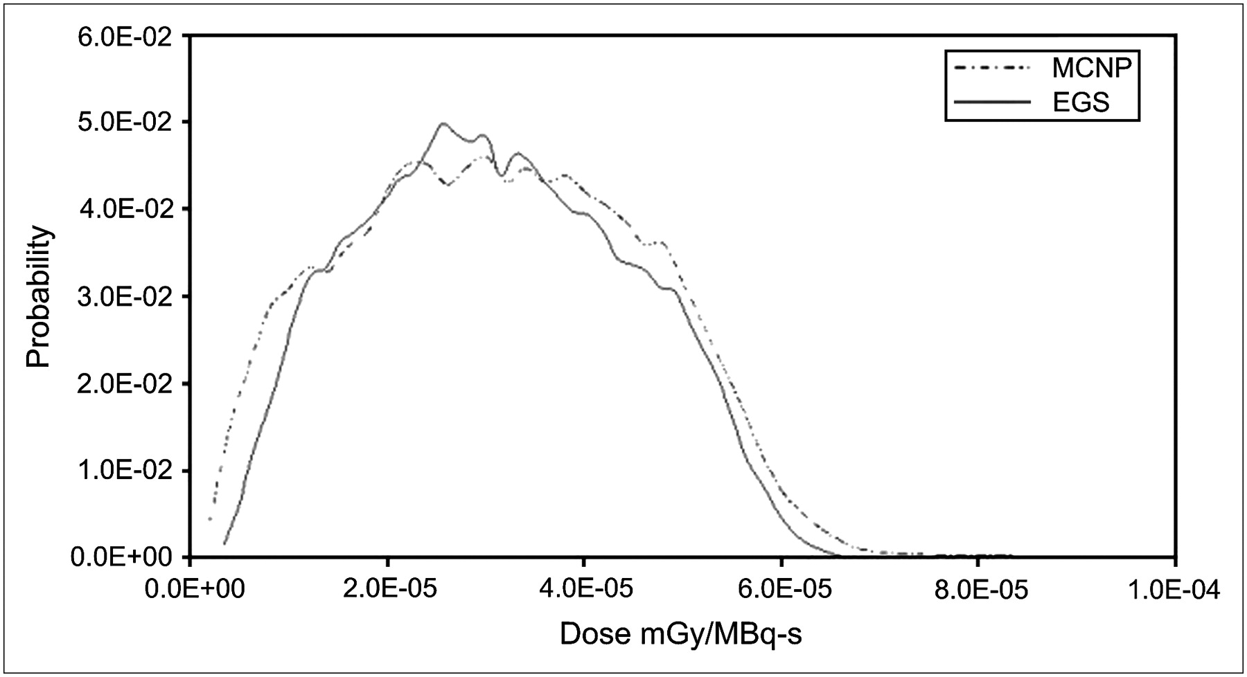
- FIGURE 5.
Comparison between MCNP-based dose volume histogram of Song et al. (32) over lung and tumor regions and results from EGS using same inputs. Mean value of MCNP method is 3.01 × 10⁻⁵ mGy/MBq-s per pixel, whereas EGS mean is 2.88 × 10⁻⁵ mGy/MBq-s per pixel.
- FIGURE 6.
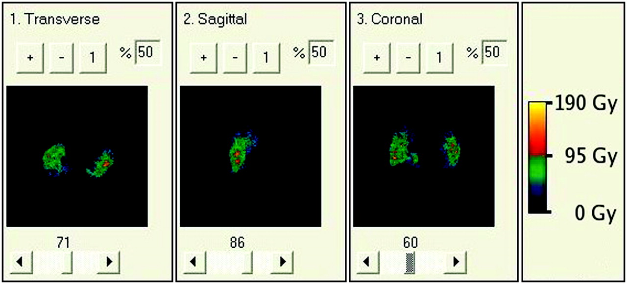
BED map resulting from 3D-RD using full patient-specific data. Although values of absorbed dose and BED are different, their relative changes from voxel to voxel are so similar that it is nearly impossible to visually differentiate the two.
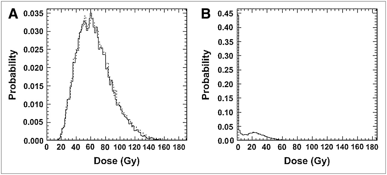
- FIGURE 7.
Differential absorbed dose (solid line) and BED (dashed line) volume histograms of tumor (A) and of lung (B) resulting from full patient-specific 3D-RD calculation.


Tables
Site α (Gy−1) β (Gy−2) μ (h−1) Lung 0.0172 0.00521 1.5 Tumor 0.365 0.028 1.3 Parameter Tumor (Gy) Lungs (Gy) Mean absorbed dose 57.7 9.5 Mean BED 58.5 9.8 EUD 25.0 8.3


Jump to section
Related Articles
Cited By...
- Dosimetry for Radiopharmaceutical Therapy: Current Practices and Commercial Resources
- Tumor Response to Radiopharmaceutical Therapies: The Knowns and the Unknowns
- PARaDIM: A PHITS-Based Monte Carlo Tool for Internal Dosimetry with Tetrahedral Mesh Computational Phantoms
- Three-Dimensional Dosimetry for Radiation Safety Estimates from Intrathecal Administration
- Recombinant Human Thyroid-Stimulating Hormone Versus Thyroid Hormone Withdrawal in 124I PET/CT-Based Dosimetry for 131I Therapy of Metastatic Differentiated Thyroid Cancer
- Radioiodine Scintigraphy with SPECT/CT: An Important Diagnostic Tool for Thyroid Cancer Staging and Risk Stratification
- Targeted Radionuclide Therapy: Proceedings of a Joint Workshop Hosted by the National Cancer Institute and the Society of Nuclear Medicine and Molecular Imaging
- Study of the Impact of Tissue Density Heterogeneities on 3-Dimensional Abdominal Dosimetry: Comparison Between Dose Kernel Convolution and Direct Monte Carlo Methods
- Radioiodine Scintigraphy with SPECT/CT: An Important Diagnostic Tool for Thyroid Cancer Staging and Risk Stratification
- Tumor Dosimetry and Response for 153Sm-Ethylenediamine Tetramethylene Phosphonic Acid Therapy of High-Risk Osteosarcoma
- Dosimetry and thyroid cancer: the individual dosage of radioiodine
- Methodology to Incorporate Biologically Effective Dose and Equivalent Uniform Dose in Patient-Specific 3-Dimensional Dosimetry for Non-Hodgkin Lymphoma Patients Targeted with 131I-Tositumomab Therapy
- The Monte Carlo Method in Nuclear Medicine: Current Uses and Future Potential
- Arterial Wall Dosimetry for Non-Hodgkin Lymphoma Patients Treated with Radioimmunotherapy
- Hybrid SPECT/CT: the end of "unclear" medicine
- 124I PET-Based 3D-RD Dosimetry for a Pediatric Thyroid Cancer Patient: Real-Time Treatment Planning and Methodologic Comparison
- SPECT/CT