


Article Figures & Data
Figures
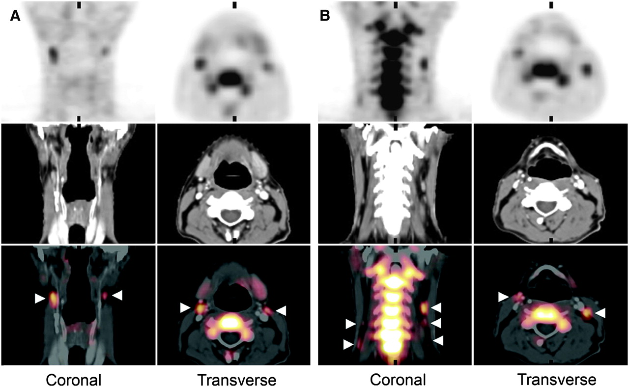
- FIGURE 1.
18F-FLT PET/CT images of patient 9 (pT2pN0M0 oral cavity carcinoma). Top panels show PET images, middle panels show CT images, and bottom panels show fusion of both image modalities. Cervical lymph nodes with increased 18F-FLT uptake are found bilaterally in level II (A, arrowheads) and in levels III and IV (B, arrowheads). All lymph nodes detected with 18F-FLT in this example were false-positive for metastasis, due to uptake in proliferating B-lymphocytes in reactive germinal centers.
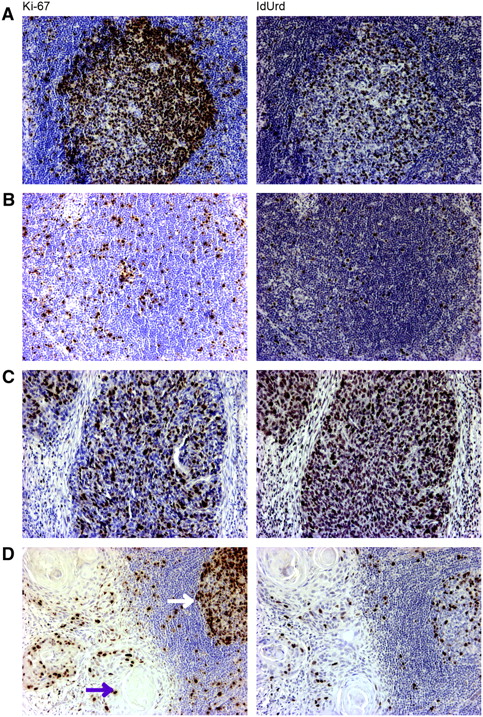
- FIGURE 2.
Ki-67 and IdUrd staining. (A) Germinal center harboring proliferating B-lymphocytes and remaining lymphoid tissue. (B) Remaining lymphoid tissue with proliferating lymphoid cells. (C) Metastasis of squamous cell carcinoma of maxillary sinus. (D) Micrometastasis with keratinization (purple arrow), fragment of a germinal center (white arrow), and surrounding lymphoid tissue. (×100)
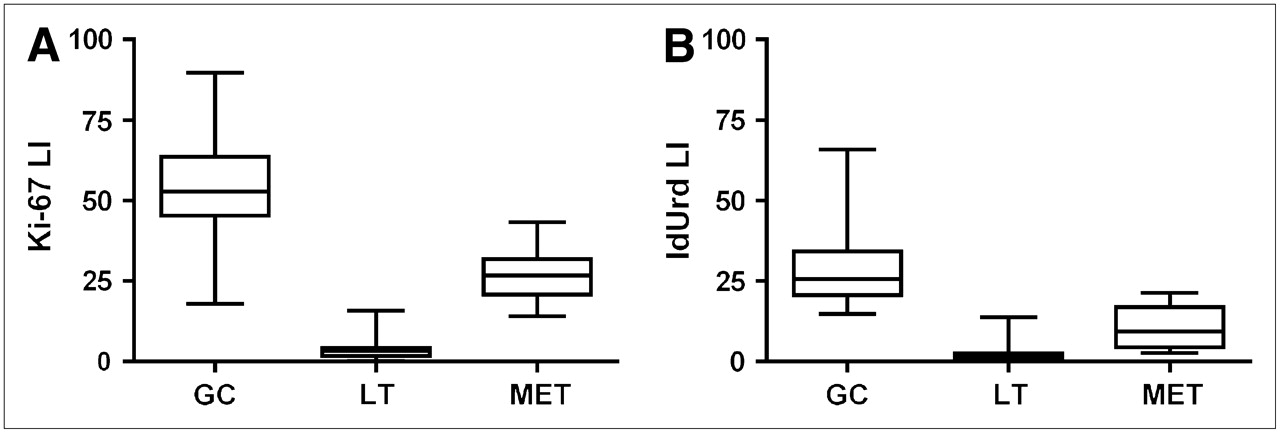
- FIGURE 3.
Ki-67 LI (A) and IdUrd LI (B) in germinal centers (GC), remaining lymphoid tissue (LT), and metastases (MET).
- FIGURE 4.
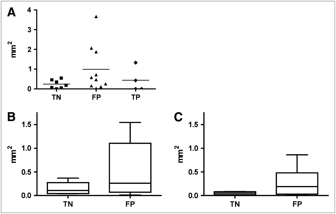
(A) Absolute area (in mm2) occupied by germinal centers in true-negative (TN; 18F-FLT-negative lymph node without metastasis), false-positive (FP; 18F-FLT-positive lymph node without metastasis), and true-positive (TP; 18F-FLT-positive lymph node with metastasis) lymph nodes. Ki-67germinal center (B) and IdUrdgerminal center (C) in TN and FP lymph nodes as measure of total proliferative activity in germinal centers (calculated as area in mm2).
- FIGURE 5.
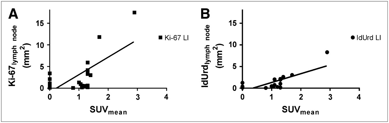
Scatter plot of Ki-67lymph node (A) and scatter plot of IdUrdlymph node (B) as measure of total proliferative activity in lymph node (calculated as area in mm2) versus SUVmean of 18F-FLT PET. Solid lines indicate linear best fit.


Tables
- TABLE 1
Patient Characteristics, Diagnostic and Therapeutic Procedures, and Histopathology of Lymph Nodes
Patient no. US and FNA cytology (side) Clinical stage Pathologic stage Pathology LN (no. of Pathologic LN) LN positive on PET* (no. of LN indicated) LN positive on PET† (no. and side of LN indicated) Site CT MRI Procedure L R L R 1 Maxillary sinus POS (L) NP POS T4N1M0 pT4pN1M0 TE + MRND, L POS (1) NA 1 NA 1, R 2 Laryngeal NP NEG NP T3N0M0 pT3pN0M0 TLE + bilateral NS NEG NEG 1 1 2, R 3 Laryngeal NP POS NP T4N2cM0 pT4pN2cM0 TLE + bilateral RND POS (7) POS (5) 2 2 1, R 4 Lower alveolar ridge NEG (L) NEG NP T4N2bM0 pT4pN0M0 TE + MRND, R NA NEG NA 2 2, L 5 Floor of mouth NR NP NP T2N0M0 pT2pN0M0 TE + bilateral SND NEG NEG NEG NEG — 6 Tongue NEG (R) NP NP T3N0M0 pT2pN0M0 TE + SND, R NA NEG — 5 — 7 Soft palate NP NP NP T2N0M0 pT2pN0M0 TE + SND, L NEG NA NEG NA 1, R 8 Tongue NP NP NEG T3N0M0 pT3pN1M0 TE + SND, R NA POS (1) — 1 1, R 9 Tongue/floor of mouth NEG (L) NP NEG T4N0M0 pT2pN0M0 TE + SND, L NEG NA 3 NA 2, R 10 Tongue/floor of mouth NEG (L) NP NEG T2N0M0 pT2pN0M0 TE + SND, L NEG NA 2 — — ↵* Available for pathologic assessment.
↵† Not available for pathologic assessment.
LN = lymph node; FNA = fine-needle aspiration; NP = not performed; POS = positive; TE = tumor excision; MRND = modified radical neck dissection; NA = not available; NEG = negative; TLE = total laryngectomy; NS = node sampling; RND = radical neck dissection; NR = not representative; SND = selective neck dissection (level I–III).
- TABLE 2
Histologic Lymph Node Assessment: SUVmean, Histopathology, and Mean Ki-67 and IdUrd Staining
Patient no. LN level Pathology Ki-67 and IdUrd staining 18F-FLT POS SUVmean 18F-FLT NEG Metastasis Group Metastasis Germinal centers Remaining LN tissue 1 R II 1.5 NA L III − TN − + + L II 2.9 + TP + − + 2 R II 1.2 − FP − ++ + R III 0.9 NA R IV 1.2 NA L III 1.1 − FP − ++ + 3 R II 1.2 NA R III 1.3 + TP + ++ + R IV 1.1 + TP + ++ + L II 1.7 + TP + − − L IV 1.3 + TP + + + 4 R II 1.4 − FP − ++ + R III − TN − − + R IV 1.1 − FP − − + R V − TN − ++ + L II 1.0 NA L IV 0.8 NA 5 R I − TN − + + L II − TN − + ++ 6 R I 1.4 − FP − ++ ++ R II 1.3 − FP − ++ ++ R III 0.9 − FP − + + 7 R IV 1.5 NA L I − TN − ++ + L III − TN − ++ + 8 R II 1.0 + TP + ++ + R III 0.8 NA 9 R II 2.1 NA R IV 1.0 NA L II 1.3 − FP − ++ + L III 2.0 − FP − + + L IV 1.0 − FP − + + L I − TN − − + 10 L II 1.6 − FP − + ++ L III 1.3 − FP − + ++ LN = lymph node; POS = positive; NEG = negative; II = level indicated by roman number, NA = not available; TN = true-negative 18F-FLT-negative lymph node without metastasis; TP = true-positive 18F-FLT-positive lymph node with metastasis; FP = false-positive 18F-FLT-positive lymph node without metastasis.
- TABLE 3
Ki-67 LI and IdUrd LI in Germinal Centers, Remaining Lymphoid Tissue, and Metastases
Ki-67 LI Germinal centers Remaining lymphoid tissue Metastases Parameter TN FP TP Overall TN FP TP Overall TP Mean 58.6 52.1 50.5 53.9 3.7 2.8 7.4 3.8 26.8 SD 16.6 13.2 4.2 13.8 1.9 2.4 7.5 3.6 7.7 Median 55.1 53.1 49.5 52.8 3.6 2.5 4.6 3.3 26.7 IdUrd LI Germinal centers Remaining lymphoid tissue Metastases Parameter TN FP TP Overall TN FP TP Overall TP Mean 33.5 27.4 23.0 28.8 1.4 1.8 4.2 2.1 10.4 SD 15.7 9.4 4.5 11.7 0.7 1.3 6.4 2.8 6.1 Median 29.6 23.9 24.9 25.7 1.6 1.6 1.5 1.6 9.3 TN = true-negative (18F-FLT-negative lymph node without metastasis); FP = false-positive (18F-FLT-positive lymph node without metastasis); TP = true-positive (18F-FLT positive lymph node with metastasis).


Jump to section
Related Articles
Cited By...
- Exploring molecular imaging to investigate immune checkpoint inhibitor-related toxicity
- 18F-FLT PET/CT Adds Value to 18F-FDG PET/CT for Diagnosing Relapse After Definitive Radiotherapy in Patients with Lung Cancer: Results of a Prospective Clinical Trial
- Using Radiolabeled 3'-Deoxy-3'-18F-Fluorothymidine with PET to Monitor the Effect of Dexamethasone on Non-Small Cell Lung Cancer
- Single-Cell Characterization of 18F-FLT Uptake with Radioluminescence Microscopy
- 18F-FLT PET/CT in the Evaluation of Pheochromocytomas and Paragangliomas: A Pilot Study
- Differential 18F-FDG and 18F-FLT Uptake on Serial PET/CT Imaging Before and During Definitive Chemoradiation for Non-Small Cell Lung Cancer
- PET Imaging of Proliferation with Pyrimidines
- PET Imaging During Radiotherapy of Head and Neck Cancer
- 3'-Deoxy-3'-18F-Fluorothymidine PET-Derived Proliferative Volume Predicts Overall Survival in High-Grade Glioma Patients
- 18F-FDG PET Detects Inflammatory Infiltrates in Spinal Cord Experimental Autoimmune Encephalomyelitis Lesions
- Early identification of antigen-specific immune responses in vivo by [18F]-labeled 3'-fluoro-3'-deoxy-thymidine ([18F]FLT) PET imaging
- Novel Positron Emission Tomography Tracer Distinguishes Normal from Cancerous Cells
- Predictive Value of Initial 18F-FLT Uptake in Patients with Aggressive Non-Hodgkin Lymphoma Receiving R-CHOP Treatment
- Can Evaluation of Targeted Therapy in Oncology Be Improved by Means of 18F-FLT?
- 18F-FLT PET/CT for Early Response Monitoring and Dose Escalation in Oropharyngeal Tumors
- Monitoring Tumor Response to Therapy with 18F-FLT PET
- Histopathologic Validation of 3'-Deoxy-3'-18F-Fluorothymidine PET in Squamous Cell Carcinoma of the Oral Cavity
- Innovations in Radiotherapy Planning of Head and Neck Cancers: Role of PET
- Kinetic Analysis of 3'-Deoxy-3'-18F-Fluorothymidine (18F-FLT) in Head and Neck Cancer Patients Before and Early After Initiation of Chemoradiation Therapy
- 18F-FDG and 18F-FLT Uptake Early After Cyclophosphamide and mTOR Inhibition in an Experimental Lymphoma Model
- PET Monitoring of Therapy Response in Head and Neck Squamous Cell Carcinoma
- 18F-FDG and 18F-FLT Do Not Discriminate Between Reactive and Metastatic Lymph Nodes in Oral Cancer
- In Vivo Characterization of Proliferation for Discriminating Cancer from Pancreatic Pseudotumors
- Imaging of Cell Proliferation: Status and Prospects