


Abstract
Non–small cell lung cancer (NSCLC) is a leading cause of cancer mortality in the United States, and pemetrexed-based therapies are regularly used to treat nonsquamous NSCLC. Despite widespread use, pemetrexed has a modest effect on progression-free survival, with varying efficacy between individuals. Recent work has indicated that dexamethasone, given to prevent pemetrexed toxicity, is able to protect a subset of NSCLC cells from pemetrexed cytotoxicity by temporarily suppressing the S phase of the cell cycle. Therefore, dexamethasone might block treatment efficacy in a subpopulation of patients and might be contributing to the variable response to pemetrexed. Methods: Differences in retention of the experimental PET tracer 3′-deoxy-3′-fluorothymidine (FLT) were used to monitor S-phase suppression by dexamethasone in NSCLC cell models, animals with tumor xenografts, and patients with advanced cancer. Results: Significant reductions in tracer uptake were observed after 24 h of dexamethasone treatment in NSCLC cell lines and xenograft models expressing high levels of glucocorticoid receptor α, coincident with pemetrexed resistance visualized by attenuation of the flare effect associated with pemetrexed activity. Two of 4 patients imaged in a pilot study with 18F-FLT PET after dexamethasone treatment demonstrated reductions in tracer uptake from baseline, with a variable response between individual tumor lesions. Conclusion: 18F-FLT PET represents a useful method for the noninvasive monitoring of dexamethasone-mediated S-phase suppression in NSCLC and might provide a way to individualize chemotherapy in patients receiving pemetrexed-based regimens.
Lung cancer is the leading cause of cancer-related mortality in the United States, accounting for 27% of cancer deaths (1). Non–small cell lung cancer (NSCLC) comprises 83% of lung cancers, and most patients have advanced disease at the time of diagnosis (1). Several randomized clinical trials have shown that pemetrexed, given as a monotherapy or in combination with a platinum-containing compound, is a preferred chemotherapeutic agent for the treatment of advanced nonsquamous NSCLC (2–4). Pemetrexed is a folate antimetabolite that causes cytotoxicity primarily through inhibition of thymidylate synthase (5,6). Although widely used, pemetrexed has a modest but variable effect in patients, with a median increase in progression-free survival of 5.3 mo in the front-line setting when combined with cisplatin, and only 3.3 mo when used as monotherapy (4,7). Furthermore, the 5-y survival for patients with metastatic NSCLC remains dismal, at less than 4% (8). Given this paradigm, it is critical to identify factors that can be used to predict and optimize the clinical benefit of pemetrexed in order to maximize efficacy and spare nonresponders the toxicity of ineffective chemotherapy. Several studies have observed an association between low tumor expression of thymidylate synthase and better outcomes in patients treated with pemetrexed (9). However, this association has not been shown to be a powerful independent predictor of patient response to pemetrexed and is not used clinically (10).
Despite its relatively mild toxicity profile, a major adverse effect of pemetrexed is the manifestation of a generalized, painful, pruritic skin rash (11). To protect against this rash, patients are routinely administered 4 mg of dexamethasone twice daily starting the day before therapy and continuing until the day after therapy (12). In addition, dexamethasone is frequently used as an antiemetic for patients receiving cisplatin or carboplatin (13). Dexamethasone is a synthetic glucocorticoid that, on binding to glucocorticoid receptor α (GRα), modulates genes involved in cell proliferation and apoptosis, as well as inflammation and the inflammatory response (14,15). Recently, dexamethasone, in a GRα-dependent fashion, has been found to cause reversible G1 cell cycle arrest in NSCLC cells, resulting in protection of cells from pemetrexed cytotoxicity (16). This may represent a significant clinical problem given that the biological half-life of dexamethasone (36–54 h) is substantially longer than that of pemetrexed (2.5 h). Histologic examination of NSCLC lesions has suggested there is an approximately equal distribution of tumors with high and low GRα expression (17). It is possible that the protective effect of dexamethasone, combined with differential tumor GRα expression, may explain, in part, the variable efficacy of pemetrexed in clinical practice. It is therefore desirable to develop a technique that can be used to ascertain the GRα level and potential pemetrexed resistance in patients. Analysis of biopsy specimens is likely inadequate for this task given the enormous clonal heterogeneity observed within solid tumors and between metastatic foci (18,19). Such heterogeneity is especially relevant in this case, as NSCLC patients receiving pemetrexed typically have advanced disease.
Monitoring of retention of 3′-deoxy-3′-fluorothymidine (FLT), developed for use with PET, would represent a rapid and noninvasive technique to functionally image the effect of dexamethasone on NSCLC over a patient’s entire cancer burden. Radiolabeled FLT is taken up by tumor cells and trapped intracellularly via phosphorylation by the S-phase–specific enzyme thymidine kinase 1 (20). Changes in FLT retention can therefore be used to monitor the effect of compounds such as dexamethasone, which alter cell cycle progression (21). Uptake of FLT is reproducible and has been shown to correlate with the proliferation marker Ki-67 in NSCLC (22–24). In the current study, FLT retention was applied as a direct functional probe to measure dexamethasone-mediated S-phase suppression in NSCLC. Additionally, the study sought to use 18F-FLT accumulation to monitor the effect of pemetrexed in vivo. The basis for this goal was the observation that compounds that inhibit de novo thymidine biosynthesis, such as pemetrexed, elicit a transient increase in FLT uptake due to upregulation of thymidine salvage, termed the flare phenomenon (25,26). This effect was recently shown in cell lines, as well as mice bearing NSCLC tumors, and may provide a way to visualize the interference of dexamethasone with pemetrexed activity in vivo (27).
MATERIALS AND METHODS
3H-FLT Uptake Measurements
All cell lines were authenticated using the Promega PowerPlex 16 System. Technical details concerning tissue culture and generation of recombinant H1299-GRα cells have been described elsewhere (13). NSCLC cells were seeded in 6-well plates with glucocorticoid-depleted medium (5 × 10⁵ cells per well). The cells were incubated in 100 nM dexamethasone, corresponding to the peak plasma concentration in humans after a single oral dose (28). Cells treated with pemetrexed were incubated with pemetrexed (5 μM) for 4 h. After treatment, the cells were transferred to medium containing approximately 1,600 Bq of 3H-FLT and incubated for 1 h. The cells were washed, lysed with 1 M KOH, and mixed with 5 mL of Ultima Gold XR scintillation cocktail (PerkinElmer). Sample activity was measured by a liquid scintillation analyzer (Packard Tricarb) and normalized to cell number using a parallel experiment and cell counting via the trypan blue exclusion method. All experiments were performed in triplicate.
Establishment of NSCLC Xenografts and Animal Imaging Protocol
Subcutaneous xenografts were established by implanting cubic fragments (∼2 by 2 by 2 mm) of tumor tissue bilaterally into the axilla of 4- to 6-wk-old female SCID/NCr mice (Charles River), and the animals were included in the study when tumors reached an average volume of 250 mm3. The mice were treated intraperitoneally with veterinary-grade dexamethasone (15 mg/kg twice daily), which allowed for steady-state serum concentrations within the pharmacologically effective range (Supplemental Table 1; supplemental materials are available at http://jnm.snmjournals.org) (28,29). Animals treated with pemetrexed were administered 10 mg/kg intravenously and were fed a low-folate diet starting 10 d before treatment. The animals were injected intravenously with approximately 9.5 MBq of 18F-FLT, synthesized as previously published (30). One hour after tracer injection, the mice underwent a 10-min emission scan using a microPET R4 scanner (Siemens). Six to 8 mice were used for each treatment condition. Each animal was imaged at baseline and after the specified treatments and served as its own control for pretreatment-to-posttreatment comparisons. Reconstructed images were evaluated with PMOD, and tracer activity within tumors was corrected for decay and converted to SUVs. Data are expressed as SUVmax, which reflects the activity of the hottest pixel within the tumor. Supplemental Figure 1 illustrates the dexamethasone treatment and animal imaging protocol.
Patient Imaging
To determine whether dexamethasone-mediated effects on FLT retention could be observed in a clinical setting, a small pilot study was conducted on patients at the Karmanos Cancer Institute. Patients were considered eligible for the study if they were diagnosed with nonsquamous NSCLC, were scheduled to receive a chemotherapy regimen that included dexamethasone, had no history of prior pemetrexed or docetaxel chemotherapy, had measurable disease with at least one lesion 2 cm or larger on CT or MRI, and could tolerate 2 PET scans. We planned to image 5 patients who consented to join the study, but only 4 of these patients completed the evaluation. The patients were injected with 18F-FLT (range, 167–265 MBq; mean, 226 MBq) over 60 s, and images were collected 1 h later using a Discovery STE PET/CT scanner (GE Healthcare). Imaging took place at baseline and again 24 h after the start of oral dexamethasone treatment (4 mg twice daily), as is standard practice in patients receiving pemetrexed. Reconstructed images were viewed using OsiriX imaging software (Pixmeo). Tumor SUVmax was obtained by drawing volumes of interest over the tumor plane with the most active pixel and the 2 adjacent planes. In patients with multiple lesions, all evaluable (>2 cm) lesions were assessed. The institutional review board at Wayne State University approved the study protocol. All patients gave written informed consent.
Statistical Considerations
All statistical tests were conducted using GraphPad Prism, version 6. For cellular tracer uptake studies, 1-way ANOVA was used. For animal studies, a paired-sample ANOVA adjusting for unequal sample size, when appropriate, was used.
RESULTS
Changes in 3H-FLT Uptake Reflect Sensitivity to Dexamethasone
The level of expression of GRα was found to be the deciding factor in whether cells would be protected from pemetrexed after dexamethasone pretreatment. To determine whether dexamethasone sensitivity is associated with changes in 3H-FLT uptake, several NSCLC cell lines with varying expression of GRα were tested (Figs. 1A and 1B). In cells with the highest relative GRα expression, there was a significant reduction in 3H-FLT accumulation after 24 h of dexamethasone treatment (Fig. 1C). 3H-FLT retention was further decreased after 48 h, consistent with the observed reduction in the S-phase fraction of cells (P < 0.01).
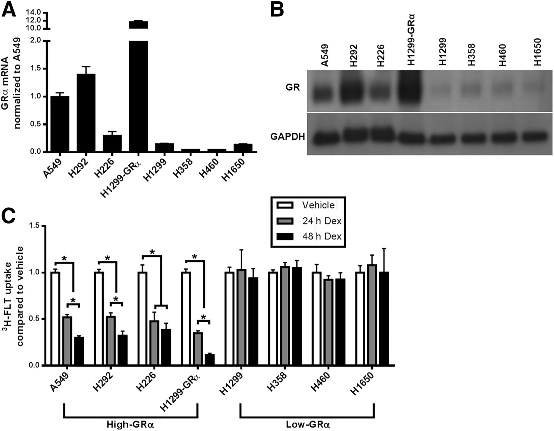
Dexamethasone reduces 3H-FLT retention most in NSCLC cell lines with highest GRα expression. (A) GRα messenger RNA measured by RT-PCR. (B) Western blot showing total GR protein expression. (C) 3H-FLT in NSCLC cell lines after 24 and 48 h of dexamethasone treatment. Dex = dexamethasone; GAPDH = glyceraldehyde 3-phosphate dehydrogenase. *P < 0.01.
Dexamethasone Reversibly Decreases 18F-FLT Retention in Human Xenografts
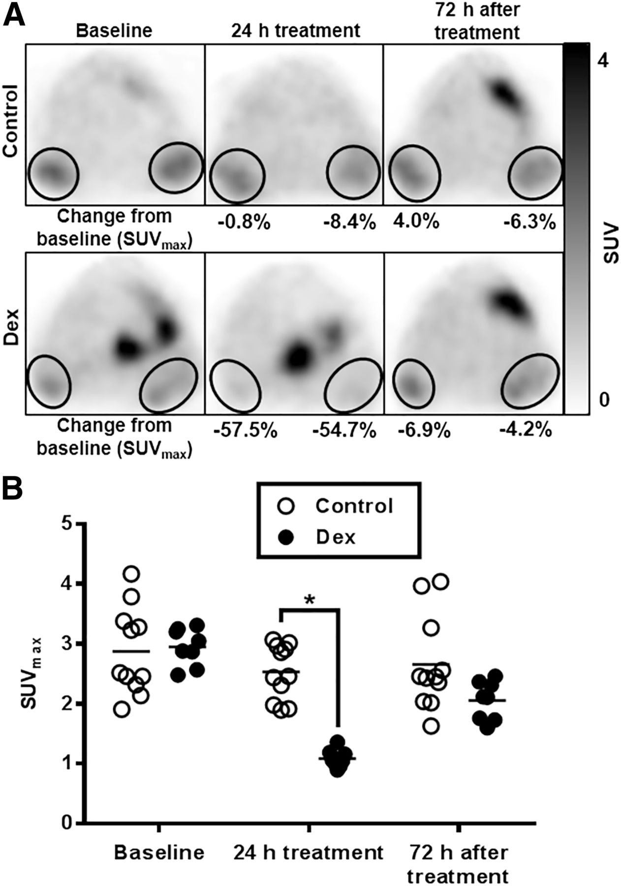
To evaluate whether 18F-FLT PET can detect dexamethasone sensitivity in vivo, mice implanted bilaterally with A549 tumors were imaged at baseline and after dexamethasone treatment. SUVmax in A549 tumors decreased by an average of 63.1% (range, −70.5 to −54.70; SD, 5.08) after 24 h of dexamethasone treatment (Fig. 2). At 72 h after treatment, the tumors regained their proliferative capacity and SUVmax was in accordance with the control tumors, indicating the reversibility of dexamethasone-mediated cell cycle arrest (P < 0.01).

Dexamethasone reduces 18F-FLT accumulation in mice bearing A549 tumors. (A) Representative images of animals treated with dexamethasone or control (saline). (B) Plot of SUVmax for tumors treated with dexamethasone or control. Dex = dexamethasone. *P < 0.01.
To further establish the ability of 18F-FLT to image the antiproliferative effect of dexamethasone, mice bearing low-GRα H1299 tumors, as well as H1299 tumors in which GRα had been lentivirally transduced, were imaged. Similar to animals with A549 tumors, mice bearing H1299-GRα tumors exhibited posttreatment reductions in tumor SUVmax (mean change, −41.3%; range, −53.0 to −11.6; SD, 14.9) that rebounded after 72 h of dexamethasone withdrawal (Fig. 3). Interestingly, in H1299 tumors, which express low levels of GRα and were unaffected by dexamethasone in cell culture, a significant decrease in 18F-FLT accumulation was observed after dexamethasone treatment (mean change, −20.8%; range, −36.4 to 2.7; SD, 9.6) (P < 0.01). Harvested tumors revealed that although GRα messenger RNA levels remained lower in H1299 tumors than in the other xenograft models used here, populations of cells within H1299 tumors stained positively for GR (Supplemental Fig. 2). Critically, the magnitude of reduction in tracer retention from baseline was associated with the relative GRα expression within the tumors.
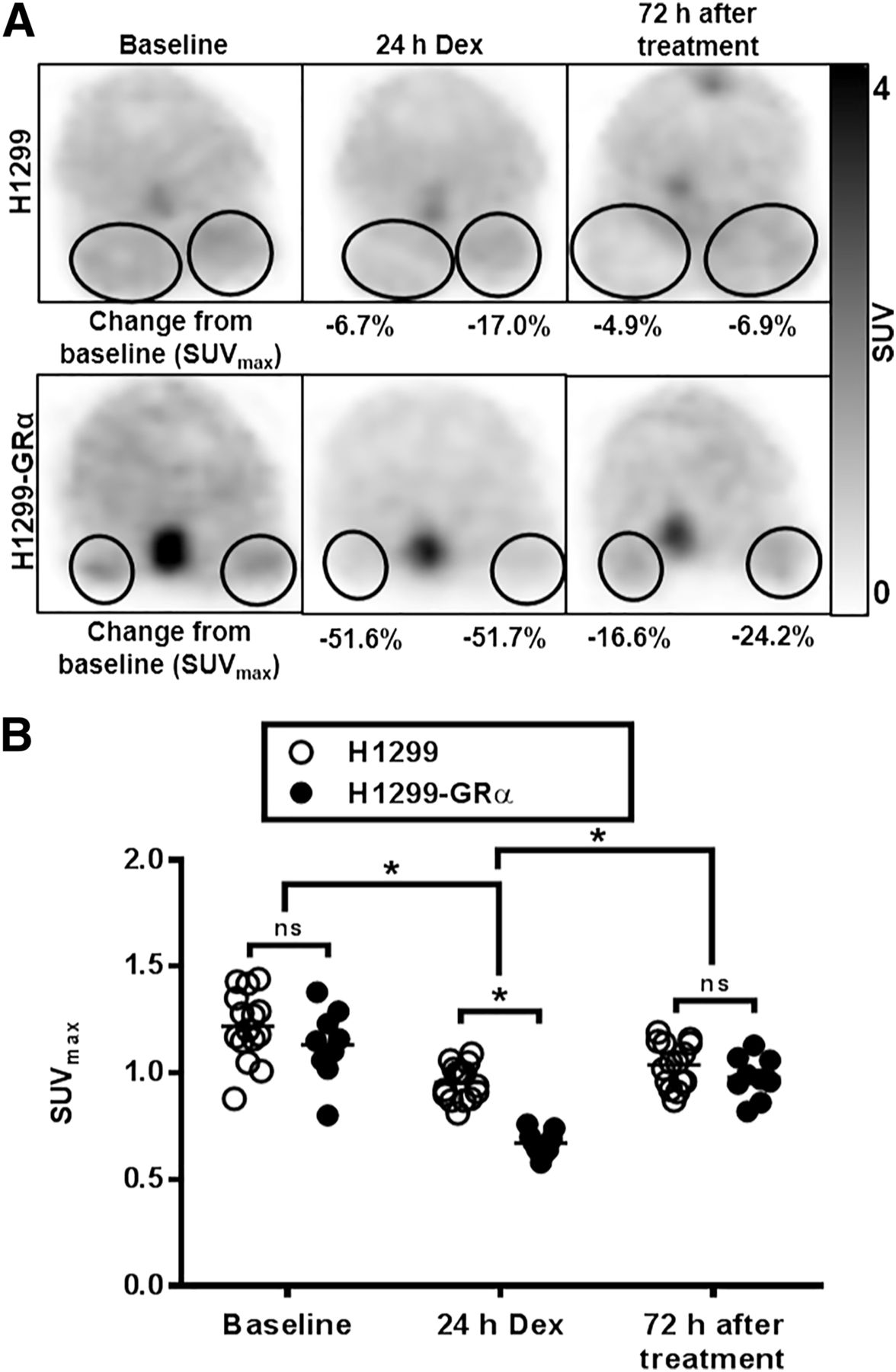
Decline in 18F-FLT retention after dexamethasone treatment depends on GRα expression. (A) Representative pre- and postdexamethasone images of animals implanted with H1299 or H1299-GRα tumors (encircled). (B) Plot comparing SUVmax between H1299 and H1299-GRα tumors. Dex = dexamethasone. *P < 0.01.
18F-FLT Visualizes Interlesion Heterogeneity in Dexamethasone Sensitivity Between Metastases in Human Tumors
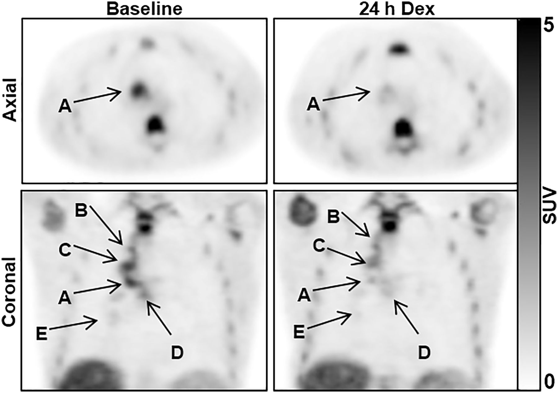
To determine whether these findings in NSCLC cell lines and xenografts were relevant to human disease, work was extended into 4 patients with advanced NSCLC. After dexamethasone treatment, tumors in patients 1 and 3 demonstrated marked reductions in tumor SUVmax from baseline (−64.7% and −54.3%, respectively). Conversely, patients 2 and 4 were largely unaffected by dexamethasone treatment, highlighting variability in the effect between patients (Table 1). Furthermore, marked heterogeneity within individual patients was observed, as the lesions of patient 1 showed a variable change in 18F-FLT uptake after dexamethasone treatment (Fig. 4) whereas patient 3 showed the effect in all lesions. Unfortunately, these results could not be compared with tumor GRα expression because of an insufficiency of available tumor tissue.
Change in Tumor 18F-FLT Uptake in NSCLC Patients After 24 Hours of Dexamethasone Treatment

18F-FLT PET captures interlesion heterogeneity. 18F-FLT PET images are shown from patient with NSCLC at baseline and after 24 h of dexamethasone treatment. In nodal metastasis A, 18F-FLT uptake decreased by 64.7%. Tracer retention in lesions B, C, D, and E decreased by 13.7%, 33.1%, 18.1%, and 34.6%, respectively. Vertebral, sternal, and rib marrow activity was unchanged. Dex = dexamethasone.
Dexamethasone Abolishes Pemetrexed-Mediated Flare in 3H-FLT Uptake in NSCLC Cell Lines
Compounds that interfere with de novo thymidine biosynthesis, such as pemetrexed, can lead to a temporary upregulation in thymidine salvage, and therefore in FLT accumulation, as cells seek to replenish intracellular thymidine exogenously. Exploiting this phenomenon may provide a method to monitor pemetrexed activity in vivo. To test this idea, 3H-FLT uptake was measured in NSCLC cells after 4 h of pemetrexed treatment (Fig. 5). 3H-FLT accumulation significantly increased in all cell lines (P < 0.01). However, 24 h of dexamethasone pretreatment abrogated this effect in sensitive cell lines.
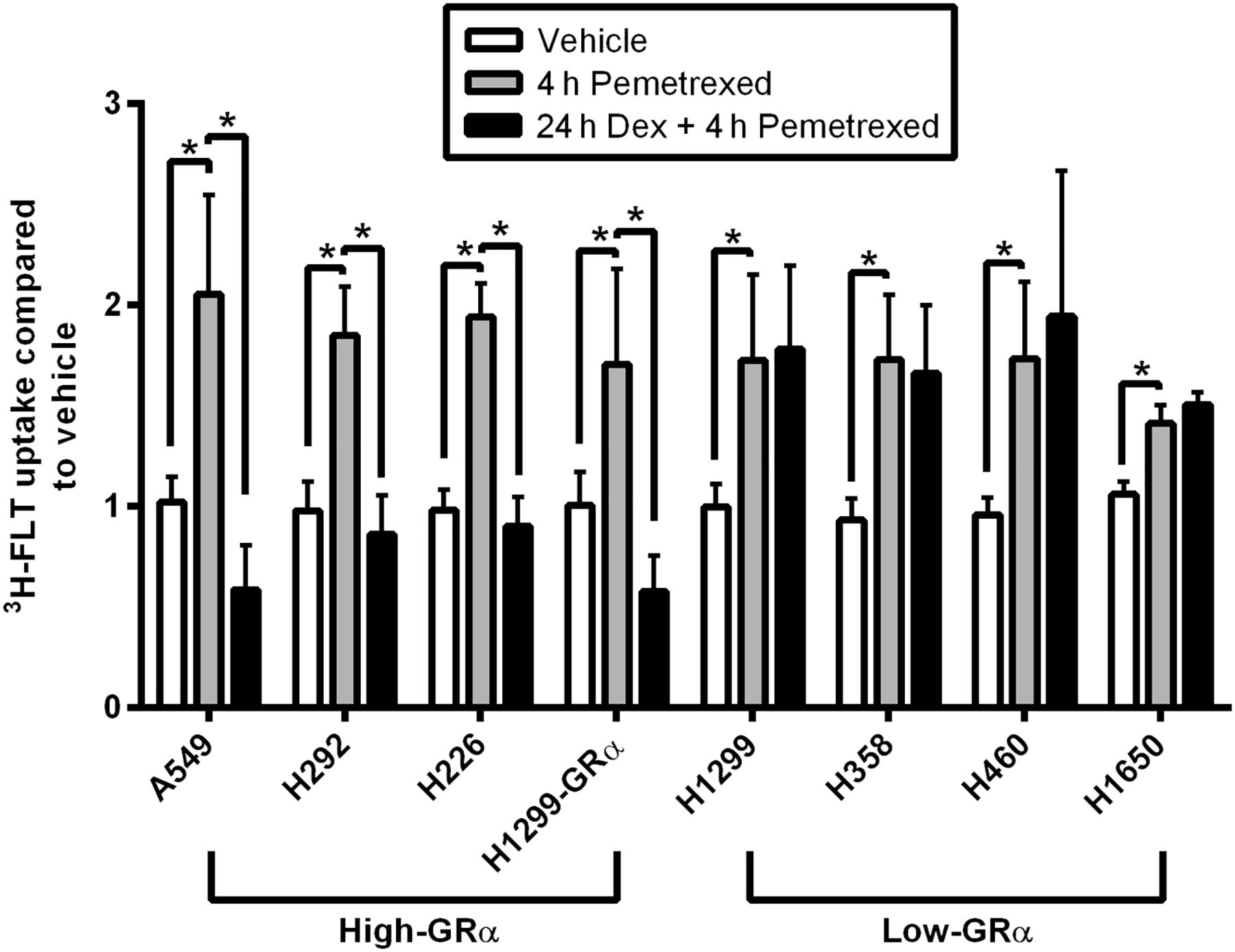
Pemetrexed increased 3H-FLT retention in NSCLC cell lines, but dexamethasone pretreatment abrogated this effect. Graph shows 3H-FLT uptake in NSCLC cells treated with vehicle (saline) or pemetrexed with and without dexamethasone pretreatment. Dex = dexamethasone. *P < 0.01.
18F-FLT Visualizes Dexamethasone Interference with Pemetrexed Activity
To evaluate the flare effect in vivo, the xenograft models described in previous experiments were used. As was observed in cell culture, mice implanted with A549 tumors showed an increase in 18F-FLT uptake by 48.9% from baseline (range, −3.5% to 102.5%; SD, 33.9) after pemetrexed treatment, but this effect was abolished if animals received dexamethasone pretreatment (mean change, −44.5% from baseline; range, −65.8 to −22.7%; SD, 15.56) (P < 0.01) (Fig. 6). After pemetrexed, H1299 tumors exhibited a greater flare from baseline than A549 tumors: SUVmax in H1299 and H1299-GRα tumors increased by 107.3% (range, 11.6%–268.5%; SD, 90.4) and 68.7% (range, 0.0%–164.5%; SD, 57.4), respectively. If animals were pretreated with dexamethasone, the flare response was completely eradicated in H1299-GRα tumors, and the change from baseline resembled that of animals treated with dexamethasone alone (mean change, −42.9% from baseline; range, −67.4 to −18.9%; SD, 15.56). H1299 tumors still exhibited a significant flare from baseline (mean change in SUVmax, 32.9%; range, −8.0% to 68.8%; SD, 30.1), but it was smaller than that produced by pemetrexed alone, potentially reflecting GR upregulation (Fig. 7).

Pemetrexed-induced flare in 18F-FLT uptake is abolished with dexamethasone pretreatment in animal xenografts. (A) Representative images of mice bearing A549 tumors (encircled). (B) Plot showing effect of pemetrexed on 18F-FLT retention with and without dexamethasone pretreatment. Dex = dexamethasone. *P < 0.01.
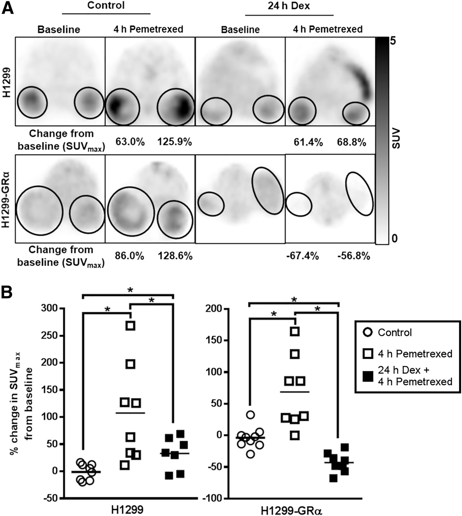
Eradication of pemetrexed-induced flare by dexamethasone occurs in GRα-dependent fashion. (A) Representative images of mice bearing H1299 or H1299-GRα tumors (encircled). (B) Plot showing effect of pemetrexed on 18F-FLT retention with and without dexamethasone pretreatment. Dex = dexamethasone. *P < 0.01.
DISCUSSION
The treatment landscape for NSCLC is rapidly evolving with the introduction of newer targeted therapies and immune-modulating agents. Despite these advances, most patients diagnosed with advanced NSCLC will at some point receive platinum-based cytotoxic chemotherapy, often with pemetrexed. Here, the use of FLT retention was explored as a biomarker to monitor dexamethasone-mediated S-phase suppression in several models of NSCLC. Studies in NSCLC cell lines indicated that treatment with dexamethasone for 24 h produced a significant reduction in 3H-FLT uptake in cell lines with relatively high GRα expression. This result was translatable to animal studies, in which mice implanted with high-GRα A549 tumors demonstrated a significant reduction in tracer uptake after dexamethasone treatment. Furthermore, when imaging isogenic H1299 and H1299-GRα tumors, the magnitude of change in 18F-FLT retention in response to dexamethasone was associated with the expression level of GRα. Of note, low-GR H1299 tumors displayed an unexpected decline in 18F-FLT uptake after dexamethasone, a result that was discordant with in vitro studies. However, although H1299 cells express relatively low amounts of GRα, they are not GRα-negative. Further, GR has been shown to be involved in numerous cellular survival pathways and can be activated by conditions such as hypoxia, nutrient deprivation, and reactive oxygen species (31,32). Because cancer cell lines in ectopic xenografts face a more hostile growth medium than those in cell culture, it is possible that stresses within the tumor microenvironment may preferentially support subpopulations of cells expressing higher levels of GRα, which could, in turn, lead to alterations in 18F-FLT retention after glucocorticoids. Indeed, immunohistochemical staining of harvested tumors demonstrated clusters of GR-positive cells.
In patients with advanced NSCLC, the changes were much more variable, with 2 of 4 patients showing some response to dexamethasone. Critically, 18F-FLT PET was able to detect heterogeneity in dexamethasone sensitivity between lesions within individual patients. The capacity to simultaneously evaluate multiple lesions in patients is a major advantage of imaging, compared with analysis of biopsy specimens, given that NSCLC patients receiving chemotherapy have advanced disease.
In addition, this study sought to use FLT accumulation to monitor pemetrexed activity through its inhibitory effect on thymidylate synthase, and the subsequent increase in FLT accumulation. In agreement with recent studies, pemetrexed treatment produced a significant increase in 3H-FLT retention compared with control (27). This effect was eradicated in cells with high GRα expression if they were pretreated with dexamethasone. Animal imaging further corroborated this finding. Mice bearing A549 and H1299-GRα tumors exhibited a significant flare from baseline after pemetrexed treatment, and this flare was prevented if the animals received dexamethasone before chemotherapy. Conversely, low-GRα H1299 tumors produced a flare regardless of dexamethasone treatment. Taken together, these data suggest that the presence of a flare in response to pemetrexed may be indicative of the activity of the drug and may be useful as an early marker for assessing response to therapy. A recent study in NSCLC patients treated with pemetrexed attempted to correlate a flare in 18F-FLT uptake with drug efficacy. The study found that only 2 of 11 subjects exhibited a flare, with the remaining individuals demonstrating either reduced or no change in tumor 18F-FLT uptake after pemetrexed treatment. Furthermore, the flare did not correlate with response to therapy (33). However, because all patients in the study received dexamethasone before their treatment, this result may have been due to dexamethasone-mediated suppression of thymidine kinase 1, which counteracts the compensatory rise in thymidine salvage due to thymidylate synthase inhibition.
An unfortunate limitation of the present work was an inability to directly correlate changes in 18F-FLT uptake after dexamethasone treatment with tumor GRα expression measured from patient biopsy samples, because of insufficient available tumor tissue. Furthermore, no conclusions can be made about the impact of dexamethasone on the efficacy of pemetrexed chemotherapy in this particular cohort, as the patients received other agents as part of their treatment regimen. Further work is needed to correlate tissue and image results regarding dexamethasone sensitivity and its impact on chemotherapy response. Despite its promise as an agent for monitoring S-phase suppression, 18F-FLT has several noteworthy limitations. Cells that are highly reliant on de novo thymidine synthesis will have low baseline 18F-FLT uptake, and therefore changes in tracer retention may be difficult to detect (34). In addition, tracer accumulation can be skewed by other factors, including inflammation, drugs that affect thymidine synthesis pathways, or increased cellular clearance of FLT-phosphate (k4) (35–37).
Ultimately, the imaging approach used here might allow for the stratification of patient tumors by dexamethasone sensitivity. Patients found to have one or more dexamethasone-sensitive lesions might be given a treatment regimen that does not require dexamethasone prophylaxis. Alternatively, this approach might facilitate adjustment of the dexamethasone treatment schedule such that the interference with therapy could be minimized while still preventing adverse events. More generally, numerous studies have found that glucocorticoids, through various mechanisms, reduce the efficacy of commonly used antineoplastic agents, including cisplatin, doxorubicin, and gemcitabine (38–40). This effect has been shown in models of breast, brain, colon, prostate, and cervical cancer, among others (41). The potential use of 18F-FLT PET as a tool to detect this phenomenon might critically affect patient outcomes, as many chemotherapeutic agents are given with glucocorticoids as part of supportive care.
CONCLUSION
18F-FLT PET represents a useful method for the noninvasive monitoring of dexamethasone-mediated S-phase suppression in NSCLC and might provide a way to individualize chemotherapy in patients receiving pemetrexed-based regimens.
DISCLOSURE
This work was partially funded by NIH grant P30 CA22453 and Department of Defense contract W81XWH-11-1-0068. No other potential conflict of interest relevant to this article was reported.
Acknowledgments
We acknowledge Dr. Larry Matherly for his helpful guidance on the use of antifolates in rodents and Dr. Mugdha Patki for her assistance with optimizing the cellular studies.
Footnotes
Published online Apr. 19, 2018.
- © 2018 by the Society of Nuclear Medicine and Molecular Imaging.
REFERENCES
- Received for publication December 19, 2017.
- Accepted for publication April 9, 2018.


Jump to section
Related Articles
Cited By...
- No citing articles found.