


Article Figures & Data
Figures
- FIGURE 1.
Cell-binding assay and functional assay. (A) Cell-binding assay of VEGF121 and DOTA-VEGF121 using PAE/KDR cells. IC50 values are 1.02 and 1.66 nmol/L for VEGF121 and DOTA-VEGF121, respectively. (B) Functional assay of VEGF121 and DOTA-VEGF121. Tubulin was used as loading control.
- FIGURE 2.
MicroPET of 64Cu-DOTA-VEGF121 in U87MG tumor-bearing mice. (A) Serial microPET scans of large and small U87MG tumor-bearing mice injected intravenously with 5–10 MBq of 64Cu-DOTA-VEGF121 (∼2–4 μg of VEGF121). Mice injected with 64Cu-DOTA-VEGF121 30 min after injection of 100 μg VEGF121 are also shown (denoted as “Small tumor + block”). (B) Two-dimensional whole-body projection of the 3 mice shown in A at 16 h after injection of 64Cu-DOTA-VEGF121. Tumors are indicated by arrows.
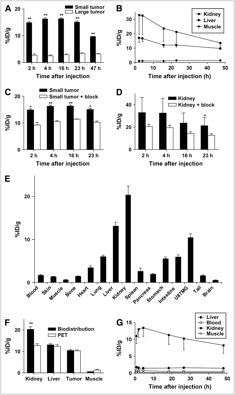
- FIGURE 3.
MicroPET and biodistribution results. (A) Comparison of 64Cu-DOTA-VEGF121 uptake in small and large U87MG tumors (3 mice per group). (B) Time–activity curves of 64Cu-DOTA-VEGF121 uptake in the kidney, liver, and muscle (n = 6). (C) Comparison of 64Cu-DOTA-VEGF121 uptake in small U87MG tumors with those injected previously with 100 μg of VEGF121 (3 mice per group). (D) Comparison of 64Cu-DOTA-VEGF121 uptake in kidneys (n = 6) with those injected previously with 100 μg of VEGF121 (n = 3). (E) Biodistribution at 23 h after injection of 64Cu-DOTA-VEGF121 in mice injected previously with 100 μg of VEGF121 (n = 3). (F) Comparison of quantification results obtained from biodistribution and microPET studies (n = 3). (G) Time–activity curves of liver, blood, kidney, and muscle in Sprague–Dawley rats injected with 64Cu-DOTA-VEGF121 (n = 3). *P < 0.05; **P < 0.01.
- FIGURE 4.
Immunofluorescence staining of VEGFR1, VEGFR2, and CD31 for kidney, small U87MG tumor, and large U87MG tumor. For VEGFR1 staining, frozen tissue slices (5-μm thick) were stained with a rabbit antimouse VEGFR1 primary antibody and a Cy3-conjugated donkey antirabbit secondary antibody. For VEGFR2 staining, tissue slices were stained with a rat antimouse VEGFR2 primary antibody and a Cy3-conjugated donkey antirat secondary antibody. For CD31 staining, slices were stained with a rat antimouse CD31 primary antibody and a Cy3-conjugated donkey antirat secondary antibody.
- FIGURE 5.
MVD analysis and western blot. (A) MVD analysis of small and large U87MG tumor. **P < 0.01. (B) Western blot of VEGFR2 in small and large U87MG tumor. Tubulin was used as loading control.


Tables
- TABLE 1
Estimated Radiation-Absorbed Doses to Adult Human After Intravenous Injection of 64Cu-DOTA-VEGF121 Based on microPET Imaging Data Obtained in Female Sprague–Dawley Rats (n = 3)
Organ mGy/MBq (SD) rad/mCi (SD) Adrenals 3.62E−02 (1.87E−03) 1.34E−01 (6.56E−03) Brain 1.61E−02 (1.13E−03) 5.96E−02 (4.08E−03) Breasts 1.68E−02 (9.64E−04) 6.22E−02 (3.76E−03) Gallbladder 3.43E−02 (8.02E−04) 1.27E−01 (3.00E−03) LLI wall 2.02E−02 (1.04E−03) 7.49E−02 (3.93E−03) Stomach 2.48E−02 (3.06E−04) 8.95E−02 (1.79E−03) ULI wall 2.45E−02 (5.20E−04) 9.06E−02 (1.82E−03) Heart 2.20E−02 (1.07E−03) 8.14E−02 (3.93E−03) Kidneys 1.05E+00 (2.72E−01) 3.87E+00 (1.01E+00) Liver 1.17E−01 (1.88E−02) 4.33E−01 (6.89E−02) Lungs 2.03E−02 (1.03E−03) 7.51E−02 (3.73E−03) Muscle 1.96E−02 (7.51E−04) 7.28E−02 (2.77E−03) Ovaries 2.12E−02 (9.81E−04) 7.84E−02 (3.72E−03) Pancreas 3.26E−02 (7.09E−04) 1.21E−01 (2.52E−03) Skin 1.63E−02 (8.39E−04) 6.05E−02 (3.09E−03) Spleen 8.45E−02 (9.79E−03) 3.13E−01 (3.63E−02) Testes 1.72E−02 (1.16E−03) 6.35E−02 (4.19E−03) Thymus 1.86E−02 (1.14E−03) 6.87E−02 (4.19E−03) Thyroid 1.77E−02 (1.18E−03) 6.56E−02 (4.37E−03) Urinary 1.95E−02 (1.18E−03) 7.22E−02 (4.39E−03) Uterus 2.11E−02 (1.04E−03) 7.82E−02 (3.81E−03) Effective dose 5.03E−02 (5.50E−03) 1.86E−01 (2.07E−02) LLI = lower large intestine; ULI = upper large intestine.


Jump to section
Related Articles
Cited By...
- Radiolabeling Molecular Biomarkers of Invasive Pituitary Neuroendocrine Tumors: A Systematic Review
- PET Imaging of Receptor Tyrosine Kinases in Cancer
- 64Cu-Labeled Repebody Molecules for Imaging of Epidermal Growth Factor Receptor-Expressing Tumors
- PET Imaging of Tissue Factor in Pancreatic Cancer Using 64Cu-Labeled Active Site-Inhibited Factor VII
- PET Imaging of VEGFR-2 Expression in Lung Cancer with 64Cu-Labeled Ramucirumab
- A Tyrosine Kinase Inhibitor-Based High-Affinity PET Radiopharmaceutical Targets Vascular Endothelial Growth Factor Receptor
- Immuno-PET of Tissue Factor in Pancreatic Cancer
- Specific biomarkers of receptors, pathways of inhibition and targeted therapies: pre-clinical developments
- Targeted Systemic Radiotherapy with scVEGF/177Lu Leads to Sustained Disruption of the Tumor Vasculature and Intratumoral Apoptosis
- Approaches to Multimodality Imaging of Angiogenesis
- Molecular Imaging: 18F-FDG PET and a Whole Lot More
- Serial Noninvasive Targeted Imaging of Peripheral Angiogenesis: Validation and Application of a Semiautomated Quantitative Approach
- Positron Emission Tomography Imaging of Poststroke Angiogenesis
- Multimodality Imaging of IL-18-Binding Protein-Fc Therapy of Experimental Lung Metastasis
- Multimodality Molecular Imaging of Tumor Angiogenesis
- Integrin-targeted imaging and therapy with RGD4C-TNF fusion protein
- Imaging of VEGF Receptor in a Rat Myocardial Infarction Model Using PET
- Advances in Anatomic, Functional, and Molecular Imaging of Angiogenesis
- Monitoring of the Biological Response to Murine Hindlimb Ischemia With 64Cu-Labeled Vascular Endothelial Growth Factor-121 Positron Emission Tomography
- Immuno-PET: A Navigator in Monoclonal Antibody Development and Applications
- Dual-Function Probe for PET and Near-Infrared Fluorescence Imaging of Tumor Vasculature
- In Vivo VEGF Imaging with Radiolabeled Bevacizumab in a Human Ovarian Tumor Xenograft
- Small-Animal PET of Tumor Angiogenesis Using a 76Br-Labeled Human Recombinant Antibody Fragment to the ED-B Domain of Fibronectin
- Multimodality Molecular Imaging of Glioblastoma Growth Inhibition with Vasculature-Targeting Fusion Toxin VEGF121/rGel