


Abstract
The epidermal growth factor receptor (EGFR) is a member of the erbB family of receptors and is overexpressed in many tumor types. A repebody is a newly designed nonantibody protein scaffold for tumor targeting that contains leucine-rich repeat modules. In this study, 3 64Cu-labeled anti-EGFR repebodies with different chelators were synthesized, and their biologic characteristics were assessed in cultured cells and tumor-bearing mice. Methods: Repebodies were synthesized with the chelators 2-(p-isothiocyanatobenzyl)-1,4,7-triazacyclononane-N,N′,N,″-triacetic acid trihydrochloride ([p-SCN-Bn]-NOTA), 2,2′,2″-(10-(2-(2,5-dioxopyrrolidin-1-yloxy)-2-oxoethyl)-1,4,7,10-tetraazacyclododecane-1,4,7-triyl) triacetic acid (DOTA-N-hydroxysuccinimide ester), or 1-(p-isothiocyanatobenzyl)diethylenetriamine pentaacetic acid trihydrochloride ([p-SCN-Bn]-DTPA) in 1.0 M NaHCO3 buffer (pH 9.2) for 24 h. Purified NOTA-, DOTA-, and DTPA-conjugated repebody were radiolabeled with 64Cu in 0.1 M NH4OAc buffer (pH 5.5). To compare the EGFR-binding affinities of the repebodies, cellular uptake studies were performed with the human non–small cell lung cancer cell line H1650 (high expression of EGFR) and the human colon adenocarcinoma cell line SW620 (low expression of EGFR). Biodistribution and small-animal PET imaging studies were performed using H1650 tumor–bearing mice. Results: Radiochemical yields of the 64Cu-labeled repebodies were approximately 70%–80%. Cellular uptake of the NOTA-, DOTA-, and DTPA-repebodies was over 4-fold higher in H1650 cells than in SW620 cells at 1 h. The 3 repebodies had accumulated specifically in H1650 tumor–bearing nude mice by 1 h after intravenous injection and were retained for over 24 h, as measured by the percentage injected dose per gram of tissue (%ID/g). Tumor uptake of all repebodies increased from 1 to 6 h (at 1 h, 6.28, 8.46, and 6.91 %ID/g for NOTA-, DOTA-, and DTPA-repebody, respectively; at 6 h, 9.4, 8.28, and 10.1 %ID/g, respectively). H1650 tumors were clearly visible after injection of each repebody, with high tumor-to-background ratios (at 1 h, 3.43, 4.89, and 2.38 for NOTA-, DOTA-, and DTPA-repebody, respectively; at 6 h, 3.05, 4.36, and 2.08; at 24 h, 3.81, 4.58, and 2.86). Conclusion: The 3 64Cu-repebody complexes demonstrated specific and rapid uptake in EGFR-expressing tumors within 1 h and may have potential as novel EGFR imaging agents for PET.
Cancer is a leading cause of human morbidity and mortality worldwide (1). Overexpression of genes that regulate proliferation, cell survival, metastasis, and angiogenesis has an important role in the development of cancer. One such gene, the epidermal growth factor receptor (EGFR, also known as erbB1 and HER1), has been implicated in the development of a wide range of tumors (2). EGFR is a member of the erbB (avian erythroblastosis oncogene B) family of receptor tyrosine kinases, which includes HER2/neu (erbB2), HER3 (erbB3), and HER4 (erbB4) (3,4). EGFR has been a target of considerable interest for cancer diagnosis and therapy because it is expressed at a high level in head and neck, breast, bladder, prostate, colorectal, kidney, lung, and brain cancer (5–9). However, the EGFR expression level is determined at biopsy, which is invasive and does not allow repeated measurements of EGFR expression (10).
To address this unmet clinical need, radiolabeled antibodies for PET imaging were developed to enable noninvasive, repeatable assessment of EGFR expression (11–13). These studies allowed visualization of tumoral EGFR expression in humans and mice. However, antibody-based imaging agents have technical limitations, such as a long residence time in the circulation and a slow penetration rate into the tumor, that result in a low tumor-to-blood ratio, translating into low contrast and, therefore, low-sensitivity imaging (10).
Through modular engineering approaches, a novel, nonantibody protein scaffold called a repebody, which comprises leucine-rich repeat modules, has been developed (14–18). Repebodies can be easily developed as high-affinity protein binders against a variety of epitopes through phage display and modular engineering, and this platform offers some advantages over immunoglobulin antibodies or artificial protein binders in terms of binding affinity, ease of engineering, and specificity. In this study, instead of using a monoclonal antibody as the protein binder, we used a repebody with high affinity for EGFR. A synthetic repebody library was constructed for a phase display, and a repebody that could target the human soluble EGFR ectodomain was selected. This repebody (rEgA) has a nanomolar affinity (dissociation constant, 9.18 nM) (14).
Herein, we report the synthesis and characterization of 3 64Cu-labeled rEgA complexes (rEgA with NOTA, DOTA, and DTPA) for noninvasive and repeated PET assessment of EGFR expression in tumors. Biologic studies, such as cellular uptake assays, biodistribution studies, and small-animal PET imaging, were performed, and the radiolabeled repebodies were compared to identify the best candidate for an imaging tool for companion diagnosis of EGFR expression.
MATERIALS AND METHODS
Repebody Conjugation with Bifunctional Chelators (BFCs) and Radiolabeling
rEgA was incubated at a 5:1 molar ratio with 2-(p-isothiocyanatobenzyl)-1,4,7-triazacyclononane-N,N′,N,″-triacetic acid trihydrochloride ([p-SCN-Bn]-NOTA), 2,2′,2″-(10-(2-(2,5-dioxopyrrolidin-1-yloxy)-2-oxoethyl)-1,4,7,10-tetraazacyclododecane-1,4,7-triyl) triacetic acid (DOTA-N-hydroxysuccinimide ester), or 1-(p-isothiocyanatobenzyl)diethylenetriamine pentaacetic acid trihydrochloride ([p-SCN-Bn]-DTPA) in 1.0 M NaHCO3 buffer (pH 9.2) for 24 h. The resulting products (NOTA-, DOTA-, and DTPA-rEgA) were purified using a disposable PD MiniTrap G-10 desalting column (GE Healthcare). All compounds were analyzed by MALDI-TOF mass spectroscopy to confirm their identities. 64CuCl2 was produced at the Korea Institute of Radiologic and Medical Sciences by 50 MeV cyclotron irradiation using a previously reported method (19). 64Cu was complexed with NOTA-, DOTA-, and DTPA-rEgA in 0.1 M NH4OAc buffer (pH 5.5) at 40°C for 1 h, and then 64Cu-rEgA variants were purified using G-10 columns. Radiochemical purity was determined by high-performance liquid chromatography with analytic size-exclusion chromatography (mobile phase starting from 70% solvent A [0.1% trifluoroacetic acid in water] and 30% solvent B [0.1% trifluoroacetic acid in acetonitrile] to 30% solvent A and 70% solvent B for 30 min; retention time: 12.1 min).
BFC-to-rEgA Ratio, Stability Assays, and Enzyme-Linked Immunosorbent Assay
The number of chelator groups per rEgA molecule was calculated as previously reported (20). In vitro and in vivo stability tests and enzyme-linked immunosorbent assays were performed as previously described (14).
Cell Lines, Culture Conditions, and Cellular Uptake Studies
Cellular uptake studies were performed to determine whether the addition of the various chelators to the rEgA would alter its specificity. Human non–small cell lung carcinoma (H1650) and human colorectal carcinoma (SW620) cell lines were obtained from the American Type Culture Collection. They were cultured in high-glucose RPMI medium. H1650 and SW620 cells (1 × 10⁶) were plated in 12-well plates and incubated at 37°C for 1, 2, or 6 h with 0.74 MBq of 64Cu-rEgA variants. The wells were washed 3 times with phosphate-buffered saline and harvested, and the cell-associated radioactivity was determined on a γ-counter. Data are expressed as the accumulation ratio (%) ± SD per 106 cells, which was calculated by dividing the radioactivity in the pellet by the radioactivity in the supernatant and pellet combined.
Small-Animal PET Imaging of 64Cu-rEgA Variants
Animal care, experiments, and euthanasia were performed in accordance with protocols approved by the Chonnam National University Animal Research Committee and the Guide for the Care and Use of Laboratory Animals (21). Male nude mice (5–6 wk old) were inoculated with H1650 or SW620 cells (106) subcutaneously in the right shoulder and were used for biodistribution, EGFR targeting, pharmacokinetic, and small-animal PET studies when the tumors reached 150–200 mm3 (∼21 d). The biodistribution in various organs was assessed 1, 6, and 24 h after injection of 7.4 MBq of each 64Cu-labeled rEgA radiotracer (n = 9 mice/group) into the tail veins of the mice, and images were acquired over a period of 10 min at 1, 6, and 24 h after injection (Inveon small-animal PET scanner; Siemens).
RESULTS
64Cu Radiolabeling and Stability Assay
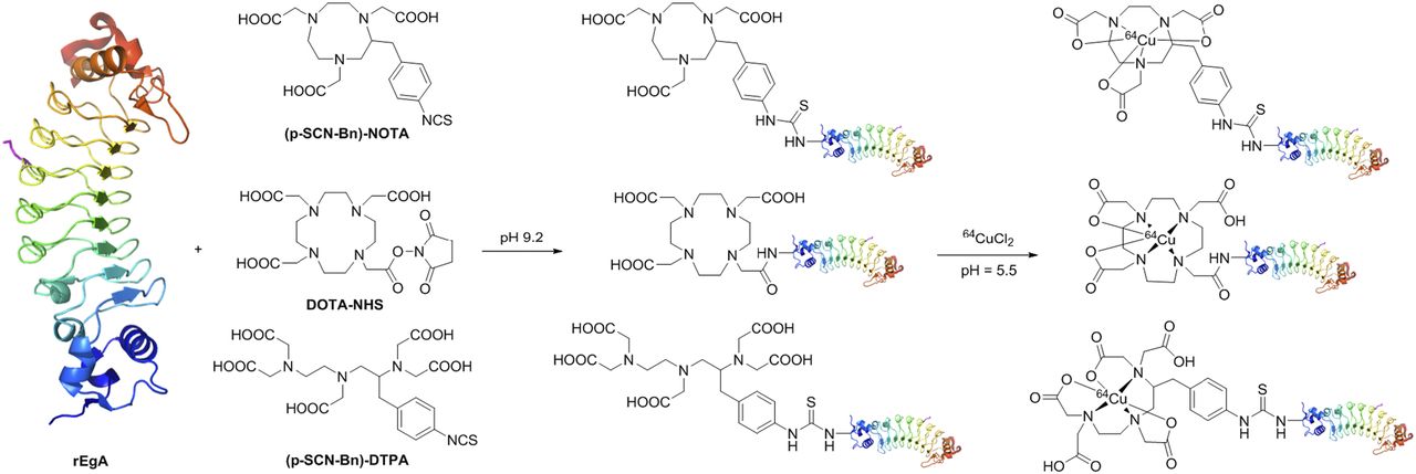
The scheme for the conjugation of rEgA with BFCs and 64Cu labeling is shown in Figure 1. Conjugation of the BFCs through the N-hydroxysuccinimide ester or p-SCN-Bn linker proved highly amenable to the aqueous conditions required for solubilizing the rEgA. The results of MALDI-TOF mass spectroscopy are presented in Supplemental Figures 1–4 (supplemental materials are available at http://jnm.snmjournals.org). The overall decay-corrected radiochemical yield was approximately 70%–80%. Identification of the radioproduct through comparison of the collected high-performance liquid chromatography fraction with rEgA labeled with nonradioactive copper is shown in Supplemental Figures 5–7.

Synthesis scheme of NOTA-, DOTA-, and DTPA-conjugated rEgA and radiolabeling. NHS = N-hydroxysuccinimide.
When the 64Cu-rEgA variants were incubated in human serum at 37°C for 48 h, the amount remaining (Rf, 0.05–0.1) was greater than 95%, indicating a relatively high in vitro stability (Fig. 2A). Furthermore, no metabolites were detected in the serum of mice 6 h after intravenous injection of 64Cu-rEgA variants (64Cu-NOTA-rEgA: 97.12 ± 2.16; 64Cu-DOTA-rEgA: 97.05 ± 1.22; 64Cu-DTPA-rEgA: 98.45 ± 0.51 [n = 9]) (Fig. 2B).
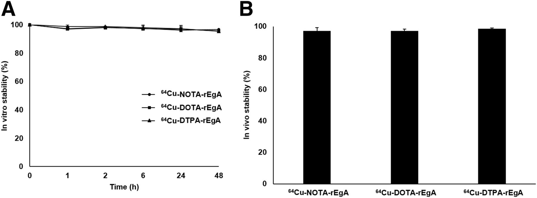
In vitro serum stability (A) and in vivo stability (B) of 64Cu-labeled NOTA-, DOTA-, and DTPA-rEgA. In vitro and in vivo stability is expressed as mean ± SD.
Optimized BFC Number and Cellular Uptake
To optimize EGFR affinity and biocompatibility, various ratios of (p-SCN-Bn)-NOTA to rEgA were used in the conjugation reaction, ranging from 0.2:1 stoichiometry to 50:1. When the 0.2:1 equivalent of NOTA was used, the average number of NOTA groups per rEgA was 0.75 ± 0.18, excluding nonconjugated rEgA. The average numbers of NOTA groups after conjugation with the 1:1, 10:1, and 50:1 ratios were 0.58 ± 0.38, 4.97 ± 0.21, and 12.1 ± 2.31, respectively. Subsequently, rEgA with various numbers of NOTA groups were radiolabeled and used in cellular uptake assays in the H1650 cell line (EGFR-positive) (22,23). The highest accumulation in the H1650 cell line after 6 h of treatment (51.84% ± 6.01%) was achieved when rEgA was conjugated with (p-SCN-Bn)-NOTA at the 0.2 equivalent (Supplemental Fig. 8). Therefore, we adopted the 0.2 ratio of BFC for chelation of rEgA, and we prepared 64Cu-NOTA-rEgA, 64Cu-DOTA-rEgA (DOTA groups per rEgA: 0.66 ± 0.15), and 64Cu-DTPA-rEgA (DTPA groups per rEgA: 0.69 ± 0.19) for in vitro and in vivo application. Furthermore, enzyme-linked immunosorbent assay revealed that the binding affinity of each of the 3 types of natCu-rEgA for the EGFR was similar to that of native rEgA (Supplemental Fig. 9).
Cellular uptake values for 64Cu-rEgA in H1650 cells and SW620 cells (EGFR-negative) (24,25) over incubation periods of 1, 2, and 6 h are shown in Figure 3. Importantly, specific accumulation was observed with the 64Cu-labeled rEgA conjugated to each chelator. The uptake of 64Cu-labeled NOTA-, DOTA-, and DTPA-rEgA increased from 5.21% ± 0.10%, 15.18% ± 1.97%, and 19.50% ± 7.69%, respectively, at 1 h to 50.82% ± 4.23%, 45.76% ± 1.08%, and 59.34% ± 8.17%, respectively, at 6 h. Furthermore, the uptake of each 64Cu-rEgA was more than 9-fold higher in H1650 cells than in SW620 cells at 6 h (P < 0.05). Specifically, the uptake of 64Cu-NOTA-rEgA was over 14-fold higher in H1650 cells than in SW620 cells (50.82% ± 4.23% vs. 3.61% ± 0.50%, P < 0.05). Overall, these results suggest that the specificity of the rEgA for EGFR was well maintained after chelator-based radiolabeling.

Cellular uptake of 64Cu-NOTA-rEgA (A), 64Cu-DOTA-rEgA (B), and 64Cu-DTPA-rEgA (C) in SW620 and H1650 cells. All values are expressed as mean ± SD per 106 cells. *P < 0.05 (Mann–Whitney U test).
Biodistribution Studies and Small-Animal PET Imaging
The biodistribution of the 3 types of 64Cu-rEgA is shown in Table 1. All exhibited rapid, high EGFR binding and prolonged tumor retention. The tumor uptake of 64Cu-labeled NOTA-, DOTA-, and DTPA-rEgA (expressed as the percentage injected dose per gram of tissue [%ID/g]) was 8.89 ± 1.04, 7.58 ± 0.31, and 6.97 ± 1.50, respectively, 1 h after intravenous injection. All types of 64Cu-rEgA showed the highest tumor uptake at 6 h after injection (NOTA, 14.47 ± 1.68 %ID/g; DOTA, 14.33 ± 0.98 %ID/g; and DTPA, 13.28 ± 2.22). Even at 24 h after injection, the distribution of each type of 64Cu-rEgA in the tumor was approximately 10.0%. High and rapid tumor uptake and prolonged retention coupled with rapid clearance and low nonspecific binding in normal organs resulted in high tumor-to-blood ratios (NOTA, 5.00 ± 0.77 %ID/g; DOTA, 4.25 ± 2.12 %ID/g; and DTPA, 6.01 ± 2.57 %ID/g at 6 h after injection) and high tumor–to–normal-muscle ratios (NOTA, 16.38 ± 1.79 %ID/g; DOTA, 13.35 ± 2.32 %ID/g; and DTPA, 13.95 ± 6.22 %ID/g at 6 h after injection) (Table 2). Furthermore, although hepatic uptake of DTPA was higher than that of NOTA and DOTA, there was no statistically significant difference among the 3 groups according to the results of the Mann–Whitney U test. Thus, hepatic uptake of 64Cu-rEgA variants may be related to the biodistribution properties of rEgA (26).
Biodistribution of 64Cu-NOTA-rEgA, 64Cu-DOTA-rEgA, and 64Cu-DTPA-rEgA in H1650 Tumor–Bearing Nude Mice at 1, 6, and 24 Hours After Injection
Comparison of Tumor-to-Blood and Tumor-to-Muscle Ratios at 1, 6, and 24 Hours After Administration of 64Cu-NOTA-rEgA, 64Cu-DOTA-rEgA, or 64Cu-DTPA-rEgA to H1650 Tumor–Bearing Mice
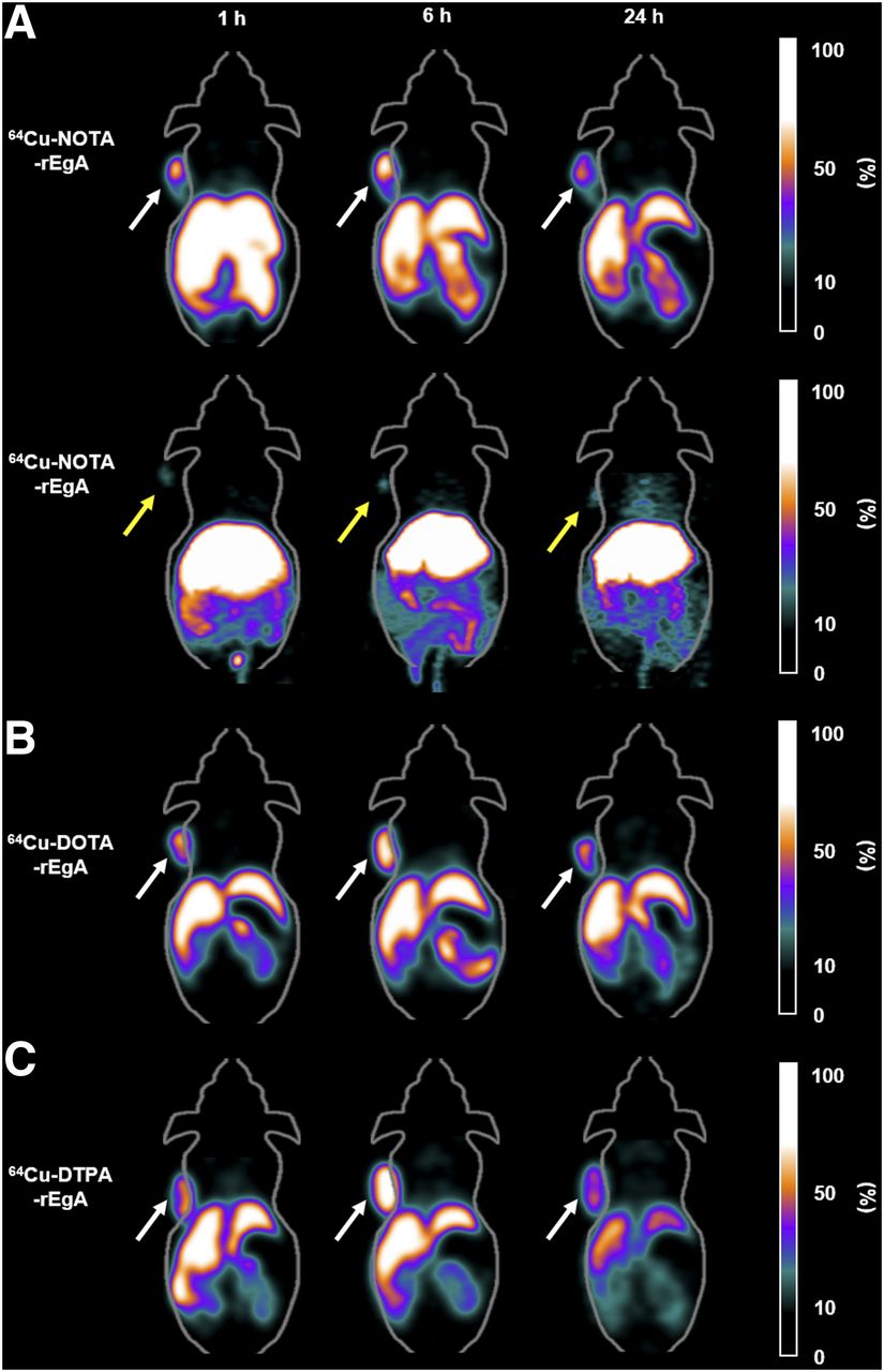
Static PET images of H1650 or SW620 tumor–bearing mice at 1, 6, and 24 h after intravenous injection of the 64Cu-rEgA are shown in Figure 4. The EGFR-expressing H1650 tumors could be clearly visualized at 1 h after injection, and tumor uptake was retained for 24 h. However, tumor uptake of 64Cu-rEgA was the highest at 6 h after intravenous injection. To verify the specificity of the 64Cu-rEgA for EGFR-expressing tumors, H1650 tumor–bearing mice were injected with cold rEgA (50 μM), and a small-animal PET study was performed with 64Cu-NOTA-rEgA after 24 h. At 6 h, tumor uptake decreased from 100% without blocking to 39%, indicating the specificity of rEgA binding in vivo. Furthermore, free 64Cu showed very low uptake in H1650 tumor–bearing mice, indicating that 64Cu-rEgA has high specificity for the EGFR (Fig. 5). The quantified tumor, blood, and muscle uptake of the 64Cu-rEgA are shown in Supplemental Figure 10, and the tumor-to-blood and -muscle ratios are compared in Figure 6. The 3 64Cu-rEgA cleared rapidly from the blood, with less than 2 %ID/g remaining 1 h after injection. Small-animal PET images obtained with 64Cu-labeled NOTA-, DOTA-, and DTPA-rEgA exhibited high tumor-to-blood (>5 at 6 h after injection) and tumor-to-muscle (>10 at 6 h after injection) ratios, as was consistent with the biodistribution results.

Coronal small-animal PET images of nude mice bearing H1650 (white arrows) or SW620 (yellow arrows) tumors at 1, 6, and 24 h after tail veil injection of 64Cu-labeled NOTA-, DOTA-, or DTPA-rEgA (each at 7.4 MBq).

Coronal small-animal PET images of H1650 tumor–bearing nude mice (arrows) that were pretreated with phosphate-buffered saline (A) or 50 μM cold rEgA (B) 24 h before 64Cu-NOTA-rEgA administration and coronal small-animal PET images of free 64Cu in H1650 tumor–bearing nude mice. Images at 1, 6, and 24 h are shown.

Comparison of tumor-to-blood (A) and tumor-to-muscle (B) ratios among 64Cu-NOTA-rEgA, 64Cu-DOTA-rEgA, and 64Cu-DTPA-rEgA.
DISCUSSION
In this study, we generated 3 versions of 64Cu-rEgA, which is a small (28.3 kDa) protein scaffold with high affinity and rapid penetration, to determine their utility as PET imaging agents for EGFR-expressing tumors. These molecules were found to be appropriate for the quantification of EGFR expression using PET. Furthermore, the present strategy can be widely applied to target different receptors for the detection of other cancers. Finally, as a molecular probe, the repebody scaffold has advantages over other formats because it is easy to engineer and highly stable over a wide range of pH and temperature values (18).
To synthesize PET molecular probes with rEgA, we chose 64Cu because of its favorable decay characteristics (half-life, 12.7 h; β+, 7.8%; β−, 38.4%). 64Cu was produced with a high yield and high specificity by the 64Ni(p,n)64Cu nuclear reaction using an MC50 cyclotron (Korea Institute of Radiologic and Medical Sciences). The rEgA was then 64Cu-labeled through BFCs (NOTA, DOTA, and DTPA). Furthermore, the longer half-life of 64Cu among positron emitters allows for imaging at later time points than 18F (half-life, 109.7 min) and is more appropriate for imaging of protein binders (27). Two conjugation strategies (DOTA via the N-hydroxysuccinimide ester; NOTA and DTPA via the p-SCN-Bn linker group) were used, and the BFC-conjugated rEgA variants were efficiently radiolabeled with 64Cu.
Our cellular and animal experiments indicate that the rEgA is a potential molecular binder for the development of imaging probes to evaluate the presence and quantity of targets in tumor tissue. The cellular uptake study demonstrated that the specificity of the rEgA was not influenced by chelation with NOTA, DOTA, or DTPA. In vivo biodistribution studies showed a rapid accumulation of activity in the tumor, with stable retention for at least 24 h. Low uptake in blood and normal muscle resulted in high tumor-to-blood and tumor-to-muscle ratios. The biodistribution results were confirmed by small-animal PET imaging. EGFR specificity was also confirmed by a blocking study using nonlabeled rEgA. Although the biodistribution results showed a trend similar to small-animal PET data, the absolute values for tissue uptake obtained in the biodistribution study were higher. This discrepancy might have resulted from the way organ weight is calculated in small-animal PET. Although, in the ex vivo biodistribution studies, the weight of each organ was precisely measured using an electric balance, tissue weight was estimated in small-animal PET from the average weight of a single voxel, which is calculated from the whole-body weight divided by the total activity of voxels. Because whole-body weight includes heavy components such as bone in addition to soft tissue such as tumor, tissue weight can be overestimated in PET imaging, potentially explaining why the %ID/g of PET imaging was lower than that of the ex vivo biodistribution study.
Development of imaging probes for visualization of EGFR expression has been reported, including a 111In-labeled murine antibody (clone 225), 64Cu-labeled peptides, and a 64Cu-labeled chimeric cetuximab molecule for SPECT or PET imaging (28–31). Furthermore, in terms of in vivo performance, cetuximab variants labeled with 89Zr (78.41 h) or 177Lu (6.65 d) have a longer physical half-life than the 64Cu-labeled molecule (28,32,33). However, EGFR expression with these radiolabeled antibodies could not be visualized in various solid tumors until after 24 h, because of low clearance and tissue penetration. In the present studies, EGFR expression could be visualized 1 h after injection of 64Cu-rEgA, suggesting that radionuclides with short half-lives, such as 18F (109.8 min) and 68Ga (68 min), might also be useful as rEgA conjugates.
CONCLUSION
The 3 64Cu-rEgA variants were synthesized easily under mild conditions, and their in vitro and in vivo characteristics were evaluated. The rapid and prolonged retention of 64Cu-rEgA in the tumor but not the blood or muscle suggests that these molecules can be used as imaging agents to obtain high-contrast PET images of EGFR expression at tumor sites shortly after injection. Our results demonstrate that the specific nature of the rEgA for targeting was not influenced by the various BFCs. The favorable in vivo kinetics and specific tumor uptake of 64Cu-rEgA warrant its further investigation for imaging of EGFR-positive tumors. Therefore, rEgA has potential as a novel scaffold for the development of imaging agents for companion diagnosis.
DISCLOSURE
This study was supported by a grant from the Bio and Medical Technology Development Program of the National Research Foundation (NRF) of Korea (NRF-2014M3A9B5073747) and by the Pioneer Research Center Program of the NRF of Korea (2015M3C1A3056410), funded by the Ministry of Science, Communications Technology (ICT), and Future Planning. Hyeon Sik Kim was supported by the Basic Science Research Program through the NRF of Korea, funded by the Ministry of Education (NRF-2016R1D1A3B01006631), and Hee-Seung Bom was supported by the Basic Science Research Program through the NRF of Korea, funded by the Ministry of Education (NRF-2016R1D1A3B01006631). Dong-Yeon Kim was supported by the Basic Science Research Program through the NRF of Korea, funded by the Ministry of Education (17R1D1A1B03029055). No other potential conflict of interest relevant to this article was reported.
Acknowledgments
We thank Gyeongmin Kim for his excellent research assistance.
Footnotes
Published online Sep. 15, 2017.
- © 2018 by the Society of Nuclear Medicine and Molecular Imaging.
REFERENCES
- Received for publication June 11, 2017.
- Accepted for publication August 24, 2017.


