


Article Figures & Data
Figures
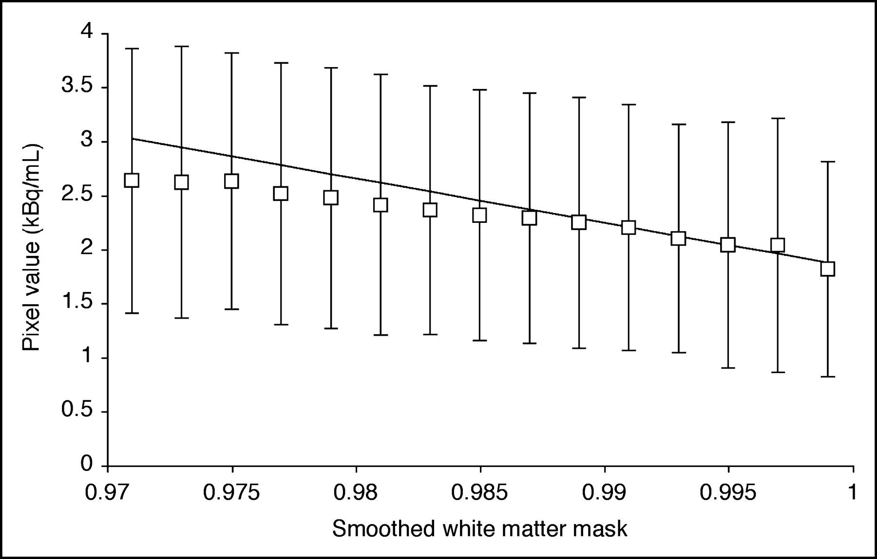
- FIGURE 1.
Estimation of white matter activity for 3S-PVC (Eq. 2) from a 11C-AA PET frame (25–30 min after injection). Activity values for pure white matter were obtained from voxels with smoothed white matter mask value (sWM) close to 1.0. Each point represents mean ± SD of activity in voxels plotted vs. smoothed white matter mask. Activity values of voxels with sWM from 0.99 to 1.0 were fitted to straight line and value at sWM = 1.0 was used as white matter value.
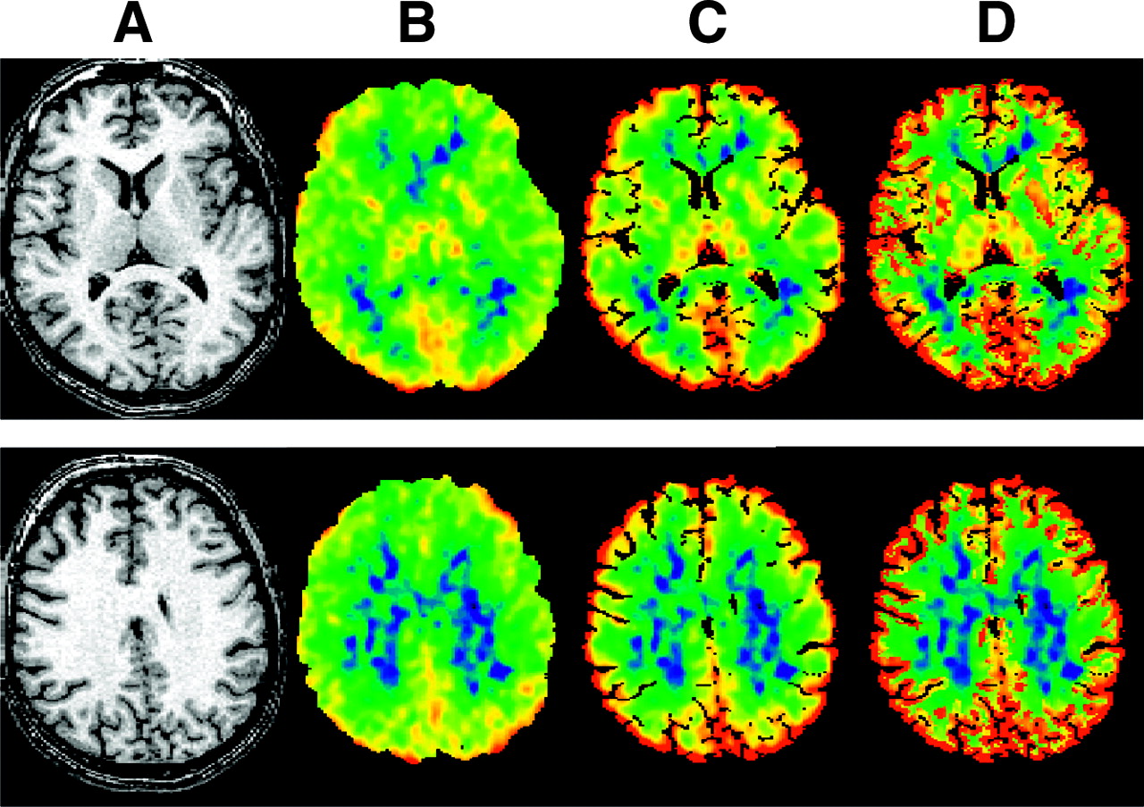
- FIGURE 2.
Transverse images derived from 1 typical subject at level of subcortical nuclei (top) and centrum semiovale (bottom). (A) MR image. (B) Original image of incorporation rate for 11C-AA (K*). (C) K* image after 2S-PVC. (D) K* image after 3S-PVC. K* images are scaled to maximum of 12 μL/min/mL.
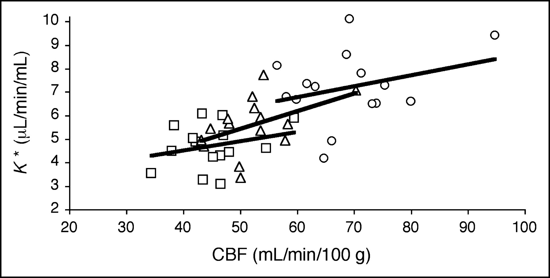
- FIGURE 3.
Relationship between CBF and 11C-AA incorporation rate (K*) before PVC (□), after 2S-PVC (▵), and after 3S-PVC (○). Each point represents global gray matter value for 1 subject (n = 15). No significant relation between K* and CBF was detected. Regression equations were as follows: Uncorrected, K* = 2.93 + 0.039 CBF (r = 0.26; P > 0.05); 2S, K* = 1.65 + 0.075 CBF (r = 0.45; P > 0.05); 3S, K* = 3.98 + 0.047 CBF (r = 0.30; P > 0.05).


Tables
Region Uncorrected 2S-PVC 3S-PVC Young Old P Young Old P Young Old P Absolute values (μL/min/mL) Frontal 4.76 (25) 4.21 (14) 0.3 5.71 (30) 5.36 (9) 0.5 7.40 (31) 6.83 (7) 0.5 Temporal 4.46 (23) 4.23 (11) 0.6 5.06 (25) 5.09 (9) 0.8 6.42 (27) 6.25 (8) 0.8 Parietal 4.79 (24) 4.40 (15) 0.5 5.91 (26) 5.77 (10) 1.0 8.06 (29) 8.07 (7) 1.0 Occipital 5.26 (26) 4.99 (15) 0.6 5.96 (28) 5.87 (10) 0.9 8.16 (29) 8.34 (9) 0.9 Caudate 4.29 (29) 3.81 (19) 0.4 4.86 (29) 4.53 (16) 0.4 5.75 (29) 5.18 (18) 0.4 Putamen 5.43 (26) 4.88 (12) 0.4 5.45 (26) 4.99 (12) 0.2 7.33 (26) 6.33 (13) 0.2 Thalamus 5.02 (26) 4.74 (13) 0.6 5.31 (26) 5.18 (11) 0.7 6.10 (27) 5.87 (11) 0.7 Cerebellum 5.41 (24) 5.16 (13) 0.6 5.93 (29) 5.74 (13) 0.5 7.75 (26) 7.16 (11) 0.5 Gray matter 4.86 (24) 4.51 (14) 0.5 5.65 (27) 5.45 (10) 0.5 7.35 (28) 7.03 (8) 0.5 Values normalized to global gray matter Frontal 0.98 (2) 0.93 (1) 0.001* 1.00 (4) 0.98 (2) 0.2 1.00 (4) 0.97 (3) 0.2 Temporal 0.92 (3) 0.95 (8) 0.3 0.90 (5) 0.93 (3) 0.1 0.88 (4) 0.89 (3) 0.4 Parietal 0.99 (2) 0.97 (4) 0.5 1.05 (5) 1.06 (2) 0.7 1.10 (5) 1.15 (3) 0.052 Occipital 1.08 (4) 1.10 (3) 0.2 1.06 (4) 1.08 (2) 0.3 1.11 (5) 1.19 (4) 0.014* Caudate 0.87 (8) 0.85 (11) 0.5 0.86 (10) 0.83 (11) 0.5 0.78 (11) 0.74 (13) 0.3 Putamen 1.12 (4) 1.09 (7) 0.4 0.97 (6) 0.92 (5) 0.046* 1.01 (8) 0.90 (7) 0.013* Thalamus 1.03 (4) 1.06 (5) 0.3 0.94 (4) 0.95 (4) 0.7 0.83 (6) 0.83 (5) 1.0 Cerebellum 1.12 (3) 1.15 (2) 0.08 1.05 (4) 1.05 (4) 0.9 1.06 (8) 1.02 (5) 0.2 ↵* Statistical significance for uncorrected P < 0.05 (unpaired t test between young and old).
Mean (% CV) for young (n = 8) and old (n = 7) healthy subjects of K* for 11C-AA in uncorrected images and after 2S-PVC and 3S-PVC.
Region Uncorrected 2S-PVC 3S-PVC Young Old P Young Old P Young Old P Absolute values (mL/mL) Frontal 0.041 (12) 0.038 (7) 0.2 0.049 (15) 0.048 (6) 0.8 0.057 (18) 0.059 (8) 0.5 Temporal 0.045 (13) 0.045 (5) 0.8 0.051 (14) 0.053 (8) 0.6 0.061 (15) 0.066 (6) 0.2 Parietal 0.040 (16) 0.039 (16) 0.8 0.048 (15) 0.051 (20) 0.6 0.059 (20) 0.067 (15) 0.2 Occipital 0.052 (9) 0.046 (12) 0.3 0.058 (7) 0.052 (11) 0.4 0.073 (8) 0.073 (9) 0.9 Caudate 0.040 (12) 0.037 (8) 0.2 0.044 (13) 0.040 (11) 0.1 0.050 (13) 0.049 (10) 0.6 Putamen 0.041 (14) 0.041 (7) 0.9 0.042 (14) 0.041 (8) 0.8 0.050 (16) 0.052 (9) 0.6 Thalamus 0.050 (14) 0.052 (14) 0.7 0.053 (14) 0.056 (13) 0.5 0.060 (17) 0.065 (14) 0.3 Cerebellum 0.052 (22) 0.051 (18) 0.9 0.057 (29) 0.058 (20) 1.0 0.069 (28) 0.069 (17) 1.0 Gray matter 0.045 (12) 0.043 (8) 0.6 0.051 (13) 0.052 (8) 0.9 0.061 (16) 0.065 (6) 0.4 Values normalized to global gray matter Frontal 0.92 (5) 0.88 (1) 0.014* 0.95 (5) 0.93 (4) 0.5 0.93 (7) 0.89 (3) 0.2 Temporal 1.02 (4) 1.04 (9) 0.6 1.00 (5) 1.03 (10) 0.5 1.00 (4) 1.03 (10) 0.5 Parietal 0.90 (8) 0.90 (10) 0.9 0.95 (9) 0.98 (12) 0.5 0.97 (9) 1.03 (10) 0.2 Occipital 1.17 (14) 1.06 (11) 0.1 1.15 (15) 1.02 (12) 0.1 1.22 (15) 1.13 (11) 0.3 Caudate 0.90 (10) 0.86 (7) 0.3 0.85 (12) 0.79 (10) 0.2 0.83 (12) 0.75 (12) 0.2 Putamen 0.94 (15) 0.89 (3) 0.9 0.82 (14) 0.80 (7) 0.7 0.82 (13) 0.80 (10) 0.7 Thalamus 1.12 (7) 1.19 (10) 0.2 1.04 (8) 1.08 (12) 0.5 0.98 (8) 1.00 (14) 0.6 Cerebellum 1.15 (13) 1.17 (13) 0.8 1.11 (18) 1.12 (17) 0.9 1.11 (16) 1.06 (13) 0.6 ↵* Statistical significance for uncorrected P < 0.05 (unpaired t test between young and old).
Mean (% CV) for young (n = 8) and old (n = 7) healthy subjects of CBV in uncorrected images and after 2S-PVC and 3S-PVC.
Region Uncorrected 2S-PVC 3S-PVC Young Old P Young Old P Young Old P Absolute values (mL/min/100 g) Frontal 49 (14) 41 (12) 0.025* 56 (15) 50 (12) 0.1 75 (16) 66 (13) 0.1 Temporal 43 (14) 39 (15) 0.2 48 (14) 46 (14) 0.5 63 (15) 59 (16) 0.4 Parietal 46 (13) 40 (13) 0.057 55 (13) 51 (13) 0.3 77 (14) 74 (17) 0.7 Occipital 49 (10) 44 (13) 0.081 55 (12) 51 (12) 0.2 77 (14) 74 (13) 0.6 Caudate 46 (17) 39 (11) 0.068 51 (17) 45 (12) 0.1 62 (17) 53 (12) 0.086 Putamen 54 (14) 49 (12) 0.2 54 (13) 49 (13) 0.2 75 (11) 66 (13) 0.059 Thalamus 54 (15) 48 (11) 0.1 57 (15) 52 (12) 0.2 66 (16) 60 (12) 0.2 Cerebellum 52 (14) 44 (10) 0.027* 57 (16) 48 (11) 0.040* 75 (19) 62 (12) 0.054 Gray matter 48 (13) 42 (12) 0.053 54 (13) 49 (12) 0.1 72 (14) 66 (13) 0.1 Values normalized to global gray matter Frontal 1.01 (3) 0.97 (2) 0.011* 1.03 (3) 1.02 (2) 0.3 1.03 (3) 1.01 (3) 0.055 Temporal 0.90 (4) 0.94 (5) 0.1 0.88 (5) 0.93 (5) 0.1 0.86 (4) 0.89 (6) 0.3 Parietal 0.96 (3) 0.95 (4) 0.8 1.01 (3) 1.03 (3) 0.2 1.06 (3) 1.13 (6) 0.028* Occipital 1.03 (5) 1.06 (5) 0.4 1.01 (5) 1.03 (4) 0.3 1.06 (4) 1.13 (7) 0.032* Caudate 0.95 (11) 0.94 (7) 0.8 0.93 (11) 0.92 (4) 0.7 0.86 (11) 0.82 (4) 0.3 Putamen 1.13 (13) 1.16 (5) 0.6 1.00 (12) 1.00 (6) 0.9 1.04 (11) 1.00 (4) 0.4 Thalamus 1.12 (9) 1.15 (7) 0.5 1.04 (7) 1.05 (6) 0.7 0.91 (7) 0.92 (8) 0.8 Cerebellum 1.09 (5) 1.07 (9) 0.6 1.04 (6) 0.99 (10) 0.2 1.05 (10) 0.92 (8) 0.3 ↵* Statistical significance for uncorrected P < 0.05 (unpaired t test between young and old).
Mean (% CV) for young (n = 8) and old (n = 7) healthy subjects of CBF in uncorrected images and after 2S-PVC and 3S-PVC.
Region K* Vb CBF Young Old P Young Old P Young Old P 2S-PVC Frontal 19 ± 9 27 ± 8 0.082 18 ± 8 24 ± 7 0.1 16 ± 7 24 ± 6 0.056 Temporal 13 ± 6 18 ± 7 0.1 13 ± 6 19 ± 4 0.041* 11 ± 6 17 ± 4 0.042* Parietal 23 ± 9 32 ± 10 0.080 21 ± 8 32 ± 9 0.038* 20 ± 8 27 ± 7 0.081 Occipital 13 ± 6 19 ± 9 0.2 13 ± 6 16 ± 7 0.4 11 ± 6 15 ± 8 0.3 Caudate 13 ± 3 18 ± 6 0.051 12 ± 3 16 ± 4 0.1 12 ± 2 16 ± 4 0.029* Putamen 0 ± 1 1 ± 1 0.1 0 ± 1 1 ± 2 0.2 0 ± 1 1 ± 1 0.13 Thalamus 6 ± 1 8 ± 2 0.018* 7 ± 2 9 ± 1 0.006* 5 ± 1 7 ± 1 0.011* Cerebellum 9 ± 9 10 ± 5 0.7 10 ± 10 14 ± 6 0.4 9 ± 14 9 ± 4 0.9 Gray matter 16 ± 6 21 ± 7 0.2 15 ± 6 20 ± 5 0.091 14 ± 6 18 ± 5 0.2 3S-PVC Frontal 54 ± 14 64 ± 19 0.2 37 ± 12 52 ± 13 0.036* 55 ± 11 63 ± 13 0.2 Temporal 43 ± 9 48 ± 11 0.3 34 ± 8 48 ± 7 0.004* 45 ± 7 49 ± 6 0.3 Parietal 67 ± 18 86 ± 22 0.1 48 ± 15 72 ± 11 0.005* 67 ± 16 85 ± 17 0.054 Occipital 54 ± 10 69 ± 17 0.1 42 ± 6 60 ± 13 0.004* 55 ± 8 68 ± 11 0.022* Caudate 34 ± 4 36 ± 11 0.6 26 ± 4 31 ± 9 0.2 36 ± 5 37 ± 9 0.8 Putamen 35 ± 6 30 ± 9 0.2 20 ± 7 26 ± 6 0.071 39 ± 7 35 ± 6 0.3 Thalamus 22 ± 5 24 ± 5 0.4 19 ± 5 26 ± 4 0.007* 22 ± 5 24 ± 3 0.4 Cerebellum 42 ± 6 40 ± 16 0.7 31 ± 9 36 ± 8 0.3 42 ± 10 40 ± 14 0.7 Gray matter 50 ± 10 57 ± 15 0.3 37 ± 9 50 ± 9 0.015* 51 ± 7 57 ± 9 0.1 ↵* Statistical significance for uncorrected P < 0.05 (unpaired t test between young and old).
Mean ± SD for young (n = 8) and old (n = 7) healthy subjects of percentage increase of K* for 11C-AA, CBF, and Vb after 2S-PVC and 3S-PVC.
In this issue


Jump to section
Related Articles
Cited By...
- Temporal Cognitive and Brain Changes in Korsakoff Syndrome
- Age-Related Change in 5-HT6 Receptor Availability in Healthy Male Volunteers Measured with 11C-GSK215083 PET
- The Synthesis and In Vivo Pharmacokinetics of Fluorinated Arachidonic Acid: Implications for Imaging Neuroinflammation
- Brain phospholipid arachidonic acid half-lives are not altered following 15 weeks of N-3 polyunsaturated fatty acid adequate or deprived diet
- Using Cerebral White Matter for Estimation of Nondisplaceable Binding of 5-HT1A Receptors in Temporal Lobe Epilepsy
- Imaging incorporation of circulating docosahexaenoic acid into the human brain using positron emission tomography
- Whole-body synthesis-secretion rates of long-chain n-3 PUFAs from circulating unesterified {alpha}-linolenic acid in unanesthetized rats
- Arachidonic Acid and the Brain
- Imaging Neuroinflammation in Alzheimer's Disease with Radiolabeled Arachidonic Acid and PET
- 5-HT1A Receptors Are Reduced in Temporal Lobe Epilepsy After Partial-Volume Correction