


Article Figures & Data
Figures
- FIGURE 1.
Paired nucleoside accumulation studies in RG2TK+ transduced cells. (A) 18F-FHBG-14C-FIAU (⊞) and 18F-FHPG-14C-FIAU (○) accumulation data (mL medium/g cells) are shown. (B) 18F-FHBG-14C-FIAU and 18F-FHPG-14C-FIAU comparisons with 3H-TdR are shown: 14C-FIAU (•), 18F-FHBG (⊞), and 18F-FHPG (□). Each point represents paired comparison of uptake results obtained from single triple-label experiment; experimental times varied between 10 and 180 min.
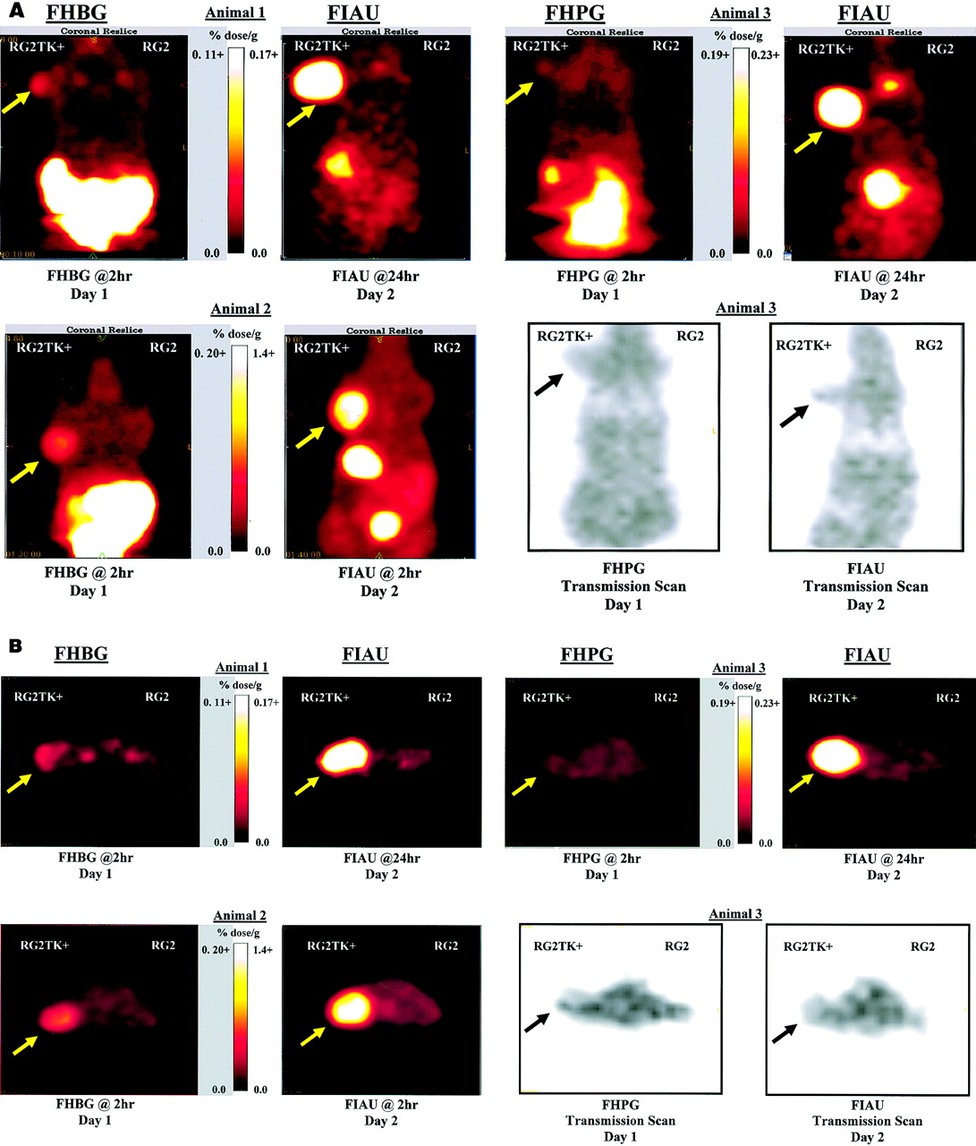
- FIGURE 2.
Paired PET imaging studies comparing 124I-FIAU and 18F-FHBG or 18F-FHPG were performed. Coronal (A) and axial (B) PET images are shown for 3 different rats. Each animal had 2 imaging studies within 24 h, and comparable image pairs are shown in each quadrant of A and B. Location of RG2TK+ xenograft is indicated by arrow. Linear scaling of image intensity was performed to achieve similar background intensity on all images; background radioactivity in thorax was 0.013 and 0.027 %dose/g for animal 1, 0.024 and 0.18 %dose/g for animal 2, and 0.23 and 0.24 %dose/g for animal 3 (FHBG or FHPG and FIAU, respectively). No radioactivity above background is visualized in control RG2 xenograft. Several tissues had substantially higher values than indicated on intensity scale. For example, FIAU levels in RG2TK+ xenografts in animals 1–3 were 0.55, 1.7, and 1.2 %dose/g, respectively. Also note high values of radioactivity in stomach (6.0 %dose/g) and bladder (3.9 %dose/g) in 2-h study (animal 2). FHBG levels in abdominal viscera (2.8–3.2 %dose/g) and bladder (11 and 80 %dose/g) were also substantial at 2 h (animals 1 and 2); similar values were measured for FHPG in abdomen (3.4 %dose/g) and bladder (63 %dose/g) of animal 3. Note that FIAU bladder radioactivity had essentially cleared by 24 h (animals 1 and 3).
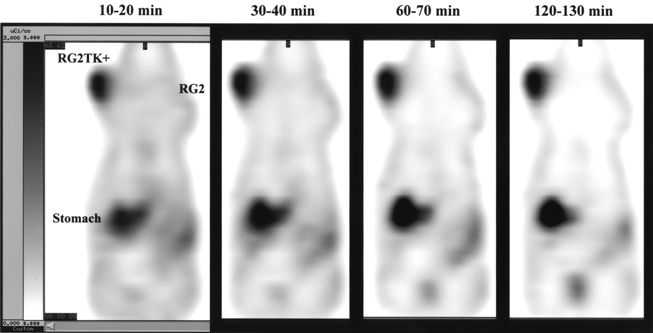
- FIGURE 3.
Comparison of 10-min imaging frames of FIAU accumulation. Ten-minute coronal image frames through RG2TK+ and RG2 xenografts are shown at various times after intravenous injection of 124I-FIAU. Image intensity scale is same for all frames.
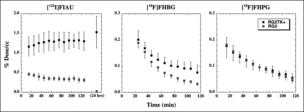
- FIGURE 4.
Radioactivity-time profiles in RG2TK+ and RG2 xenografts after intravenous injection of 124I-FIAU, 18F-FHBG, and 18F-FHPG. Values (%dose/mL) are the mean ± SD (n = 6) of maximum pixel value measured in each xenograft. FHBG and FIAU data were obtained from same set of animals imaged on consecutive days.
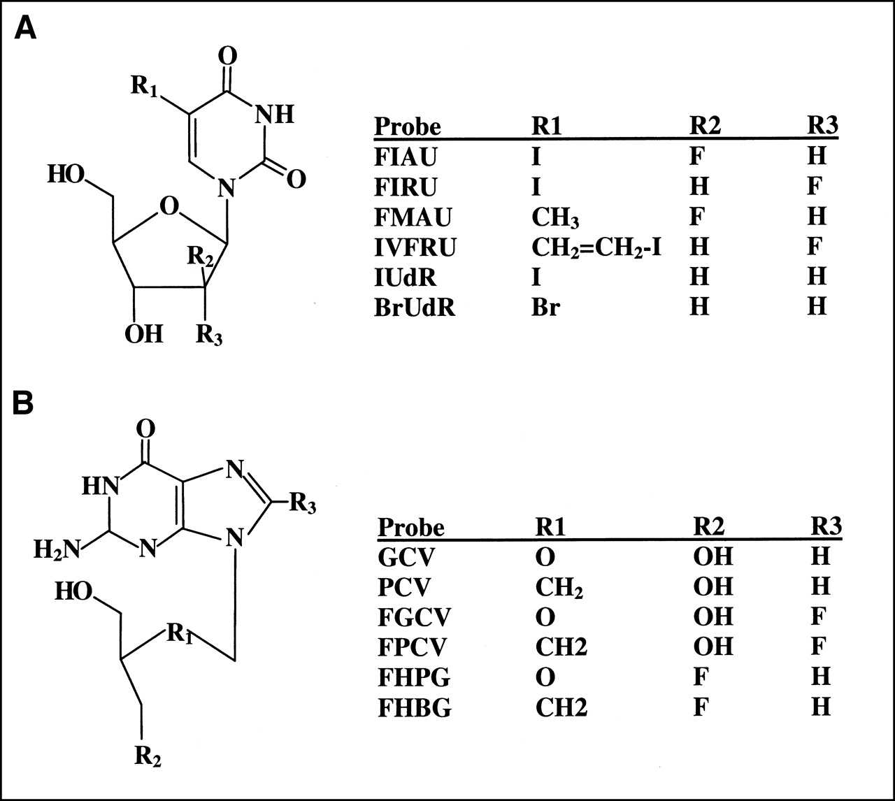
- FIGURE 5.
Chemical structures of pyrimidine (A) and acycloguanosine (B) nucleoside probes. IUdR = 2′-deoxy-5-iodo-1-β-d-ribofuranosyl-uracil; BrUdR = 2′-deoxy-5-bromo-1-β-d-ribofuranosyl-uracil.


Tables
Tissue FIAU, 2 h FHBG, 2 h* FHPG, 2 h* FIAU, 24 h† Mean ± SD n Mean ± SD n Mean ± SD n Mean ± SD n RG2TK+ 1.217 ± 0.170 12 0.074‡ ± 0.049 6 0.023‡ ± 0.008 6 1.527§ ± 0.399 11 RG2 0.112 ± 0.042 12 0.013¶ ± 0.006 6 0.018¶ ± 0.005 6 0.026‡ ± 0.015 11 Plasma 0.244 ± 0.071 12 0.010¶ ± 0.003 6 0.043¶ ± 0.032 6 0.062‡ ± 0.035 11 Muscle 0.070 ± 0.015 12 0.016¶ ± 0.007 6 0.039‖ ± 0.025 6 0.015‡ ± 0.010 11 Liver 0.083 ± 0.028 12 0.026¶ ± 0.012 6 0.038¶ ± 0.020 6 0.018‡ ± 0.009 5 Stomach 0.137 ± 0.040 12 0.014¶ ± 0.004 6 0.016¶ ± 0.007 6 0.210§ ± 0.123 5 Heart 0.073 ± 0.017 12 0.006¶ ± 0.003 6 0.012‡ ± 0.004 6 0.011‡ ± 0.007 5 Lung 0.165 ± 0.045 12 0.013¶ ± 0.008 6 0.019‡ ± 0.005 6 0.028‡ ± 0.018 5 Kidney 0.249 ± 0.124 11 0.052‖ ± 0.061 6 0.079‖ ± 0.025 5 0.026‡ ± 0.018 5 Small intestine 0.170 ± 0.035 12 0.038¶ ± 0.025 6 0.029¶ ± 0.025 6 0.076‡ ± 0.026 5 Large intestine 0.170 ± 0.029 12 0.052¶ ± 0.027 6 0.035¶ ± 0.024 6 0.056‡ ± 0.014 5 Thyroid 0.125 ± 0.039 12 0.010¶ ± 0.004 6 0.016¶ ± 0.004 6 0.022‡ ± 0.014 5 Spleen 0.145 ± 0.047 12 0.021¶ ± 0.010 6 0.026‡ ± 0.009 6 0.027‡ ± 0.010 5 Brain 0.029 ± 0.007 12 0.0009¶ ± 0.0012 6 0.0027¶ ± 0.0012 6 0.0011‡ ± 0.0007 5 ↵* Comparison of FHBG or FHPG vs. FIAU at 2 h by paired t test.
↵† Comparison of FIAU at 24 h vs. 2 h by unpaired t test.
↵‡ P < 0.0001.
↵§ P = not significant.
↵¶ 0.0001 < P < 0.0015.
↵‖ 0.005 < P < 0.02.
Probability values are nominal significance levels for univariate tests unadjusted for multiple comparisons; P < 0.0001 and 0.0001 < P < 0.0015 remain significant even with conservative Bonferroni correction.
- TABLE 2
Paired Comparisons of Probe Accumulation in HSV1-tk Transduced and Wild-Type RG2 Xenografts
Measurement 2 h* 2 h* 24 h* FIAU FHBG FIAU FHPG FIAU Xenograft difference† (RG2TK+) − (RG2) 1.12 ± 0.16 0.062 ± 0.043 1.11 ± 0.19 0.006 ± 0.010 1.50 ± 0.69 Xenograft ratio‡ RG2TK+/RG2 11.3 ± 2.6 5.9 ± 2.3 8.9 ± 2.2 1.4 ± 0.7 65 ± 21 RG2TK+/muscle 18.1 ± 3.8 4.9 ± 2.2 16.9 ± 3.1 0.77 ± 0.44 124 ± 58 ↵* Paired comparison from same animal tissue sampling dataset.
↵† Background-corrected RG2TK+ value. Difference expressed as %dose/g tissue.
↵‡ Concentration ratio.
Paired 2-sample t tests of FIAU 2 h vs. FHBG 2 h or FIAU 2 h vs. FHPG 2 h for (RG2TK+) − (RG2), RG2TK+/RG2, RG2TK+/muscle: P < 0.0001. Unpaired, 2-sample t tests of FIAU 2 h vs. 24 h for RG2TK+/RG2, RG2TK+/muscle: P < 0.0001. Unpaired, 2-sample t tests of FIAU 2 h vs. 24 h for (RG2TK+) − (RG2): P = not significant.
Radiolabeled probe Chemical name References Pyrimidine derivatives FIAU (123I, 124I, 125I, 131I) 2′-Fluoro-2′-deoxy-1-β-d-arabinofuranosyl-5-iodouracil 1–7, 22–24 FIRU (125I) 2′-Fluoro-2′-deoxy-5-iodo-1-β-d-ribofuranosyl-uracil 22, 23 FMAU (11C) 2′-Fluoro-2′-deoxy-5-methyl-1-β-d-arabinofuranosyl-uracil 25, 26 IVFRU (123I, 125I, 131I) 2′-Fluoro-2′-deoxy-5-iodovinyl-1-β-d-ribofuranosyl-uracil 23, 27, 28 IUdR (3H) 2′-Deoxy-5-iodo-1-β-d-ribofuranosyl-uracil (iododeoxyuridine) 1 Acycloguanosine derivatives ACV (8-14C, 8-3H) 9-[(2-Hydroxy-1-ethoxy)methyl]guanine (acyclovir) 11, 24 GCV (8-14C, 8-3H) 9-[(2-Hydroxy-1-(hydroxymethyl)ethoxy)methyl]guanine (ganciclovir) 1, 10, 11 PCV (8-3H) 9-[4-Hydoxy-3-(hydoxymethyl)butyl]guanine (penciclovir) 14, 15 FGCV (8-18F) 8-Fluoro-9-[(2-hydroxy-1-(hydroxymethyl)ethoxy)methyl]guanine (fluoroganciclovir) 12, 13 FPCV (8-18F) 8-Fluoro-9-[4-hydoxy-3-(hydoxymethyl)butyl]guanine (fluoropenciclovir) 14, 15 FHPG (3-18F) 9-[3-Fluoro-1-hydoxy-2-propoxymethyl]guanine 16, 17, 24, 25 FHBG (4-18F) 9-[4-Fluoro-3-(hydoxymethyl)butyl]guanine 18, 19, 29 References relate primarily to probe comparisons; because of reference number limitation, complete listing of all references was not possible.
In this issue


Jump to section
Related Articles
Cited By...
- Molecular Imaging with Reporter Genes: Has Its Promise Been Delivered?
- CRISPR-enhanced engineering of therapy-sensitive cancer cells for self-targeting of primary and metastatic tumors
- Reporter gene imaging of targeted T cell immunotherapy in recurrent glioma
- Comparative Analysis of T Cell Imaging with Human Nuclear Reporter Genes
- Molecular Imaging with Bioluminescence and PET Reveals Viral Oncolysis Kinetics and Tumor Viability
- Evaluation of Prostate-Specific Membrane Antigen as an Imaging Reporter
- Imaging Expression of the Human Somatostatin Receptor Subtype-2 Reporter Gene with 68Ga-DOTATOC
- Development of a Universal Anti-Polyethylene Glycol Reporter Gene for Noninvasive Imaging of PEGylated Probes
- Titration of Variant HSV1-tk Gene Expression to Determine the Sensitivity of 18F-FHBG PET Imaging in a Prostate Tumor
- Engineered Antibody Fragments with Infinite Affinity as Reporter Genes for PET Imaging
- Escherichia coli Nissle 1917 Facilitates Tumor Detection by Positron Emission Tomography and Optical Imaging
- Imaging of HSV-tk Reporter Gene Expression: Comparison Between [18F]FEAU, [18F]FFEAU, and Other Imaging Probes
- Multimodality Molecular Imaging with Combined Optical and SPECT/PET Modalities
- A Human-Derived Reporter Gene for Noninvasive Imaging in Humans: Mitochondrial Thymidine Kinase Type 2
- Positron Emission Tomography of Herpes Simplex Virus 1 Oncolysis
- Virus-Associated Tumor Imaging by Induction of Viral Gene Expression
- Imaging Stem Cells Implanted in Infarcted Myocardium
- Tumor-Specific In Vivo Transfection with HSV-1 Thymidine Kinase Gene Using a Sindbis Viral Vector as a Basis for Prodrug Ganciclovir Activation and PET
- Molecular Imaging with 123I-FIAU, 18F-FUdR, 18F-FET, and 18F-FDG for Monitoring Herpes Simplex Virus Type 1 Thymidine Kinase and Ganciclovir Prodrug Activation Gene Therapy of Cancer
- Preclinical Safety Evaluation of 18F-FHBG: A PET Reporter Probe for Imaging Herpes Simplex Virus Type 1 Thymidine Kinase (HSV1-tk) or Mutant HSV1-sr39tk's Expression
- Imaging Chemically Modified Adenovirus for Targeting Tumors Expressing Integrin {alpha}v{beta}3 in Living Mice with Mutant Herpes Simplex Virus Type 1 Thymidine Kinase PET Reporter Gene
- The Human Norepinephrine Transporter in Combination with 11C-m-Hydroxyephedrine as a Reporter Gene/Reporter Probe for PET of Gene Therapy
- Mesenchymal Stem Cell Targeting of Microscopic Tumors and Tumor Stroma Development Monitored by Noninvasive In vivo Positron Emission Tomography Imaging
- Evaluation of 76Br-FBAU as a PET Reporter Probe for HSV1-tk Gene Expression Imaging Using Mouse Models of Human Glioma
- Myocardial Kinetics of Reporter Probe 124I-FIAU in Isolated Perfused Rat Hearts After In Vivo Adenoviral Transfer of Herpes Simplex Virus Type 1 Thymidine Kinase Reporter Gene
- Synthesis and Evaluation of 2'-Deoxy-2'-18F-Fluoro-5-Fluoro-1-{beta}-D-Arabinofuranosyluracil as a Potential PET Imaging Agent for Suicide Gene Expression
- PET of Cardiac Transgene Expression: Comparison of 2 Approaches Based on Herpesviral Thymidine Kinase Reporter Gene
- Quantitation of Pulmonary Transgene Expression with PET Imaging
- Imaging Tri-Fusion Multimodality Reporter Gene Expression in Living Subjects
- Noninvasive Imaging of Transgene Expression by Use of Positron Emission Tomography in a Pig Model of Myocardial Gene Transfer
- In Vivo High Resolution Three-Dimensional Imaging of Antigen-Specific Cytotoxic T-Lymphocyte Trafficking to Tumors