


Abstract
9-(4-18F-Fluoro-3-[hydroxymethyl]butyl)guanine (18F-FHBG) is a sensitive and specific PET reporter probe for imaging the PET reporter genes, herpes himplex 1 thymidine kinase (HSV1-tk) and its mutant HSV1-sr39tk. 18F-FHBG has suitable pharmacokinetics and dosimetry for clinical applications and imaging of HSV1-TK has been demonstrated in the livers of hepatocellular cancer patients. Methods: Male and female Sprague–Dawley rats and New Zealand White rabbits were divided into equal groups receiving either 14 μg/kg cold FHBG or carrier solution, for a 14-d acute toxicity assessment. We monitored body weight, food and water consumption, body temperature, cardiovascular electrical and functional indices, respiratory performance and oxygen saturation, comprehensive blood chemistry, complete blood count (CBC), and urinalysis. We conducted daily cage-side examinations for the detection of any clinical abnormalities. Tissues of the animals that were euthanized and necropsied on day 14 were prepared for histopathologic examination. Results: No significant differences in cardiovascular and respiratory parameters, food consumption, body weight, urine components, or clinical signs attributable to test article toxicity were observed between the treatment and control groups. Any differences noted in the blood chemistry and CBC parameters were deemed to be incidental findings unrelated to the administration of the FHBG. Conclusion: Acute toxicity evaluation of FHBG at 100 times the expected human dose does not indicate harm to organ function or tissues. The Food and Drug Administration has approved FHBG as an Investigational New Drug.
Noninvasive imaging reporter genes are useful for many medical and biologic research applications (1,2). PET reporter genes (PRG) and probes (PRP) offer potential for long-term imaging of therapeutic transgenes and cells in patients (3,4). PRG/PRP systems provide a sensitive and quantitative imaging technique and the trace amount of probes administered lessens the possibility of pharmacologic effects. Herpes simplex virus type 1 thymidine kinase (HSV1-tk) has been studied extensively and satisfies several characteristics of an ideal PRG (5). HSV1-tk gene expression can be imaged with pyrimidine nucleoside analogs, such as 5-124I-iodo-2′-fluoro-2′-deoxy-1-β-d-arabino-5-iodouracil (124I-FIAU) (6) or acycloguanosine derivatives, such as the side-chain radiolabeled analog of penciclovir 9-(4-18F-fluoro-3-[hydroxymethyl]butyl)guanine (18F-FHBG) (7).
Our aim was to use 18F-FHBG as an investigational new PET tracer for the detection of HSV1-tk or HSV1-sr39tk (8) gene expression in approved clinical trials. Since its original synthesis and evaluation (cell culture, mice, and monkeys) (9,10), several improved protocols for synthesis and purification of 18F-FHBG have been described (11–14). Furthermore, application of 18F-FHBG for imaging therapeutic transgenes in small animals has been investigated (15–18). In comparison with other 18F-labeled acycloguanosine analogs, 18F-FHBG is a better substrate for both HSV1-TK and HSV1-sr39TK (note: tk implies the gene and TK implies the enzyme protein) (19). Compared with 124I-FIAU, 18F-FHBG's sensitivity depends on the mode of HSV1-tk delivery into the cells. Retrovirally transduced RG2 rat glioma cells (RG2-HSV1-tk), stable transfected C6 rat glioma cells, and RG2-HSV1-tk subcutaneous tumors accumulate greater amounts of FIAU (19,20). However, FHBG accumulation is significantly higher in HSV1-tk adenovirus transduced (>5 × 10⁶ plaque-forming units) or HSV1-sr39tk–expressing (regardless of gene delivery technique) C6 cells (19). Furthermore, 18F-FHBG entrapment is less than that of 124I-FIAU in control cells because of less efficient phosphorylation of 18F-FHBG by mammalian TK (19). This characteristic improves 18F-FHBG's signal-to-noise ratio and may decrease the chance of toxicity in nontarget cells. Preliminary trials on normal human volunteers have demonstrated favorable pharmacokinetics, dosimetry, and safety of 18F-FHBG (11) and it has detected HSV1-TK in the livers of patients enrolled in a suicide gene therapy trial in Spain (21). Therefore, 18F-FHBG is a suitable investigational tracer for imaging HSV1-tk or HSV1-sr39tk in patients. Our preclinical safety studies supported the Food and Drug Administration (FDA) Investigational New Drug approval (IND #61,880) that allows 18F-FHBG clinical trials in the United States.
An Appendix to this article contains a supplemental Methods section, additional tables, and additional figures and is available online only at http://jnm.snmjournals.org.
MATERIALS AND METHODS
Study Subjects and Test Article
Thirty male and female Sprague–Dawley rats and 6 male and female New Zealand White rabbits (Charles River Laboratories) were used to evaluate the acute toxicity of FHBG for 14 d. UCLA Animal Research Committee approved all study protocols. All studies, performed according to good laboratory practice (GLP) regulations, were inspected and audited by an independent Quality Assurance Unit. FHBG, a generous gift from Dr. Jae Min Jeong (Seoul National University, Seoul, Korea), was reanalyzed by hydrogen NMR and further purified by high-performance liquid chromatography. Sterile purified FHBG was dissolved in 0.01 mol/L NaOH (concentration = 0.32 μg/μL), aliquoted, and stored at −20°C. The carrier solution was a sterile solution of 50 mmol/L ammonium acetate/ethanol (93:7) made isotonic by addition of NaCl (180 mmol/L final concentration).
Dosage, Study Groups, and Acute Toxicity Monitoring
The dose of test article (cold FHBG) examined for acute toxicity, 14 μg/kg, was equivalent to 100× mass of the maximum tracer dose expected for human injection. To calculate this dose we assumed a specific activity of 1.48 × 10¹⁰ Bq/μmol and a tracer-injected dose of 555 MBq. The rats were divided into 6 groups of 5 males and 5 females. Groups 2, 4, and 6 were tail-vein injected with 14 μg/kg of FHBG dissolved in 0.01 mol/L NaOH in 1 mL carrier solution. Groups 1, 3, and 5 were control animals, injected an equivalent amount of 0.01 mol/L NaOH in carrier solution. Test article injection day was day 0. Groups 1 and 2 were monitored for 1 d, groups 3 and 4 for 7 d, and groups 5 and 6 for 14 d after test article injection. Schedules and types of analysis for each group are listed in Table 1. In addition, all rats were observed once daily for any abnormal clinical signs (e.g., depression, lethargy, skin lesions, hair loss, stool irregularities, and so forth). Food and water consumption of groups 3 and 4 was monitored from day −7 through day +7. Body temperature (measured rectally, using either a Becton Dickenson digital clinical thermometer or a digital thermistor probe) was recorded on days −1, 0 (before and after test article injection), 1, 7 (groups 3–6), and 14 (groups 5 and 6).
Procedure Schedule for Rats
Rabbits were divided into 2 groups of 3 males and 3 females. Fourteen micrograms per kilogram of FHBG, added to 1 mL carrier solution, were injected into the ear vein of 1 group of rabbits on day 0, and an equivalent volume of 0.01 mol/L NaOH, added to 1 mL carrier solution, was injected into the ear vein of another (control) group of rabbits on day 0. Body temperature was recorded just before test article injection and at 10 and 60 min after test article injection. Body temperature was also recorded on day 7. Rectal body temperatures were recorded using a BAT-12 precision digital thermometer equipped with an OT-1 thermocouple Copper-Constantan sensor probe (Physitemp Instruments). All rabbits were observed for the presence of any clinical abnormalities, from the day of arrival in quarantine until euthanasia on day 14. After necropsy, tissues were prepared for histopathologic examination. Rabbits were weighed on the day of arrival, day −7, day −1, day 1, day 7, and day 14. On days −1, 1, and 14, rabbits underwent a femoral cut-down surgery to measure their blood pressure (BP) directly from the femoral artery. Electrocardiogram (ECG), oxygen saturation of hemoglobin ([by pulse oximetry), respiratory rate, and heart rates (HR) were measured and recorded just before the surgeries on those days. Also, before anesthetizing the rabbits for surgery on days −1, 1, and 14, approximately 3 mL of venous blood were collected from the ear artery for blood chemistry and hematology analysis. Urine was collected for analysis as described in the Methods section (online Appendix) on days −1, 1, and 14. Food and water consumption was monitored from day −7 to day 14.
Data Analysis
Rat Blood Chemistry and Hematology.
We analyzed these data by averaging the measurements from each group and sex. The averages were compared between group 1 day−1 (G1D−1) and group 1 day 1 (G1D1), G1D1 and group 2 day 1 (G2D1), group 3 day 7(G3D7) and group 4 day 7 (G4D7), and group 5 day 14 (G5D14) and group 6 day 14 (G6D14) to determine if there were any significant differences (P < 0.05; t test). In this manner one can discover the immediate effects of the carrier solution as well as the effects of FHBG on days 1, 7, and 14.
Rabbit Blood Chemistry and Hematology.
Because baseline measurements were available from both control and FHBG-treated rabbits on day −1, we decided to calculate the differences between the measurements on day 1 and day −1 and day 14 and day −1 to evaluate the potential changes caused by the test article in each group. This was done by subtracting the measurement of each blood parameter at day 1 or day 14 by the measurement at day −1. Then the changes in FHBG-treated groups were compared with the changes in the control groups.
RESULTS
Blood Chemistry and Hematology
Rats.
Tables 1A and 2A (online Appendix) detail the results of statistical analysis. The day after carrier solution injection, significant decreases occurred in aspartate aminotransferase (AST) (P < 0.02) and creatine kinase (CK) (P < 0.001) concentrations and significant increases occurred in lipase (P < 0.005), calcium (P < 0.001), and chloride (P < 0.01) concentrations as well as the Na/K ratio (P < 0.05) of control males. In control females, CK concentration (P < 0.005) decreased, whereas lipase (P < 0.02), calcium (P < 0.002), chloride (P < 0.02), sodium (P < 0.001), creatinine (P < 0.005), globulin (P < 0.05), and total protein (TP) (P < 0.02) concentrations increased. In both control males and females, red blood cell (RBC) (P < 0.02 and 0.01, respectively) and HGB (P < 0.05 and 0.005, respectively) concentrations as well as percentage hematocrit (Hct) (P < 0.01 and 0.005, respectively) decreased and a moderate polychromasia (P < 0.005) was observed. However, a significant decrease (P < 0.001) in percentage eosinophil (percentage of white blood cells [WBC]) and absolute eosinophil counts was observed only in control females.
There were also significant differences in some chemistry panel measures between control and FHBG-administered rats. Only alkaline phosphatase (AP) was significantly higher in FHBG males on day 1 (P < 0.01), but it was significantly lower on day 14 (P < 0.005). On day 7, both calcium and phosphorus concentrations (P < 0.05) were slightly lower in FHBG males. The calcium and phosphorus levels in FHBG males were closer to baseline than the levels in control males on day 7. On day 14, blood urea nitrogen (BUN) (P < 0.005) and chloride (P < 0.02) concentrations as well as the BUN/creatinine ratio (P < 0.02) were significantly lower in FHBG males, but these measurements were closer to baseline than those in control males. No significant differences existed between any of the blood chemistry measurements of FHBG and control females on day 1. On day 7, amylase (P < 0.005), albumin (P < 0.02), and TP (P < 0.05) concentrations were significantly lower in FHBG females, but again these measurements were closer to baseline than the measurements from female control rats. On day 14, blood samples from control females had slightly greater concentrations of AP (P < 0.05), AST (P < 0.05), and CK (P < 0.05), but lipase (P < 0.005) and calcium (P < 0.05) concentrations were significantly greater in FHBG females.
We observed a few significant differences between the complete blood count (CBC) results of control versus FHBG rats after test article administration. On day 1 (P < 0.05 and 0.05, respectively), Hct and HGB, respectively, were higher in FHBG males, but on day 7 (P < 0.002 and 0.05, respectively), they were higher in control males. On day 1, moderate and slight polychromasia, respectively, were observed in control and FHBG males. On day 7, the percentage eosinophil of FHBG males, though equal to baseline, was significantly lower (P < 0.05) than control. Although, average RBC concentration (P < 0.01) was less in FHBG males than in control males on day 7, it was higher than the average RBC of both control and FHBG males on day 1. The mean corpuscular hemoglobin concentration (MCHC) (P < 0.01) was slightly greater in FHBG males on day 7. On day 14, WBC (P < 0.02), absolute neutrophil (P < 0.05), and absolute lymphocyte (P < 0.02) concentrations and lymphocyte (%) (P < 0.005) were higher in control males. This was not observed in females and also not before day 14 in males. The MCH (P < 0.05) and percentage of segmented neutrophil numbers (P < 0.05) were higher in FHBG relative to control males on day 14. On day 1, RBC (P < 0.02) and HGB (P < 0.05) concentrations as well as Hct (P < 0.05) and eosinophil (P < 0.05) percentages were higher in FHBG females but were closer to baseline (except for Hct) than control females. On days 7 and 14, the only significant difference was a greater percentage eosinophil (P < 0.05) and lower RBC (P < 0.05) concentration, respectively, in FHBG females.
Rabbits.
From day −1 to day 1, the CK and BUN concentration increased significantly more in control than in FHBG males (P < 0.05) and the average percentage basophil and absolute basophil concentration decreased in FHBG males but increased slightly in control males (P < 0.05). From day −1 to day 1, the only change that was significantly different between FHBG and control females was the phosphorus concentration (P < 0.005), increasing in control and decreasing negligibly in FHBG females. From day −1 to day 14, there were no significant differences between the changes in the blood chemistry parameters of FHBG and control males and females. The only significant difference was the change in absolute eosinophil concentration, increasing in FHBG females and decreasing in control females (P < 0.05).
Urinalysis
Rats.
We were able to collect urine from groups 5A and 6A (control and FHBG males), allowing for a comparison on day 14. Trace amounts of blood were found in 2 FHBG and control urine samples, most likely due to contamination during sample collection. Protein was present in the urine of all but one control. Glucose was detected in 1 control and 1 FHBG urine sample. A small amount of ketones was detected in FHBG urine samples, whereas all control samples were negative for ketones. The average specific gravity and pH values of control and FHBG urine samples were not significantly different. Bilirubin and nitrite were not found in any urine samples. One FHBG urine sample contained trace amounts of leukocytes. All FHBG and control urine samples contained 0.2 mg/dL urobilogen each.
Rabbits.
We did not observe any significant differences in any of the urinalysis parameters between the control and FHBG-treated males and females on days −1, 1, and 14.
Cardiovascular Parameters
Rats.
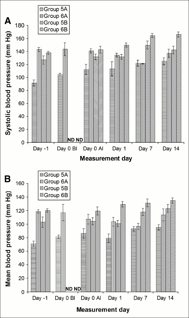
Figures 1A and 1B demonstrate the systolic and mean BP, respectively, of females and males, measured using the tail cuff method. On average, both the systolic and the mean BP of FHBG males started higher than that of control males, before the injection of test articles. However, by day 7, the BP measures converge, as the average for FHBG males decreased and that of controls increased. The BP of both FHBG and control females followed the same trend and did not converge. We also measured the BP of males, directly through the femoral artery, on day 14 and did not observe a significant difference between FHBG-treated and control animals. Furthermore, the average direct mean and systolic BP measurements are very close to those obtained using the tail cuff method (data not shown). The HR were measured using the ECG on days −1, 0 (before and after test article injection in males and only after injection in females), 1, 7, and 14 (online Appendix, Fig. 1A). There appears to be a slight downward HR trend in groups 5A (male control) and 5B (female control) from day −1 to day 14. However, the average HR of group 6A (male FHBG) and 6B (female FHBG) fluctuate slightly and do not follow either an upward or a downward trend. The experienced cardiologist, who obtained the ECG and BP measurements in all rats, certified that the results of all cardiovascular examinations were normal.

Average rat BP ± SEM before and after test article injection. Group 5A was control males, group 6A was FHBG males, group 5B was control females, and group 6B was FHBG males. n = 5 for all groups. (A) Systolic BP. Significant differences were observed between averages of FHBG and control males on day −1 (P < 0.001), day 0 before injection (P < 0.005), and day 0 after injection (P < 0.02) but not on other days. Means of FHBG and control females are significantly different on day 14 (P < 0.01) but not on any other days. (B) Mean BP. Significant differences were observed between averages of FHBG and control males on day −1 (P < 0.001), day 0 before injection (P < 0.05), and day 1 (P < 0.05) but not on other days. Averages of FHBG and control females are significantly different only on day 1 (P < 0.002). ND = not determined; BI = before injection; AI = after injection.
Rabbits.
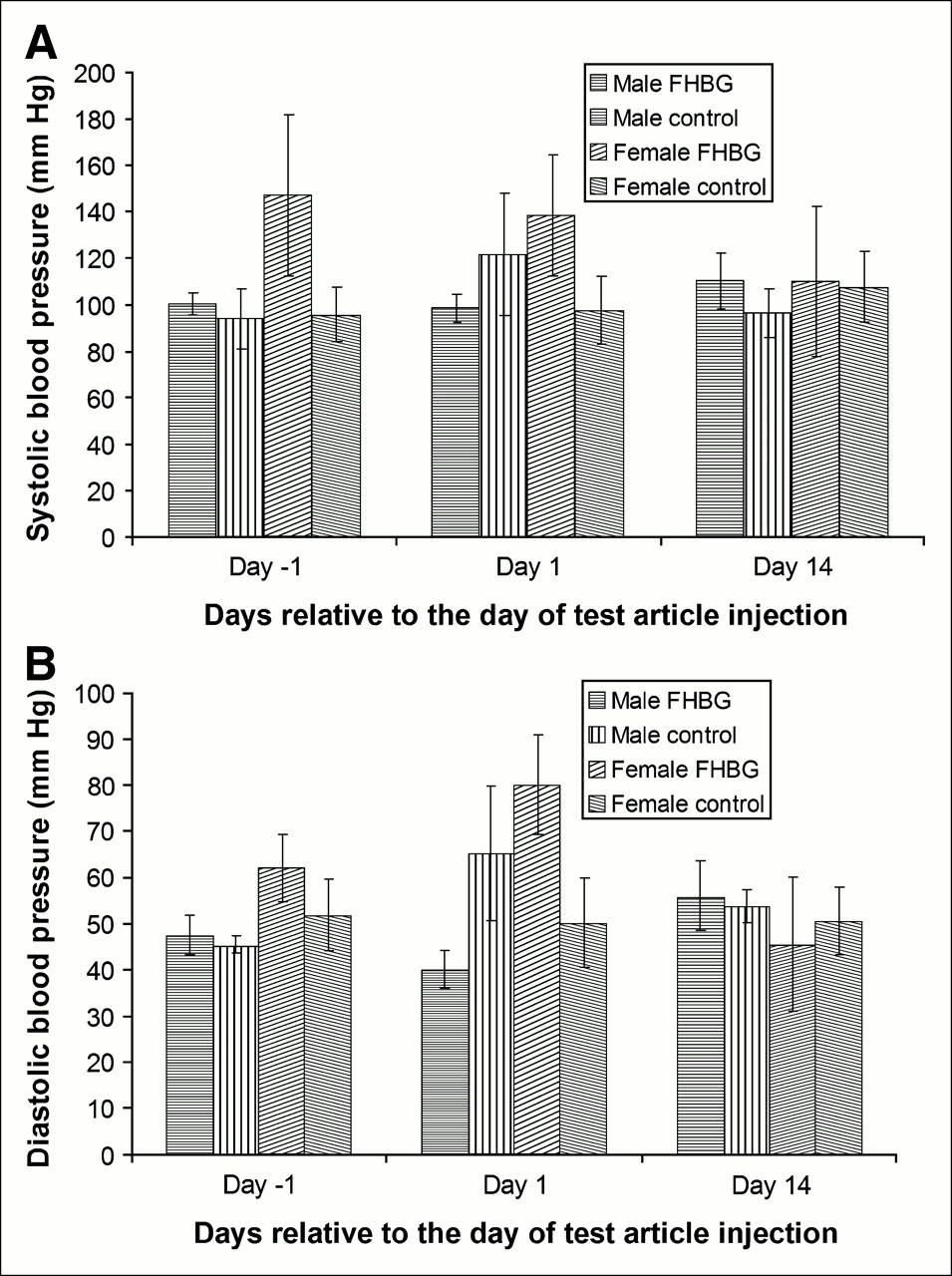
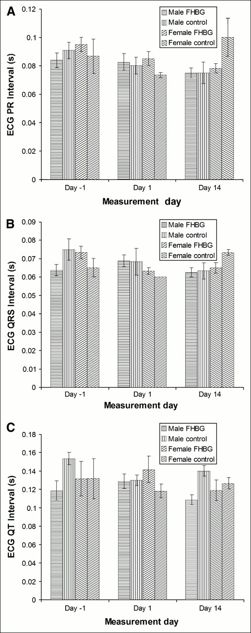
In all rabbits, the BP measurements were taken directly through femoral arterial cannulation. Figures 2A and 2B illustrate the systolic and diastolic BP of male and female rabbits on days −1, 1, and 14. There are no significant differences between the systolic and diastolic BP of both FHBG and control males and females on days −1, 1, and 14. We quantitatively analyzed the ECG recordings from each rabbit, comparing their PR intervals, QRS complexes, and QT intervals. These data are shown in Figures 3A–3C. The only significant difference was between the QT interval of FHBG and control males on days −1 (P < 0.05) and 14 (P < 0.02). However, on both days, the average QT interval of FHBG males was shorter than that of control males, indicating that FHBG did not affect the QT interval. We also recorded the HR using the ECG (online Appendix, Fig. 2A) and did not observe a significant difference between the average HR of FHBG and control males and females.

(A) Average rabbit systolic BP ± SEM on days −1, 1, and 14. (B) Average rabbit diastolic BP ± SEM on days −1, 1, and 14. No significant differences were observed between systolic or diastolic BP of FHBG and control females or males. n = 4 for FHBG males and n = 3 for all other groups.

Electrocardiograph PR interval (A), QRS complex (B), and QT interval (C) measured in seconds. Data are averages ± SEM. QT intervals of FHBG and control male rabbits are significantly different on days −1 (P < 0.05) and 14 (P < 0.02). No other significant differences were observed. n = 4 for FHBG males and n = 3 for all other groups.
Pulse Oximetry and Respiratory Rate
In both rats and rabbits, the oxygen saturation of HGB (%) and respiratory rate were measured under isoflurane anesthesia (1.5%–2.5% in oxygen) during ECG recordings. In general, the percentage oxygen saturation of both female and male rats was close to 100%. FHBG did not affect the respiratory rates of male or female rats. The blood oxygen percentages of both FHBG and control male and female rabbits were >98% (average) at all measurement times. FHBG did not affect respiratory rates of male or female rabbits.
Food and Water Consumption
Rats.
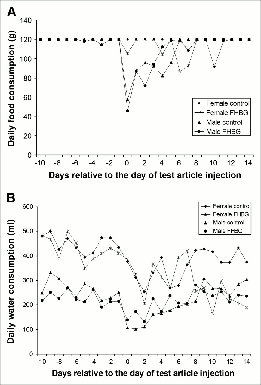
Figures 4A and 4B illustrate the food and water consumption, respectively, of both males and females, before injection of the test articles and up to 7 d after injection of the test article. The level of food and water consumption of control and FHBG rats follows similar trends.

Food (A) and water (B) consumption of rats before and after test article injections. Data are averages (n = 5).
Rabbits.
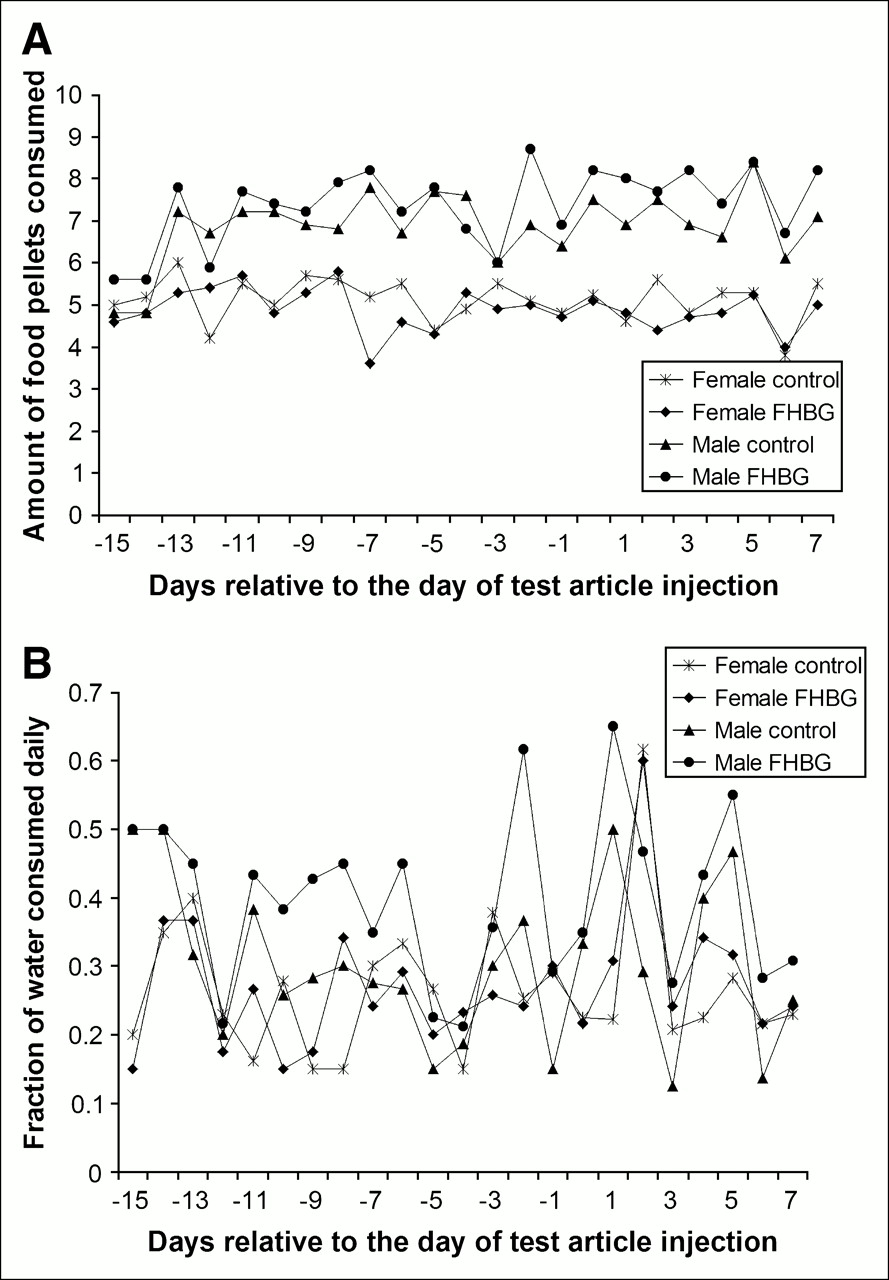
Figures 5A and 5B illustrate the average daily food and water consumption, respectively, of female and male rabbits. Food consumption seems to decline in both males and females after the surgeries, but the levels of consumption did not differ between control and FHBG- treated animals. The daily water consumption of control and FHBG-treated males followed similar trends. In the females, water consumption of FHBG-treated and control animals followed similar trends until day 8 but diverged after day 8 as FHBG-treated females reduced their consumption.

Food (A) and water (B) consumption of rabbits before and after test article injections. Data are averages (n = 4 for FHBG males and n = 3 for all other groups).
Body Weight and Temperatures
There were no significant average weight differences between FHBG and control rats or rabbits. In rats, rectal temperature was measured on day −1, day 0 before injection, day 0 within 1 h after injection, day 0 ∼1 h after injection, day 1, day 7, and day 14. The average temperature of FHBG male rats did not change significantly at other times relative to day −1. Compared with day 0 before injection, the average temperature was significantly lower on day 14 (P < 0.001). However, the average temperature on day 14 was also significantly lower within 1 h after FHBG injection (P < 0.002) or about 1 h after FHBG injection (P < 0.001). In control male rats, the average temperature on day −1 was significantly lower than that at all other times. However, compared with day 0 before injection, there was no significant change in temperatures. In FHBG-treated female rats, compared with day −1, there was a significant change less than an hour after test article injection (P < 0.002) and about 1 h after injection (P < 0.001). However, compared with average temperatures immediately before injection, there were no significant changes. In control female rats, the average temperatures were significantly higher within 1 h after injection compared with both day −1 (P < 0.02) and with day 0 before injection (P < 0.01). Also, the average temperatures on day 14 were significantly higher than those on day −1 (P < 0.05). Table 2 lists the average body temperatures of all rabbit groups before injection, <1 h after injection, ≥1 h after injection, and on day 7. With the exception of control male rabbits, whose temperatures rose significantly from before injection to <1 h after injection (P < 0.05), there were no other significant changes.
Average Rabbit Body Temperatures
Histopathology and Clinical Observations
Tables 3A and 4A (online Appendix) summarize the histopathology results and the important findings are addressed in the Discussion. No significant clinical signs were observed in any of the rats or rabbits after administration of either FHBG or the carrier solution. Animals had normal bowel movements with no evidence of diarrhea or blood in their stools. They remained alert and responsive after administration of FHBG or the carrier solution. No abnormalities of skin or adnexa were noted, except for minor hair loss in 1 of 60 rats.
DISCUSSION
FHBG is similar in chemical structure to penciclovir (PCV) and ganciclovir (GCV) and, like those antiviral drugs, can be phosphorylated by HSV1-TK and incorporated into newly synthesized DNA. Although inefficiently, these acycloguanosine analogs are also phosphorylated by mammalian thymidine kinases and carry dose-dependent side effects. On the basis of previous human dosimetry data (11), the injected dose per patient per year should not exceed 555 MBq (to keep urinary bladder wall exposure < 0.05 Sv); thus, only a trace dose of <0.14 μg/kg (specific activity = 14.8 MBq/nmol) of 18F-FHBG will be injected. This dose is about 35,000 times the daily administered dose of GCV (5 mg/kg), which is often administered over 14 d in cytomegalovirus retinitis patients (22). Furthermore, PCV is prescribed as a 1% topical cream in HSV-infected patients, exposing them to 10 mg of absorbed PCV. The specific activity of 18F-FHBG produced in our laboratory should be >37 MBq/nmol, further reducing its mass dose. Administering trace doses of 18F-FHBG should significantly reduce the chances of chemical toxicity. The possible effects of FHBG on the function of major organs, based on acute toxicity studies in rats and rabbits, will be discussed.
Central Nervous System (CNS)
FHBG was not expected to have any adverse effects on the CNS, because PET studies in humans and animals have shown that 18F-FHBG does not cross the blood–brain barrier (11). Therefore, we relied on abnormal clinical signs, such as lethargy and unusual behavior and changes in body temperature, BP, or appetite—which are not attributed to non-CNS dysfunctions—to provide clues about CNS side effects. Furthermore, histopathologic studies of the brain would provide information about anatomic changes 14 d after FHBG administration. These studies did not provide evidence indicating that, at 100× tracer doses, FHBG has any effect on the CNS of rats or rabbits.
Cardiovascular System
FHBG at 100× tracer dose did not significantly affect ECG, BP, and HR of rats and rabbits. Monitoring AST and CK, in both rats and rabbits, also did not provide evidence of an adverse effect of FHBG on the cardiovascular system. On the basis of these results and the lack of evidence of cardiac tissue damage due to FHBG, we cannot conclude that FHBG (14 μg/kg) will have an adverse effect on the cardiovascular system for up to 14 d after intravenous administration.
Respiratory System
We assessed the effect of FHBG on the function of the respiratory system by monitoring blood HGB oxygen saturation levels and the respiratory rate and on the health of lung tissue by histopathologic analysis on day 14. The results do not indicate that FHBG causes a reduction in blood oxygen levels or affects respiratory rates; thus, FHBG (14 μg/kg) may not alter the function of the respiratory system. Histopathologic analysis of rabbits found pulmonary edema, congestion, anthracosis, or nodular calcification in 5 of 6 treated rabbits. However, bronchointerstitial pneumonia, pulmonary nodular calcification, pulmonary hemorrhage, or tracheal vascular ectasia and submucosal edema were also found in 4 of 6 control rabbits. The pathologist suggested that these findings were due to endotracheal intubation and were not caused by the test article. In fact, only a single FHBG-treated female rat had pulmonary lesions, since rats did not have tracheal intubation. This rat's lesion may be due to inhalation of foreign material or antigenic stimulation. Therefore, there is no conclusive evidence that FHBG causes damage to pulmonary tissues.
Musculoskeletal
Rats.
The blood chemistry parameter AP was significantly higher on day 1 and above normal ranges in males, possibly indicating an effect on the hepatobiliary system or bones. However, this difference was absent in females and in both sexes on day 7 or day 14. The average level of phosphorus was only significantly lower in FHBG males on day 7, but without relative hypercalcemia this difference is not meaningful. Due to the lack of other evidence, we do not believe that FHBG causes bone damage. Blood chemistry monitoring and histopathologic analysis did not indicate FHBG-induced damage to the musculoskeletal system. Only lingual muscle mineralization was found in 1 of 5 FHBG females.
Rabbits.
Blood chemistry results and histopathologic analysis did not provide any evidence of FHBG-induced damage to rabbit musculoskeleton.
Hepatobiliary System
Rats.
Observation of significantly higher AP levels in FHBG males may indicate a hepatobiliary disorder. However, this was observed only on day 1 in males and was not confirmed by a significant difference in alanine aminotransferase (ALT) and γ-glutamyl transpeptidase (GGT) levels between FHBG-treated and control rats. Significant increases of average lipase occurred in both carrier solution and FHBG-treated males and females, but only on day 14 were lipase levels significantly greater in FHBG-treated versus control females. Even though lipase is a pancreatic enzyme, serum lipase levels are not an index of pancreatic function and the increase can be attributed to a carrier solution– induced diuretic effect. Other blood chemistry parameters reflective of hepatobiliary function are albumin, globulin, TP, cholesterol, glucose, calcium, and bilirubin. The serum albumin of FHBG-injected females was significantly lower than that of controls on day 7, but the values of both groups were within the normal range reported for female rats. Therefore, FHBG does not affect the hepatobiliary function parameters. Histopathologic analysis demonstrated pancreatic interstitial fibrosis in 1 of 5 FHBG-treated males, but there was bile duct hyperplasia or exocrine pancreas acinar regeneration in 3 of 5 control males. One FHBG-treated female had both bile duct hyperplasia and interstitial pancreatitis and another had only bile duct hyperplasia. Because these occurred in as many control males, these data do not suggest pancreatic damage due to FHBG at these trace doses. The pathologist did not observe liver damage in any of the rats and considered these to be incidental findings, as there was no consistent lesion pattern.
Rabbits.
We did not observe any FHBG-specific effects on AP, ALT, GGT, lipase, albumin, globulin, TP, bilirubin, cholesterol, glucose, and calcium in both females and males, on either day 1 or day 14. Histopathologic analysis in a single treated male revealed peripancreatic hemorrhage and cholangiohepatitis, which the pathologist did not attribute to FHBG treatment.
Renal Pathways
Rats.
Urinalysis did not indicate any effect of FHBG on the renal pathways. An increased blood concentration of calcium, sodium, and chloride ions in both control and FHBG-treated rats seems to indicate that the carrier solution has a short-term diuretic effect. This may also explain the increase in TP concentration on day 1. Histopathologic analysis did not indicate FHBG-induced damage to nephrons.
Rabbits.
We did not discover any evidence of FHBG-induced toxicity in the renal pathways through urinalysis and blood chemistry monitoring. We observed renal tubular mineralization in 2 of 4 FHBG-treated and 1 of 3 control males as well as in 2 of 3 control females, indicating that this effect was not FHBG specific.
Skin
All animals were monitored for skin abnormalities during daily cage-side examinations and by final histopathologic analysis. Ulcerative dermatitis was observed in a single FHBG-treated female rat. The lesion was focal and mild, likely resulting from a scratch. No skin lesions were found during clinical examination of rats or rabbits and histopathologic examination of rabbits.
Gastrointestinal
Rats.
On average, the food and water consumption trends of FHBG-treated and control rats were similar, and significant weight loss did not occur. Blood chemistry results did not provide evidence of FHBG-induced toxicity of the gastrointestinal system. We observed intestinal muscular hypertrophy in 1 of 5 FHBG-treated males and acute intercolitis in 1 of 5 FHBG-treated females.
Rabbits.
With the exception of post day-8 water consumption of FHBG-treated females, which was less than that of controls, food and water consumption averages were similar, and no significant weight loss occurred. Blood chemistry results did not provide evidence of FHBG-induced gastrointestinal toxicity. We found minimal duodenal submucosal granuloma and mild acute enteritis in 1 FHBG-treated male and moderate colonic submucosal edema in another FHBG-treated male.
Endocrine and Exocrine Glands
Rats.
Blood chemistry tests did not provide evidence of FHBG effects on the thyroid and parathyroid glands. Also, the amylase test did not indicate an effect on the salivary glands. Thyroid follicle degeneration and cystic adenitis were observed in 1 control female and thyroid follicle degeneration was observed in another control female. Histopathologic abnormalities were not observed in the glands of FHBG-treated rats.
Rabbits.
Blood chemistry tests did not provide evidence of FHBG effects on the thyroid and parathyroid glands. Also, the amylase test did not indicate an effect on the salivary glands or pancreas. We observed moderate adrenal nodular hyperplasia in 1 FHBG-treated male and no other gland-related abnormalities in other rabbits.
Sex Organs
Rats.
Uterine lumen ectasia was found in only 2 control females. Histopathology did not provide any evidence of FHBG-induced toxicity in testes, ovaries, uterus, or prostate.
Rabbits.
Mild testicular degeneration was observed in 2 FHBG-treated and 1 control male. Mild-to-moderate, chronic, and multifocal prostate epithelial hyperplasia was observed in 1 FHBG male. There was a mild ovarian mineralized nodule in 1 FHBG female, but reproductive function was not affected in females, as reflected by the large number of developing follicles within the ovaries.
Hematopoietic and Immune System
Average WBC, absolute lymphocyte, and neutrophil concentrations and lymphocyte relative percentages were significantly lower in FHBG males on day 14, but, with the exception of neutrophils, these parameters were also below baseline in control rats. It appears that a synergistic effect between the carrier solution and 100× FHBG caused an increased reduction in WBC and lymphocytes by day 14 in male rats. However, this effect was not seen in female rats or rabbits of both sexes. The carrier solution may have also affected RBC, HGB, and Hct (%) in rats. There was no evidence suggesting FHBG-induced toxicity in the hematopoietic system of rabbits, as the only effect observed was a slight increase in the basophils of control and a decrease in FHBG males, resulting in a significant difference between them on day 1. We observed mild lympholysis in 4 of 5 FHBG-treated and 1 of 5 control male rats but no cases in female rats. Thymic hemorrhage and lymphoid hyperplasia were each observed in 1 of 5 control male rats. We observed lympholysis in 1 of 4 FHBG-treated males and 1 of 3 control females, lymphoid hyperplasia in 1 of 4 FHBG-treated males, and lymph node congestion in 1 of 3 control male rabbits. Thymic hemorrhage was observed in all except for 2 of 4 FHBG-treated male rabbits. Therefore, histopathologic examination may indicate an immune system effect of FHBG in male rats but no effect in female rats and rabbits of both sexes.
Use of 18F-FHBG in Future Clinical Trials
This preclinical safety study and our previous human experience indicate that 18F-FHBG is safe for use in future cell and gene therapy trials on patients. The FDA has approved injection of up to 259 MBq (7 mCi) of 18F-FHBG for 10 initial volunteers who participate in imaging trials. Systematic safety evaluation of 18F-FHBG is still required during the initial patient trials with 18F-FHBG, including blood chemistry and hematology, temperature, HR, BP, and blood oxygen level monitoring, ECG recording, urinalysis, and neurophysiologic monitoring. Furthermore, volunteers need to return for 1-d and 1-wk follow-up.
CONCLUSION
In our preclinical safety studies we chose to assess the chemical toxicity of 100× the maximum tracer dose that we planned to administer to human patients in clinical imaging trials (14 μg/kg). This study did not provide any significant evidence suggesting that intravenously administered FHBG (14 μg/kg) had an adverse effect on the CNS, cardiovascular system, respiratory system, renal organs, gastrointestinal system, sex organs, hepatobiliary system, musculoskeletal organs, skin, or glands of rats or rabbits. The only significant effect of FHBG (14 μg/kg) was lower WBC and lymphocyte counts in male rats on day 14. We did not observe this effect in rabbits or female rats or in our previous human studies with 18F-FHBG. Therefore, we do not expect chemical toxicity of trace doses of 18F-FHBG in humans. However, during FHBG clinical trials, investigators should be aware of the observations described in this article, in case they observe an adverse effect.
Acknowledgments
We thank Dr. Mohammad Namavari for assistance in purification of cold FHBG and advice about its dissolution and storage. We thank the UCLA Department of Laboratory Animal Medicine technicians, pathologists, and cardiologists for many of the procedures. Partial funding for this project was provided by an intramural grant from the UCLA Gene Therapy Program and by a fellowship in basic radiological sciences from the RSNA Research and Education Foundation.
References
- Received for publication June 7, 2005.
- Accepted for publication January 9, 2006.


Jump to section
Related Articles
Cited By...
- Imaging of T-cell Responses in the Context of Cancer Immunotherapy
- PET Reporter Gene Imaging and Ganciclovir-Mediated Ablation of Chimeric Antigen Receptor T Cells in Solid Tumors
- Reporter gene imaging of targeted T cell immunotherapy in recurrent glioma
- Early Stem Cell Engraftment Predicts Late Cardiac Functional Recovery: Preclinical Insights From Molecular Imaging
- Titration of Variant HSV1-tk Gene Expression to Determine the Sensitivity of 18F-FHBG PET Imaging in a Prostate Tumor
- A New Acycloguanosine-Specific Supermutant of Herpes Simplex Virus Type 1 Thymidine Kinase Suitable for PET Imaging and Suicide Gene Therapy for Potential Use in Patients Treated with Pyrimidine-Based Cytotoxic Drugs
- Escherichia coli Nissle 1917 Facilitates Tumor Detection by Positron Emission Tomography and Optical Imaging
- Imaging of Gene Expression in Live Pancreatic Islet Cell Lines Using Dual-Isotope SPECT
- Virus-Associated Tumor Imaging by Induction of Viral Gene Expression