


Abstract
The utility of 5-76Br-bromo-2′-fluoro-2′-deoxyuridine (76Br-FBAU), a uracil analog, as a PET reporter probe for use with the herpes simplex virus type 1 thymidine kinase (HSV1-tk) reporter gene system for gene expression imaging was evaluated in vivo and in vitro using human and rat glioma cells. Methods: Human glioma cell lines U87 and U251 were transduced with replication-defective adenovirus constitutively expressing HSV1-tk (Ad.TK) or a control expressing green fluorescent protein (Ad.GFP). These cells were incubated with 76Br-FBAU for 20–120 min to determine the percentage of total dose uptake. In vitro uptake of equimolar concentrations (1.8 × 10⁻⁸ mol/L) of 76Br-FBAU and 2′-fluoro-2′-deoxy-5-iodouracil-β-d-arabinofuranoside (14C-FIAU) was also determined in RG2-TK rat glioma cells stably expressing HSV1-tk and in control RG2 cells at 30–120 min. In vivo uptake of 76Br-FBAU was determined in subcutaneous U87 tumor intratumorally transduced with Ad.TK by ex vivo biodistribution. Uptake in intracranial U87 tumors transduced with Ad.TK expressing HSV1-tk was measured by brain autoradiography. In vivo PET was performed on subcutaneous and intracranial U87 tumors transduced with Ad.TK and on subcutaneous and intracranial stably expressing RG2-TK tumors. Results: U87 and U251 cells transduced with Ad.TK had significantly increased uptake of 76Br-FBAU compared with cells transduced with Ad.GFP over 20–120 min. In stably expressing cells at 120 min, 14C-FIAU uptake in RG2-TK tumor cells was 11.3 %ID (percentage injected dose) and in RG2 control cells was 1.7 %ID, and 76Br-FBAU uptake in RG2-TK tumor cells was 14.2 %ID and in RG2 control cells was 1.5 %ID. Ex vivo biodistribution of subcutaneous U87 tumors transduced with Ad.TK accumulated 76Br-FBAU significantly more than in the control Ad.GFP transduced tumor and normal tissue, with the lowest uptake in brain. Autoradiography showed localized uptake in intracranial U87 and U251 cells transduced with Ad.TK. PET image analyses of mice with RG2-TK tumors resulted in an increased tumor-to-background ratio of 13 and 26 from 2 to 6 h after injection, respectively, in intracranial tumors. Conclusion: 76Br-FBAU accumulates in glioma cells constitutively expressing HSV1-tk by either adenoviral transduction or in stably expressing cell lines both in vitro and in vivo. 76Br-FBAU shows promise as a PET reporter probe for use with the HSV1-tk in vivo gene expression imaging system.
PET is an imaging modality well suited for in vivo molecular imaging. Various gene expression imaging systems have been developed for PET using various reporter gene systems, including herpes simplex virus type 1 thymidine kinase (HSV1-tk), cytosine deaminase, dopamine 2 receptor, and sodium–iodide symporter (1–3). Of these systems, HSV1-tk has been used most widely as an imaging reporter gene with various radiolabeled substrates used for reporter probes. A mutant HSV1-tk reporter gene, HSV1-sr39tk, has also been shown to have improved affinity for acycloguanosine substrates with decreased affinity for native uracil substrates (4,5).
Two main categories of substrates, uracil and acycloguanosine derivatives, have been investigated and used extensively as reporter probes with the HSV1-tk reporter gene for imaging gene expression. Ongoing studies have compared the sensitivity and specificity of 2′-fluoro-2′-deoxy-5-124I-iodo-1-β-d-arabinofuranosyluracil (124I-FIAU), a uracil substrate, with 9-([3-18F-fluoro-1-hydroxy-2-propoxy]methyl)guanine (18F-FHPG) or 18F-fluoropenciclovir (18F-PCV), acycloguanosine substrates for HSV1-tk phosphorylation. Some studies have shown that 124I-FIAU has higher sensitivity than acycloguanosine derivatives (6,7), whereas other studies have shown that 18F-FHPG may be advantageous over 124I-FIAU because of increased specificity due to its significantly lower accumulation in cells not expressing HSV1-tk (8). This comparison of 124I-FIAU, 18F-FHPG, and 18F-PCV reports increased accumulation of 124I-FIAU in HSV1-tk stably expressing cells but increased accumulation of 18F-FHPG and 18F-PCV in adenovirus-infected cells expressing HSV1-sr39tk.
Recently, a new catabolism-resistant uracil analog, 5-76Br-bromo-2′-fluoro-2′-deoxyuridine (76Br-FBAU), was developed with good in vivo stability and incorporation into DNA (9–11). With a half-life (t1/2) of 16.2 h for 76Br, 76Br-FBAU offers a potential opportunity for extended observation in PET. 76Br-FBAU was evaluated in a NFSA fibrosarcoma mouse tumor model as a potential proliferation marker, and >95% of the radioactivity in the tumor was observed to be associated with DNA (9). Pretreatment of these mice with cimetidine, an antihistamine drug and competitive inhibitor of tubular secretion of cationic drugs, was shown to increase the uptake of 76Br-FBAU in both tumor and normal proliferative tissues by prolongation of its plasma half-life. 76Br-FBAU tumor uptake in the presence of cimetidine increased the uptake to about 5 %ID/g (percentage injected dose/gram) in tumor, increased by about 3–4 times from uptake without cimetidine. Other studies also found that intracellular 76Br-FBAU incorporates predominantly into DNA (12,13).
The purpose of this study was to investigate the utility of 76Br-FBAU as a novel PET reporter probe with the HSV1-tk reporter gene system for in vivo gene expression imaging. We evaluated the uptake of 76Br-FBAU in human glioma cells by both in vitro and in vivo methods with adenoviral expression of HSV1-tk and HSV1-tk stably expressing cells. We also compared the relative sensitivity of 76Br-FBAU and 14C-FIAU for HSV1-tk–mediated retention.
MATERIALS AND METHODS
Radiochemicals
14C-FIAU was purchased from Moravek Biochemicals. It was reported to be ≥98% pure by high-performance liquid chromatography (HPLC) and had a specific activity (SA) of 2.035 GBq/mmol (0.055 Ci/mmol) and a concentration of 3,700 kBq/mL (100 μCi/mL). 76Br (t1/2 = 16.2 h) was prepared at the National Institutes of Health (NIH) cyclotron facility using natural arsenic metal targets and the nuclear reaction As(3He,2n)76Br (13).
Radiosynthesis of 76Br-FBAU
76Br-FBAU was prepared according to literature methods (13). Briefly, 1-(2-fluoro-2-deoxy-β-Δ-arabanofuranosyl)-5-trimethyl-stannyluracil (100 μg), water (30 μL), and peracetic acid (30 μL, 0.3% v/v in acetic acid) were added to dry 76Br-NH4Br. The solution was vortexed and allowed to stand at room temperature for 20 min. Then 0.1 mol/L NaH2PO4 (40 μL) was added to the reaction mixture and the entire solution was injected onto a Supelco Supecosil LC18S (4.6 × 250 mm) analytic HPLC column eluting with phosphate buffer (pH 5, 0.05 mol/L) with 9% ethanol at 1 mL/min. The wavelength for ultraviolet detection was 278 nm. The product eluting at 9 min was collected and passed through a 0.2-μm sterile filter. The SA of 76Br-FBAU was 406,408 ± 155,400 GBq/mmol (10,984 ± 4,200 Ci/mmol, n = 30) as determined by reinjection of the formulated compound onto an identical HPLC column.
Construction and Purification of Replication-Defective Adenovirus
The Ad.TK virus was previously made in Dr. Howard A. Fine’s laboratory (National Cancer Institute, NIH, Bethesda, MD). The recombinant adenovirus vector expressing HSV1-tk (Ad.TK) was constructed by inserting the HSV1-tk complementary DNA into a cytomegalovirus (CMV) early promoter adenovirus shuttle plasmid. This shuttle vector recombines with the pJM17 plasmid in 293 cells (14). The recombinant viruses were plaque purified and propagated in 293 cells. The viral stocks were purified by 2 cesium chloride ultracentrifugations. The control recombinant adenovirus vector was constructed using an empty pAdTrack plasmid recombined with pAdEasy1 and transfected in 293 cells to make recombinant adenoviruses. Virus titers were determined by spectrophotometric absorbance at 260 nm and by plaque assays.
Cell Lines and Culture Conditions
Human U87MG glioma cells (American Type Culture Collection [ATCC]) were maintained in minimal essential medium Eagle (MEM) containing 10% fetal bovine serum (FBS). Human U251 glioma cells (gift from Dr. Giovanni Melillo’s laboratory, National Cancer Institute, NIH, Bethesda, MD) were maintained in RPMI 1640 medium containing 5% FBS. 293 cells (ATCC) were maintained in MEM containing 10% horse serum (complement inactivated) and 1% nonessential amino acids. The RG2-TK and RG2 rat glioma cell lines (gift from Dr. Ronald Blasberg’s laboratory, Memorial Sloan-Kettering Cancer Center, New York, NY) were maintained in MEM containing 10% FBS with neomycin selection.
Cell Uptake Studies
Human glioma cell lines, U87 or U251, were infected at 100 multiplicity of infection (MOI) with replication-defective adenovirus constitutively expressing, via the CMV promoter, HSV1-tk (Ad.TK) or control virus expressing only green fluorescence protein (Ad.GFP). All cells were incubated at 37°C for different times (20–120 min) with 74 kBq/mL (2 μCi/mL) of 76Br-FBAU or 7.4 kBq/mL (0.2 μCi/mL) of 3H-thymidine. After incubation, the cells were rinsed twice with ice-cold phosphate-buffered saline and collected using a cell scraper for counting on a γ-counter and a β-counter.
In cell uptake studies using the RG2-TK or RG2 cells, the same number of cells was grown in 6-well plates to 80% confluence. All cells were incubated at 37°C for different times (30–120 min). Initial uptake studies used 4 × 10⁻¹⁰ mol/L of 76Br-FBAU (74 kBq/mL [2 μCi/mL] at a SA of about 185,000 GBq/mmol [5,000 Ci/mmol]) and 3.6 × 10⁻⁵ mol/L 14C-FIAU (74 kBq/mL [2 μCi/mL] at a SA of 2.035 GBq/mmol [0.055 Ci/mmol]). Subsequent experiments used equimolar concentrations (1.8 × 10⁻⁸ mol/L) of 76Br-FBAU or 14C-FIAU and the cells were collected as described.
HSV1-tk Western Blot
For western blots, protein extracts of cells were prepared in lysis buffer (20 mmol/L N-(2-hydroxyethyl)piperazine-N′-(2-ethanesulfonic acid), pH 7.4; 50 mmol/L β-glycerol phosphate; 2 mmol/L ethylene glycol-bis(β-aminoethyl ether)-N,N-tetraacetic acid; 1 mmol/L dithiothreitol; 10 mmol/L NaF; 1 mmol/L sodium orthovanadate; 1% Triton-100; and 10% glycerol) with protease and phosphatase inhibitors. The protein concentration of cell lysates was measured by the Bradford method (Bio-Rad), and 30 μg of protein were loaded onto NuPAGE gels (Invitrogen). Proteins were transferred to polyvinylidene difluoride membranes and stained with a thymidine kinase monoclonal antibody (QED Bioscience Inc., catalog no. 34003(3B3.E11)).
Genomic DNA Incorporation
Cells were homogenized by adding DNAzol (Invitrogen, Life Technologies), a genomic DNA isolation reagent, to the culture plate and lysed by agitating the culture plate. One-hundred percent ethanol was added to the supernatant and the solution was centrifuged at 4,000g for 1–2 min to precipitate genomic DNA. The DNA pellet was washed with 75% ethanol before both the supernatant and the pellet was counted for radioactivity. The percentage of genomic DNA incorporation was determined by comparing the ethanol-washed pellet with the total supernatant plus pellet radioactivity for each sample.
Ex Vivo Biodistribution Studies
Athymic nude mice (obtained from National Cancer Institute, NIH, Frederick, MD) were injected subcutaneously with 5 × 10⁶ U87 cells in the right flank. Mice bearing U87 subcutaneous tumors, at about 3 wk, were injected intraperitoneally with ∼5,550 kBq (∼150 μCi) of 76Br-FBAU and euthanized after 8 h. Organs and tumor were harvested, weighed, and counted on a γ-counter to obtain a biodistribution profile.
Ex Vivo Digital Brain Autoradiography
Athymic nude mice were stereotactically injected in the right parietal region with 1 × 10⁶ U87 cells previously transduced in vitro with 100 MOI of Ad.TK or Ad.GFP. After 7 d, to allow for tumor growth, these mice were injected intraperitoneally with ∼5,550 kBq (∼150 μCi) of 76Br-FBAU and euthanized after 8 h. The brain was removed, fresh frozen, and sectioned on a cryotome. Brain sections were exposed to Fuji Phosphor-imager plates for 16 h and read by the Fuji 5000BAS. Radioactivity retention was quantified by drawing regions of interests (ROIs) on the digital pictures.
Small-Animal PET
Athymic nude mice with subcutaneous U87 tumors were grown as described. In the intracranial mouse model, 1 × 10⁵ U87 cells were stereotactically injected into the right parietal region and allowed to grow for 1 wk. These mice were then intratumorally transduced with Ad.TK or Ad.GFP (∼5 × 10⁹ plaque-forming units) by direct stereotactic injection and allowed to grow for 3 more days. The subcutaneous or intracranial U87 tumor-bearing mice were injected intraperitoneally with 76Br-FBAU (∼5,550 kBq [∼150 μCi]) on the day of the experiment and imaged at 6 or 24 h on the NIH small-animal PET scanner.
RG2-TK or RG2 cells were grown subcutaneously by injection of 5 × 10⁶ cells subcutaneously in the right flank of athymic nude mice and allowed to grow for 2–3 wk. For intracranial tumors, 1 × 10⁵ RG2-TK or RG2 cells were injected in the right parietal region and allowed to grow for 7 d. Animals with subcutaneous or intracranial tumors were injected intraperitoneally with 76Br-FBAU (∼5,550 kBq [∼150 μCi]) before imaging at 2, 6, or 24 h after injection.
The ATLAS (Advanced Technology Laboratory Animal Scanner) PET scanner has an 11.8-cm ring diameter, an 8-cm aperture, a 6-cm effective transverse field of view, and a 2-cm axial field of view. The scanner consists of 18 “phoswich” (or depth-of-interaction) detector modules surrounding the imaging volume. The spatial resolution of this system is about 2.0-mm full width at half maximum in the central field of view when 3-dimensional ordered-subset expectation maximization (3D OSEM) reconstruction (3 iterations, 16 subsets) is used with resolution recovery and exact positioning of the lines of response (15).The radioisotope 76Br decays by positron emission accompanied by multiple γ-rays. To minimize the background in the images caused by coincident cascade γ-rays, we used a narrow energy window (400–650 keV) on the 511-keV photopeak. The central point source sensitivity for this energy window was estimated to be 1.0% (16). Scan duration was 5 min per bed position with 5 bed positions per mouse acquired with static imaging. No attenuation or other corrections were performed. Whole-body PET images of mice injected with 76Br-FBAU acquired with these acquisition parameters had approximately 2.5–5.0 × 10⁶ counts per image.
RESULTS
In Vitro Studies
Figure 1A illustrates increased 76Br-FBAU uptake in both U87 and U251 cells (human glioma cell lines) transduced in vitro with Ad.TK, a replication-defective adenovirus expressing HSV1-tk, compared with cells infected with Ad.GFP, a replication-defective adenovirus that does not express HSV1-tk. 76Br-FBAU uptake in HSV1-tk–expressing cells increased linearly with time from 20 to 120 min of incubation, whereas the negative control cells did not show any appreciable uptake of 76Br-FBAU in this time period. A western blot using a thymidine kinase antibody confirmed the expression of HSV1-tk in the cells transduced with Ad.TK (Fig. 1B).
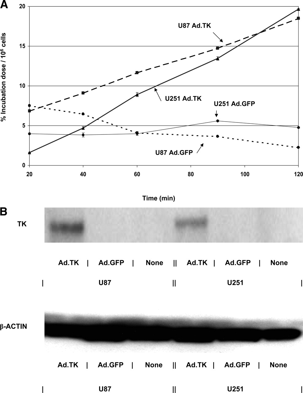
(A) In vitro 76Br-FBAU uptake over 120 min in U87 and U251 cells, each expressing Ad.TK or Ad.GFP. Average binding (n = 3) is expressed in percentage of incubation dose normalized to 1 million cells. Error bars are SDs at each time point. (B) Western blot of protein extract from U87 or U251 cells transduced with Ad.TK or Ad.GFP. Staining with monoclonal thymidine kinase antibody shows expression in Ad.TK transduced cells but not in Ad.GFP or nontransduced cells. β-Actin expression is similar for all 3 conditions.
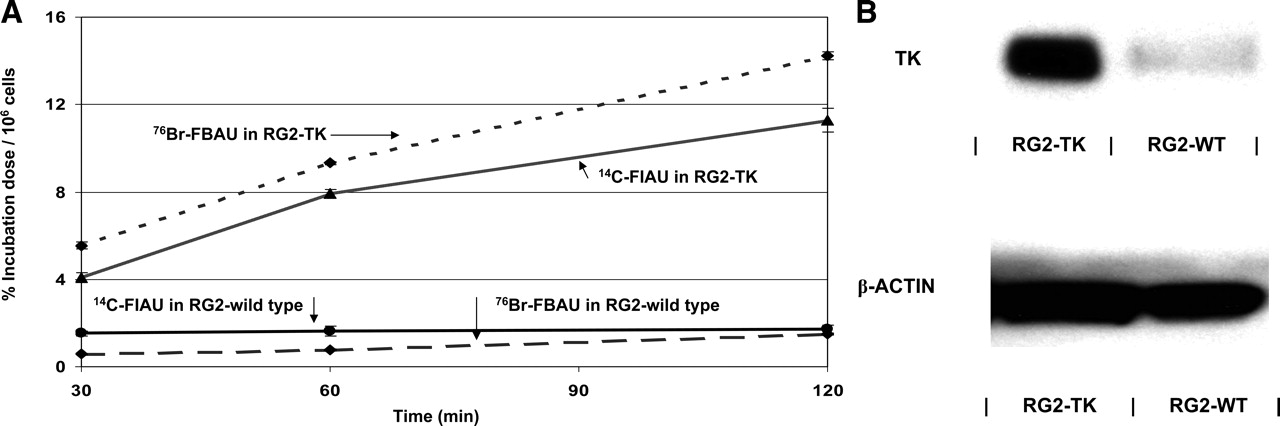
Figure 2A shows increased uptake of 76Br-FBAU compared with 14C-FIAU over 30–120 min in RG2-TK rat glioma cells. There was only minimal uptake of either reporter probe by control RG2 cells during the same period of time. At 120 min, 14C-FIAU uptake in RG2-TK cells was 11.3 %ID and was 1.7 %ID in the control RG2 cells. 76Br-FBAU uptake in RG2-TK cells at 120 min was 14.2 %ID and was 1.5 %ID in the control RG2 cells. To better compare the affinities of these reporter probes to HSV1-tk, we used equimolar concentrations of 76Br-FBAU or 14C-FIAU (1.8 × 10⁻⁸ mol/L). Similarly, a western blot using a thymidine kinase antibody confirmed the expression of HSV1-tk in the stably expressing RG2-TK cells (Fig. 2B).

(A) In vitro uptake of 76Br-FBAU and 14C-FIAU uptake in RG2-TK and RG2 wild-type (RG2-WT) cells with equimolar concentrations of substrate (1.8 × 10⁻⁸ mol/L). Curves represent average uptake (n = 3) of radiolabeled reporter probes, expressed in percentage of incubation dose and normalized to 1 million cells. (B) Western blot of protein extract from RG2-TK and RG2-WT cells. Staining with monoclonal thymidine kinase antibody shows expression in RG2-TK cells but not in RG2-WT cells. β-Actin expression is similar for all 3 conditions.
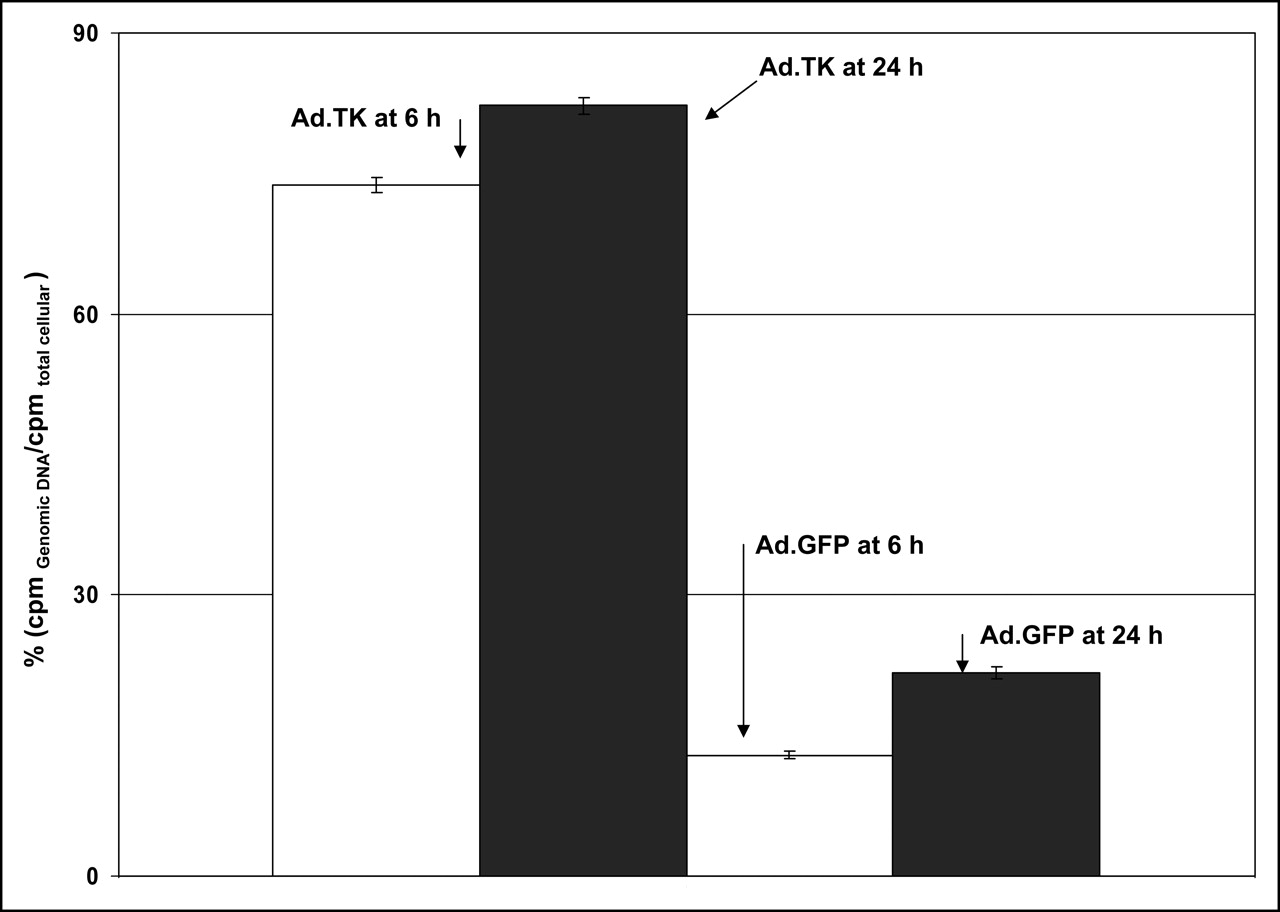
We wanted to confirm that 76Br-FBAU once trapped intracellularly after phorphorylation with HSV1-tk was incorporated into genomic DNA. In vitro U87 cells were harvested after 6 and 24 h of incubation with 76Br-FBAU and the amount of radioactivity in the genomic DNA was compared with total intracellular radioactivity, which was derived from cytoplasmic and genomic radioactivity (Fig. 3). The percentage of radioactivity in genomic DNA over the total cellular radioactivity was significantly greater in Ad.TK transduced U87 cells as compared with Ad.GFP transduced U87 cells. The percentage incorporation in genomic DNA in Ad.TK transduced U87 cells at 6 and 24 h was 73.8% ± 0.7% and 82.2% ± 0.9%, respectively. U87 cells transduced with Ad.GFP had genomic DNA incorporation of 12.9% ± 0.4% and 21.6% ± 0.6% at 6 and 24 h, respectively.

76Br-FBAU incorporation in genomic DNA in U87 cells transduced with Ad.TK or Ad.GFP in cell culture in vitro after 6 and 24 h of incubation.
Ex Vivo Studies
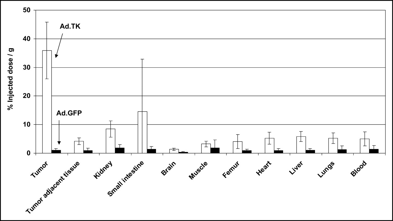
Ex vivo 76Br-FBAU biodistribution experiments were conducted in a mouse model bearing a subcutaneous U87 tumor, which was then intratumorally transduced with Ad-TK or Ad-GFP. 76Br-FBAU was injected intraperitoneally followed by euthanasia 8 h later. We obtained the tumor and various organs and tissues (brain, lung, heart, liver, kidney, small intestine, muscle, femur, blood, and soft tissue adjacent to the tumor) (Fig. 4). Ad.TK tranduced U87 subcutaneous tumor had a significantly higher uptake of 76Br-FBAU compared with other tissues, followed by the small intestine. The amount of uptake in the small intestine is thought to be due to the proliferation of intestinal cells. U87 tumor transduced with Ad.GFP had only minimal uptake of 76Br-FBAU.

Average biodistribution of 76Br-FBAU, 8 h after injection, in nude mice with subcutaneous U87 tumor injected intratumorally with Ad.TK or Ad.GFP (n = 5).
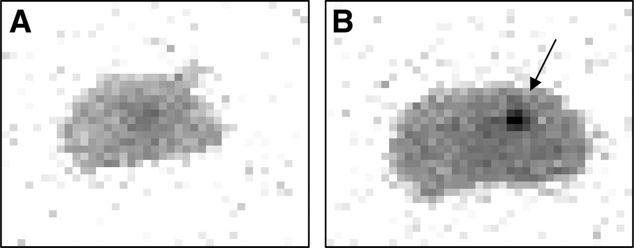
We also found that mice bearing intracranial U87 tumors expressing HSV1-tk was able to focally accumulate 76Br-FBAU on brain autoradiographs compared with control Ad.GFP transduced intracranial tumors (Fig. 5). The area of uptake corresponds to the area of stereotactic injection of the U87 cells in the right parietal region.

Autoradiography of intracranial U87 tumor pretransduced with Ad.GFP (A) or Ad.TK (B) at 8 h after injection of 76Br-FBAU. Arrow indicates radioactivity uptake in region of tumor injection site.
In Vivo ATLAS Imaging Studies
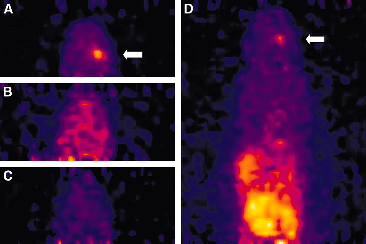
We also obtained in vivo PET images of mice bearing intracranial or subcutaneous U87 tumors. Figure 6 shows a focus of uptake in the right brain in a mouse with intracranial U87 tumor expressing HSV1-tk by intratumoral Ad.TK transduction. Neither Ad.GFP vector injected into intracranial U87 tumor nor Ad.TK injected into normal brain showed focal accumulation of 76Br-FBAU (Fig. 6). Although there are no anatomic coregistration data to definitely confirm that the focus of uptake is in the right parietal region, this is consistent with the anatomic site of our stereotactic injection of the tumor and adenovirus. Note that there is uptake of 76Br-FBAU in the intestinal tract at 6 h after reporter probe injection, which is probably uptake by highly proliferating intestinal cells expressing mammalian thymidine kinase (9,12,13).

Focal uptake of 76Br-FBAU (arrow) in mouse bearing right parietal intracranial U87 tumor stereotactically injected with Ad.TK in right parietal region (A). Control mouse bearing right parietal intracranial U87 tumors injected with Ad.GFP (B) or mouse with normal brain injected with Ad.TK (C) do not show focal reporter probe uptake. Whole-body scan shows reporter probe uptake in gastrointestinal tract (D).
To quantify the radioactivity retention in the mice bearing tumor expressing HSV1-tk, we analyzed the PET images for 76Br-FBAU tumor-to-background ratio with ROI comparison using the ATLAS software. The ratio of uptake in the intracranial RG2-TK tumor to the uptake in the normal brain tissue was 10 ± 1.8 at 2 h after 76Br-FBAU injection and 22 ± 9.0 at 6 h after injection. Although not statistically significant, it demonstrates a persistent, and possibly increasing, high tumor-to-background ratio in tumors expressing HSV1-tk in background tissue with low endogenous mammalian thymidine kinase activity. We also analyzed subcutaneous RG2-TK tumor-to-background ratios using regional bowel, brain, and air for background, but no differences were seen.
DISCUSSION
In this article we report the use of 76Br-FBAU, a thymidine analog, for use as a reporter probe with the HSV1-tk reporter gene. Our initial proof-of-principle experiments in vitro and in vivo used a replication-defective adenovirus constitutively expressing HSV1-tk and a control adenovirus constitutively expressing GFP. Adenoviral vectors have been widely used in previously reported PET gene expression imaging experiments (17–20) and were optimal for high expression of HSV-tk in our initial experiments. The advantage of adenovirus includes its broad tropism, allowing for efficient transduction of a wide variety of cell types that are either dividing or nondividing (21,22). The resulting target cells transduced at a high MOI with an adenovirus containing a highly expressing promoter will result in increased expression of our reporter gene of interest for proof-of-principle experiments. One of the disadvantages of adenovirus is that the virus does not integrate into the genome and has only transient expression.
Human glioma cell lines U87 and U251 were able to demonstrate increased in vitro uptake after infection with Ad.TK versus Ad.GFP under the same conditions. A western blot verified HSV1-tk protein expression from Ad.TK–infected cells.
Ex vivo biodistribution studies were conducted with subcutaneous U87 tumors grown in athymic nude mice. We introduced Ad.tk into preexisting subcutaneous tumors via intratumoral injection and saw a very high level of distribution of 76Br-FBAU to the tumor site versus selected organs. Intratumor injection is a published method of delivery of high copies of adenoviral vectors into a desired target organ or tumor (23,24). A disadvantage of this method is the heterogeneous distribution within the tumor because of variability in the physical delivery. In our subcutaneous and intracranial in vivo experiments, we were able to demonstrate by ex vivo biodistribution and PET that increased intratumoral expression of HSV1-tk was able to accumulate 76Br-FBAU.
Other methods of adenovirus delivery include intravenous delivery with selective uptake within tumors or preinfection of adenovirus into the cells in vitro before introducing them in vivo. We did not try the preinfection method for subcutaneous tumors because of the extended time (3–4 wk) needed to grow the tumor.
In the intracranial tumor models we initially preinfected the cells with adenovirus before implanting the cells stereotactically into the brain because of the relatively shorter time needed to detect tumor growth (1 wk) and were able to detect this on autoradiographs of histologic brain sections. However, we were not able to detect a signal on subsequent ATLAS PET images of the brain at the tumor location by this method (data not included). The inability to detect reporter probe uptake by PET was attributed to the transient nature of adenoviral gene expression with decreased adenoviral HSV1-tk levels in the 7–10 d needed for the growth of the intracranial tumor. Also, autoradiographs of thin histologic sections of brain are more sensitive at picking up signal than whole-brain PET images. Subsequently, we were able to demonstrate intracranial tumor uptake of 76Br-FBAU on ATLAS PET images with direct stereotactic intratumoral injection of Ad.TK into preimplanted U87 tumor cells. We used 2 different controls to show that this was actually attributed to successful Ad.TK transduction of U87 cells. A control Ad.GFP vector injected into intracranial U87 tumor did not show accumulation of 76Br-FBAU. When Ad.TK was injected into normal brain there was no focal accumulation of the reporter probe at the injection site. This was attributed to the poor penetration of 76Br-FBAU past the blood–brain barrier in normal brain versus a more porous blood–tumor barrier (13). These controls imply that only when Ad.TK was expressed in U87 intracranial tumors with a disrupted blood–tumor barrier could 76Br-FBAU be taken up at the tumor or injection site.
To compare in vitro tumor uptake of 76Br-FBAU with an established HSV1-tk radiolabeled probe we needed to establish consistent homogeneous tumor HSV1-tk expression. We used the RG2-tk rat glioma cell line that stably expresses HSV1-tk. This cell line was originally used with 124I-FIAU for the first HSV1-tk gene expression imaging experiments (25,26). In our in vitro experiments we were now able to assume equal expression of HSV1-tk per RG2-tk cell, allowing for increasing HSV1-tk expression with increasing cell number. Another important factor that we controlled in our in vitro experiments with RG2-tk was to take into consideration the mass equivalence of the substrates being compared. Previously published HSV1-tk gene expression studies did not closely control for parameter, which can potentially lead to erroneous conclusions regarding the sensitivity of a particular reporter probe (6–8). Initially the same microcurie amount of 76Br-FBAU and 14C-FIAU was used. Incubation of RG2-tk cells with 4 × 10⁻¹⁰ mol/L 76Br-FBAU (74 kBq/mL [2 μCi/mL] at a SA of about 185,000 GBq/mmol [5,000 Ci/mmol]) showed 30.1% uptake at 2-h incubation versus 9.6% of 3.6 × 10⁻¹⁰ mol/L 14C-FIAU (74 kBq/mL [2 μCi/mL] at a SA of 2.035 GBq/mmol [0.055 Ci/mmol]) (unpublished data). Incubation of RG2-tk cells at an equimolar concentration (1.8 × 10⁻⁸ mol/L) of 76Br-FBAU or 14C-FIAU did not reveal very different percentage uptake values (76Br-FBAU uptake of 21.5% vs. 14C-FIAU uptake of 17.1% at 2-h incubation) although we were still able to demonstrate higher uptake of 76Br-FBAU compared with 14C-FIAU under the same conditions. Uptake in RG2 wild-type cells expressing only mammalian thymidine kinase was low (∼1–2 %ID) for both tracers and did not cause significant background uptake from endogenous thymidine kinase activity. Although radiolabeled FBAU is under active investigation as a potential cell proliferation radiotracer (9,12,27,28), in our experiments 76Br-FBAU did not have significant uptake from endogenous mammalian thymidine kinase in proliferating glioma tumor cells. In our particular application this was advantageous because glioma cell endogenous mammalian thymidine kinase activity would not significantly interfere with its use for HSV1-tk gene expression imaging.
Our experiments confirm previous reports that 76Br-FBAU incorporates into DNA of proliferating cells (9,12). These reports investigated the potential use of 76Br-FBAU for cell proliferation imaging using endogenous cellular mammalian thymidine kinase. 3′-Deoxy-3′-18F-fluorothymidine, a cell proliferation radiotracer currently under active investigation for clinical applications, remains trapped primarily in the cytoplasm and does not incorporate significantly into genomic DNA. 76Br-FBAU was found to incorporate predominantly into the DNA of proliferating cells, which may correlate more directly with proliferation. In our in vitro experiments we were able to show that most of total intracellular 76Br-FBAU incorporated into the genomic DNA of U87 human glioma cells with HSV1-tk expression. Control U87 cells expressing only endogenous mammalian thymidine kinase had genomic DNA incorporation of 12.9% ± 0.4% and 21.6% ± 0.6% at 6 and 24 h, respectively, which is lower DNA incorporation than previously recorded in proliferating cells.
Finally, 76Br-FBAU may have a particular advantage over the most commonly used uracil analog, 124I-FIAU, for PET of HSV1-tk expression because the cyclotron production of 76Br yields higher millicurie amounts. Both the production factor and its 16.2-h half-life may be important factors in its potential future clinical use. Another practical advantage of 76Br-FBAU is the dehalogenation of iodine from 124I-FIAU and the need to block thyroid uptake before its application in vivo. A disadvantage of 76Br-FBAU is its higher Eβ+ of 3.68 MeV with decreased image resolution, although some of these effects may be compensated for by reconstruction techniques. Recent studies used 18F-FBAU (27,28), which allows for better image resolution, although its shorter half-life may preclude longer imaging times and the synthesis is more complicated.
CONCLUSION
In this study we were able to demonstrate that 76Br-FBAU shows promise as a PET reporter probe for use with the HSV1-tk in vivo gene expression imaging system. We showed that 76Br-FBAU accumulates in cells constitutively expressing HSV1-tk by either adenoviral transduction or in a stably expressing cell line. In the glioma cell lines we studied (U87, U251, and RG2) there is low-background mammalian thymidine kinase–specific uptake of 76Br-FBAU, which allows for more specific imaging of HSV1-tk gene expression. The cellular accumulation of 76Br-FBAU appears to be partially via incorporation into DNA, which may be significant for delayed images.
Acknowledgments
We thank Wei Chen for her help with the Ad.GFP and Joji Tokugawa for his help with the in vivo imaging. This work was produced by the authors as part of their official duties as U.S. Government employees. Copyright protection is not available for any work of the U.S. Government. This work is in the public domain and is freely available for publication.
Footnotes
Received Dec. 8, 2004; revision accepted Jul. 25, 2005.
For correspondence or reprints contact: Laura Ravasi, MD, PET Radiochemistry Group, National Institute of Biomedical Imaging and Bioengineering, National Institutes of Health, 10 Center Dr., Bldg. 10, Room 1C401, Bethesda, MD 20982.
E-mail: laura_ravasi{at}nih.gov


