
Main menu

  • Home
  • Content
    • Current
    • Ahead of print
    • Past Issues
    • JNM Supplement
    • SNMMI Annual Meeting Abstracts
    • Continuing Education
    • JNM Podcasts
  • Subscriptions
    • Subscribers
    • Institutional and Non-member
    • Rates
    • Journal Claims
    • Corporate & Special Sales
  • Authors
    • Submit to JNM
    • Information for Authors
    • Assignment of Copyright
    • AQARA requirements
  • Info
    • Reviewers
    • Permissions
    • Advertisers
  • About
    • About Us
    • Editorial Board
    • Contact Information
  • More
    • Alerts
    • Feedback
    • Help
    • SNMMI Journals
  • SNMMI
    • JNM
    • JNMT
    • SNMMI Journals
    • SNMMI

User menu

  • Subscribe
  • My alerts
  • Log in
  • My Cart

Search

  • Advanced search
Journal of Nuclear Medicine
  • SNMMI
    • JNM
    • JNMT
    • SNMMI Journals
    • SNMMI
  • Subscribe
  • My alerts
  • Log in
  • My Cart
Journal of Nuclear Medicine

Advanced Search

  • Home
  • Content
    • Current
    • Ahead of print
    • Past Issues
    • JNM Supplement
    • SNMMI Annual Meeting Abstracts
    • Continuing Education
    • JNM Podcasts
  • Subscriptions
    • Subscribers
    • Institutional and Non-member
    • Rates
    • Journal Claims
    • Corporate & Special Sales
  • Authors
    • Submit to JNM
    • Information for Authors
    • Assignment of Copyright
    • AQARA requirements
  • Info
    • Reviewers
    • Permissions
    • Advertisers
  • About
    • About Us
    • Editorial Board
    • Contact Information
  • More
    • Alerts
    • Feedback
    • Help
    • SNMMI Journals
  • View or Listen to JNM Podcast
  • Visit JNM on Facebook
  • Join JNM on LinkedIn
  • Follow JNM on Twitter
  • Subscribe to our RSS feeds
Research ArticleOncology

Tumor-to-Blood Ratio for Assessment of Somatostatin Receptor Density in Neuroendocrine Tumors Using 68Ga-DOTATOC and 68Ga-DOTATATE

Ezgi Ilan, Irina Velikyan, Mattias Sandström, Anders Sundin and Mark Lubberink
Journal of Nuclear Medicine February 2020, 61 (2) 217-221; DOI: https://doi.org/10.2967/jnumed.119.228072
Ezgi Ilan
1Section of Nuclear Medicine and PET, Department of Surgical Sciences, Uppsala University, Uppsala, Sweden
2Medical Physics, Uppsala University Hospital, Uppsala, Sweden; and
  • Find this author on Google Scholar
  • Find this author on PubMed
  • Search for this author on this site
Irina Velikyan
1Section of Nuclear Medicine and PET, Department of Surgical Sciences, Uppsala University, Uppsala, Sweden
3PET Centre, Medical Imaging Centre, Uppsala University Hospital, Uppsala, Sweden
  • Find this author on Google Scholar
  • Find this author on PubMed
  • Search for this author on this site
Mattias Sandström
1Section of Nuclear Medicine and PET, Department of Surgical Sciences, Uppsala University, Uppsala, Sweden
2Medical Physics, Uppsala University Hospital, Uppsala, Sweden; and
  • Find this author on Google Scholar
  • Find this author on PubMed
  • Search for this author on this site
Anders Sundin
1Section of Nuclear Medicine and PET, Department of Surgical Sciences, Uppsala University, Uppsala, Sweden
3PET Centre, Medical Imaging Centre, Uppsala University Hospital, Uppsala, Sweden
  • Find this author on Google Scholar
  • Find this author on PubMed
  • Search for this author on this site
Mark Lubberink
1Section of Nuclear Medicine and PET, Department of Surgical Sciences, Uppsala University, Uppsala, Sweden
2Medical Physics, Uppsala University Hospital, Uppsala, Sweden; and
  • Find this author on Google Scholar
  • Find this author on PubMed
  • Search for this author on this site
  • Article
  • Figures & Data
  • Supplemental
  • Info & Metrics
  • PDF
Loading

Article Figures & Data

Figures

  • Tables
  • Additional Files
  • FIGURE 1.
    • Download figure
    • Open in new tab
    • Download powerpoint
    FIGURE 1.

    (A and B) Box plots of SUV in blood at 40–45 min after injection for 68Ga-DOTATOC (A) and 68Ga-DOTATATE (B) for high and low Ki values. One tumor per patient is included in plots. Boxes are median and interquartile range, and whiskers are full range of data. Significant differences (P < 0.05) were found in SUVblood between high and low Ki for 68Ga-DOTATOC, however, not for 68Ga-DOTATATE (P > 0.05). (C and D) Relation between Ki and SUV in blood for 68Ga-DOTATOC (C) and 68Ga-DOTATATE (D). Solid line represents exponential fit (y = a/x) for visual illustration.

  • FIGURE 2.
    • Download figure
    • Open in new tab
    • Download powerpoint
    FIGURE 2.

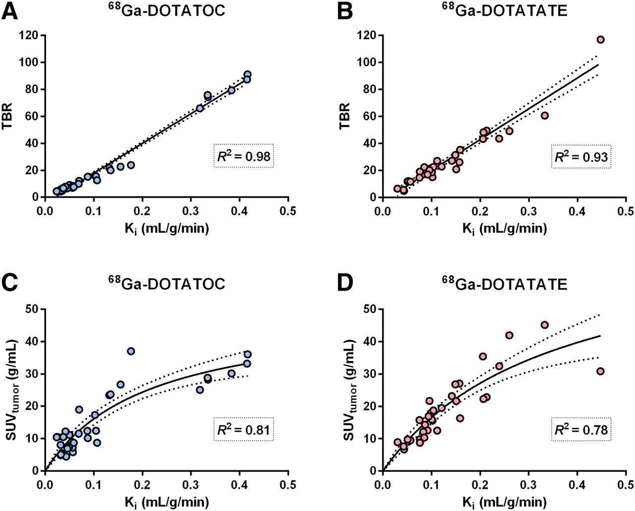
    Correlation between Ki and TBR for 68Ga-DOTATOC (A) and 68Ga-DOTATATE (B) and between Ki and SUV in tumors for 68Ga-DOTATOC (C) and 68Ga-DOTATATE (D). Solid lines represent linear regression fits (A and B) and fits to hyperbolic line (C and D), and dashed lines are 95% confidence band of these fits.

  • FIGURE 3.
    • Download figure
    • Open in new tab
    • Download powerpoint
    FIGURE 3.

    Comparison of Ki, SUVtumor, and TBR between 68Ga-DOTATOC and 68Ga-DOTATATE. Significant difference was found between 68Ga-DOTATOC and 68Ga-DOTATATE for TBR (P = 0.019, Wilcoxon matched-pairs test) but not for Ki or SUVtumor (P = 0.083 and 0.413, respectively).

Tables

  • Figures
  • Additional Files
    • View popup
    TABLE 1

    Demographics and Clinical Characteristics of Study Patients

    SexAge (y)NET typeTracerPeptide (μg)Ki-67 indexPrevious surgeryHistory and previous therapyMetastasesOngoing therapy
    F67pNET/NEC (glucagonoma)TOC/TATE23/233%—SSA, streptozotocin-fluorouracil, PRRT, transformation to NEC and carboplatine-etopsideLiver—
    F63SI NETTOC/TATE17/291%Primary tumorLiver trpl 1999 because of cyst diseaseLiver, mesenteric lgl—
    M67SI NETTOC/TATE18/301%Primary tumor—Liver, mesenteric lglSSA
    M50SI NETTOC/TATE20/3318%Primary tumor mesenteric lgll—Liver, mesenteric lgl, retroperitoneal lgllSSA
    M64pNECTOC/TATE26/2530%—Avastin, temozolomideLiver—
    F73pNETTOC/TATE22/223%—Streptozotocin-fluorouracilLiver, abdominal lgllSSA
    M57SI NETTOC253%—SSAAbdominal lgll, mesenteric lglSSA
    M53pNET (malignant insulinoma)TOC183%Primary tumorStreptozotocin-fluorouracil, SirtexLiver, mesenteric lglEverolimus
    F72pNET (MEN-1, gastrin-producing)TOC15No biopsy——Retroperitoneal lgll—
    M51pNETTOC223%Primary tumor—Retroperitoneal lgll—
    M74SI NETTOC231%——Mesenteric lgll—
    F67pNETTOC252%—Streptozotocin-fluorouracilLiver—
    M50SI NETTOC474%——Liver, mesenteric lgl, peritoneal carcinomatosisSSA
    F*52SI NETTOC255%——Liver, mesenteric thoracic neck lgll, bone, breast, ovarySSA
    F69SI NETTOC279%——Liver, boneSSA
    F47SI NETTOC419%——Liver, mesenteric lgl, abdominal and retroperitoneal lgllSSA
    M72Rectal NETTATE1330%——Liver, pararectal lgll—
    F69SI NETTATE2212%——Liver, peritoneal carcinomatosisSSA
    M67pNETTATE817%——Liver, abdominal lgll, boneSSA
    M75Rectal NETTATE1410%——Liver, abdominal lgll, peritoneal carcinomatosis—
    F*53SI NETTATE165%Primary tumor—Liver, abdominal lgll, bone, breast, lungSSA
    F58Duodenal NET (gastrinoma)TATE223%Primary tumor liver resection, RF—LiverSSA
    F75Atypical lung NETTATE396%SSA
    • ↵* Same patient.

    • pNET = pancreatic NET; NEC = neuroendocrine carcinoma; TOC = 68Ga-DOTATOC; TATE = 68Ga-DOTATATE; SSA = long acting somatostatin analog; PRRT = peptide receptor radionuclide therapy; SI = small intestine; trpl = transplantation; lgl = single lymph node; lgll = multiple lymph nodes; Sirtex = transarterial liver embolization with 90Y-spheres; MEN-1 = multiple endocrine neoplasia type 1; RF = radiofrequency ablation.

    • View popup
    TABLE 2

    Reconstruction Settings for the 3 Different Scanners

    Reconstruction settingDiscovery STDiscovery IQDiscovery MI
    Reconstruction algorithmOSEMOSEM with PSF modelingToF OSEM with PSF modeling
    Iterations/subsets2/284/123/16
    Postprocessing filter (mm)545
    Matrix size128 × 128256 × 256256 × 256
    Pixel size (mm)3.91 × 3.91 × 3.271.95 × 1.95 × 3.261.95 × 1.95 × 2.79
    • OSEM = ordered-subset expectation maximization; PSF = point-spread function; ToF = time of flight.

Additional Files

  • Figures
  • Tables
  • Supplemental Data

    Files in this Data Supplement:

    • Supplemental Data
PreviousNext
Back to top

In this issue

Journal of Nuclear Medicine: 61 (2)
Journal of Nuclear Medicine
Vol. 61, Issue 2
February 1, 2020
  • Table of Contents
  • Table of Contents (PDF)
  • About the Cover
  • Index by author
Print
Download PDF
Article Alerts
Sign In to Email Alerts with your Email Address
Email Article

Thank you for your interest in spreading the word on Journal of Nuclear Medicine.

NOTE: We only request your email address so that the person you are recommending the page to knows that you wanted them to see it, and that it is not junk mail. We do not capture any email address.

Enter multiple addresses on separate lines or separate them with commas.
Tumor-to-Blood Ratio for Assessment of Somatostatin Receptor Density in Neuroendocrine Tumors Using 68Ga-DOTATOC and 68Ga-DOTATATE
(Your Name) has sent you a message from Journal of Nuclear Medicine
(Your Name) thought you would like to see the Journal of Nuclear Medicine web site.
Citation Tools
Tumor-to-Blood Ratio for Assessment of Somatostatin Receptor Density in Neuroendocrine Tumors Using 68Ga-DOTATOC and 68Ga-DOTATATE
Ezgi Ilan, Irina Velikyan, Mattias Sandström, Anders Sundin, Mark Lubberink
Journal of Nuclear Medicine Feb 2020, 61 (2) 217-221; DOI: 10.2967/jnumed.119.228072

Citation Manager Formats

  • BibTeX
  • Bookends
  • EasyBib
  • EndNote (tagged)
  • EndNote 8 (xml)
  • Medlars
  • Mendeley
  • Papers
  • RefWorks Tagged
  • Ref Manager
  • RIS
  • Zotero
Share
Tumor-to-Blood Ratio for Assessment of Somatostatin Receptor Density in Neuroendocrine Tumors Using 68Ga-DOTATOC and 68Ga-DOTATATE
Ezgi Ilan, Irina Velikyan, Mattias Sandström, Anders Sundin, Mark Lubberink
Journal of Nuclear Medicine Feb 2020, 61 (2) 217-221; DOI: 10.2967/jnumed.119.228072
Twitter logo Facebook logo LinkedIn logo Mendeley logo
  • Tweet Widget
  • Facebook Like
  • Google Plus One
Bookmark this article

Jump to section

  • Article
    • Abstract
    • MATERIALS AND METHODS
    • RESULTS
    • DISCUSSION
    • CONCLUSION
    • DISCLOSURE
    • KEY POINTS
    • Acknowledgments
    • Footnotes
    • REFERENCES
  • Figures & Data
  • Supplemental
  • Info & Metrics
  • PDF

Related Articles

  • This Month in JNM
  • PubMed
  • Google Scholar

Cited By...

  • Cardiac Function Consequences of the Persistence of Acute Myocarditis Somatostatin-PET Criteria Four Months Post-Acute Phase
  • Factors Associated with Myocardial Uptake on Oncologic Somatostatin PET Investigations and Differentiation from Myocardial Uptake of Acute Myocarditis
  • Google Scholar

More in this TOC Section

Oncology

  • Role of PET/CT in the management of multiple myeloma
  • FDG-PET/CT and NaF-PET/CT in the diagnosis and assessment of radiation therapy-induced vascular complications in patients with head and neck cancer
  • 18F-FDG PET/CT manifestations of three Cases of Female Desmoplastic Small Round Cell Tumor
Show more Oncology

Clinical

  • Role of PET/CT in the management of multiple myeloma
  • FDG-PET/CT and NaF-PET/CT in the diagnosis and assessment of radiation therapy-induced vascular complications in patients with head and neck cancer
  • 18F-FDG PET/CT manifestations of three Cases of Female Desmoplastic Small Round Cell Tumor
Show more Clinical

Similar Articles

Keywords

  • 68Ga-DOTATOC
  • 68Ga-DOTATATE
  • neuroendocrine tumors, tumor-to-blood ratio
  • SUV
  • net influx rate
SNMMI

© 2025 SNMMI

Powered by HighWire