
Main menu

  • Home
  • Content
    • Current
    • Ahead of print
    • Past Issues
    • JNM Supplement
    • SNMMI Annual Meeting Abstracts
    • Continuing Education
    • JNM Podcasts
  • Subscriptions
    • Subscribers
    • Institutional and Non-member
    • Rates
    • Journal Claims
    • Corporate & Special Sales
  • Authors
    • Submit to JNM
    • Information for Authors
    • Assignment of Copyright
    • AQARA requirements
  • Info
    • Reviewers
    • Permissions
    • Advertisers
  • About
    • About Us
    • Editorial Board
    • Contact Information
  • More
    • Alerts
    • Feedback
    • Help
    • SNMMI Journals
  • SNMMI
    • JNM
    • JNMT
    • SNMMI Journals
    • SNMMI

User menu

  • Subscribe
  • My alerts
  • Log in
  • My Cart

Search

  • Advanced search
Journal of Nuclear Medicine
  • SNMMI
    • JNM
    • JNMT
    • SNMMI Journals
    • SNMMI
  • Subscribe
  • My alerts
  • Log in
  • My Cart
Journal of Nuclear Medicine

Advanced Search

  • Home
  • Content
    • Current
    • Ahead of print
    • Past Issues
    • JNM Supplement
    • SNMMI Annual Meeting Abstracts
    • Continuing Education
    • JNM Podcasts
  • Subscriptions
    • Subscribers
    • Institutional and Non-member
    • Rates
    • Journal Claims
    • Corporate & Special Sales
  • Authors
    • Submit to JNM
    • Information for Authors
    • Assignment of Copyright
    • AQARA requirements
  • Info
    • Reviewers
    • Permissions
    • Advertisers
  • About
    • About Us
    • Editorial Board
    • Contact Information
  • More
    • Alerts
    • Feedback
    • Help
    • SNMMI Journals
  • View or Listen to JNM Podcast
  • Visit JNM on Facebook
  • Join JNM on LinkedIn
  • Follow JNM on Twitter
  • Subscribe to our RSS feeds
Research ArticlePhysics And Instrumentation

89Zr-Immuno-PET: Toward a Noninvasive Clinical Tool to Measure Target Engagement of Therapeutic Antibodies In Vivo

Yvonne W.S. Jauw, Joseph A. O’Donoghue, Josée M. Zijlstra, Otto S. Hoekstra, C. Willemien Menke-van der Houven van Oordt, Franck Morschhauser, Jorge A. Carrasquillo, Sonja Zweegman, Neeta Pandit-Taskar, Adriaan A. Lammertsma, Guus. A.M.S. van Dongen, Ronald Boellaard, Wolfgang A. Weber and Marc C. Huisman
Journal of Nuclear Medicine December 2019, 60 (12) 1825-1832; DOI: https://doi.org/10.2967/jnumed.118.224568
Yvonne W.S. Jauw
1Department of Hematology, Cancer Center Amsterdam, Amsterdam UMC, Vrije Universiteit Amsterdam, Amsterdam, The Netherlands
2Department of Radiology and Nuclear Medicine, Cancer Center Amsterdam, Amsterdam UMC, Vrije Universiteit Amsterdam, Amsterdam, The Netherlands
  • Find this author on Google Scholar
  • Find this author on PubMed
  • Search for this author on this site
Joseph A. O’Donoghue
3Department of Medical Physics, Memorial Sloan Kettering Cancer Center, New York, New York
  • Find this author on Google Scholar
  • Find this author on PubMed
  • Search for this author on this site
Josée M. Zijlstra
1Department of Hematology, Cancer Center Amsterdam, Amsterdam UMC, Vrije Universiteit Amsterdam, Amsterdam, The Netherlands
  • Find this author on Google Scholar
  • Find this author on PubMed
  • Search for this author on this site
Otto S. Hoekstra
2Department of Radiology and Nuclear Medicine, Cancer Center Amsterdam, Amsterdam UMC, Vrije Universiteit Amsterdam, Amsterdam, The Netherlands
  • Find this author on Google Scholar
  • Find this author on PubMed
  • Search for this author on this site
C. Willemien Menke-van der Houven van Oordt
4Department of Medical Oncology, Cancer Center Amsterdam, Amsterdam UMC, Vrije Universiteit Amsterdam, Amsterdam, The Netherlands
  • Find this author on Google Scholar
  • Find this author on PubMed
  • Search for this author on this site
Franck Morschhauser
5EA7365-GRITA-Groupe de Recherche sur les forms Injectables et les Technologies Associées, Université de Lille, and Department of Hematology, CHU Lille, Lille, France
  • Find this author on Google Scholar
  • Find this author on PubMed
  • Search for this author on this site
Jorge A. Carrasquillo
6Department of Radiology, Memorial Sloan Kettering Cancer Center, New York, New York; and
7Molecular Imaging and Therapy Service, Memorial Sloan Kettering Cancer Center, New York, New York
  • Find this author on Google Scholar
  • Find this author on PubMed
  • Search for this author on this site
Sonja Zweegman
1Department of Hematology, Cancer Center Amsterdam, Amsterdam UMC, Vrije Universiteit Amsterdam, Amsterdam, The Netherlands
  • Find this author on Google Scholar
  • Find this author on PubMed
  • Search for this author on this site
Neeta Pandit-Taskar
6Department of Radiology, Memorial Sloan Kettering Cancer Center, New York, New York; and
7Molecular Imaging and Therapy Service, Memorial Sloan Kettering Cancer Center, New York, New York
  • Find this author on Google Scholar
  • Find this author on PubMed
  • Search for this author on this site
Adriaan A. Lammertsma
2Department of Radiology and Nuclear Medicine, Cancer Center Amsterdam, Amsterdam UMC, Vrije Universiteit Amsterdam, Amsterdam, The Netherlands
  • Find this author on Google Scholar
  • Find this author on PubMed
  • Search for this author on this site
Guus. A.M.S. van Dongen
2Department of Radiology and Nuclear Medicine, Cancer Center Amsterdam, Amsterdam UMC, Vrije Universiteit Amsterdam, Amsterdam, The Netherlands
  • Find this author on Google Scholar
  • Find this author on PubMed
  • Search for this author on this site
Ronald Boellaard
2Department of Radiology and Nuclear Medicine, Cancer Center Amsterdam, Amsterdam UMC, Vrije Universiteit Amsterdam, Amsterdam, The Netherlands
  • Find this author on Google Scholar
  • Find this author on PubMed
  • Search for this author on this site
Wolfgang A. Weber
6Department of Radiology, Memorial Sloan Kettering Cancer Center, New York, New York; and
7Molecular Imaging and Therapy Service, Memorial Sloan Kettering Cancer Center, New York, New York
  • Find this author on Google Scholar
  • Find this author on PubMed
  • Search for this author on this site
Marc C. Huisman
2Department of Radiology and Nuclear Medicine, Cancer Center Amsterdam, Amsterdam UMC, Vrije Universiteit Amsterdam, Amsterdam, The Netherlands
  • Find this author on Google Scholar
  • Find this author on PubMed
  • Search for this author on this site
  • Article
  • Figures & Data
  • Supplemental
  • Info & Metrics
  • PDF
Loading

Article Figures & Data

Figures

  • Tables
  • Additional Files
  • FIGURE 1.
    • Download figure
    • Open in new tab
    • Download powerpoint
    FIGURE 1.

    Biodistribution of 89Zr-labeled mAb: physiologic components. (A) Reversible nonspecific uptake due to antibody in vascular tissue compartment and antibody entering tissue interstitium through paracellular pores, and through endothelial cells mediated by neonatal Fc-receptor, leaving tissue by convective transport through lymph flow. (B) Irreversible nonspecific uptake due to mAb degradation in lysosome, followed by residualization of 89Zr. (C) Specific uptake due to target engagement (target binding and internalization of mAb-target antigen). (Adapted from Lobo et al. (8) and Chen et al. (10).)

  • FIGURE 2.
    • Download figure
    • Open in new tab
    • Download powerpoint
    FIGURE 2.

    Transfer constants for 89Zr-anti-HER2 in kidney. (A and B) Example for 1 patient in 89Zr-anti-HER2 study, with measured activity concentrations in serum (A) and measured activity concentrations in kidney (B). (C) Patlak linearization to determine offset (VT) and slope (Ki) of linear fit to last 3 time points (same data). (D) Reversible and irreversible contributions to total measured signal. No target expression has been reported for HER2 in normal kidney. Therefore, we hypothesize that total signal consists of nonspecific uptake. After 100 h after injection, total uptake predominantly consists of irreversible nonspecific uptake due to 89Zr-residualization after mAb degradation. p.i. = after injection.
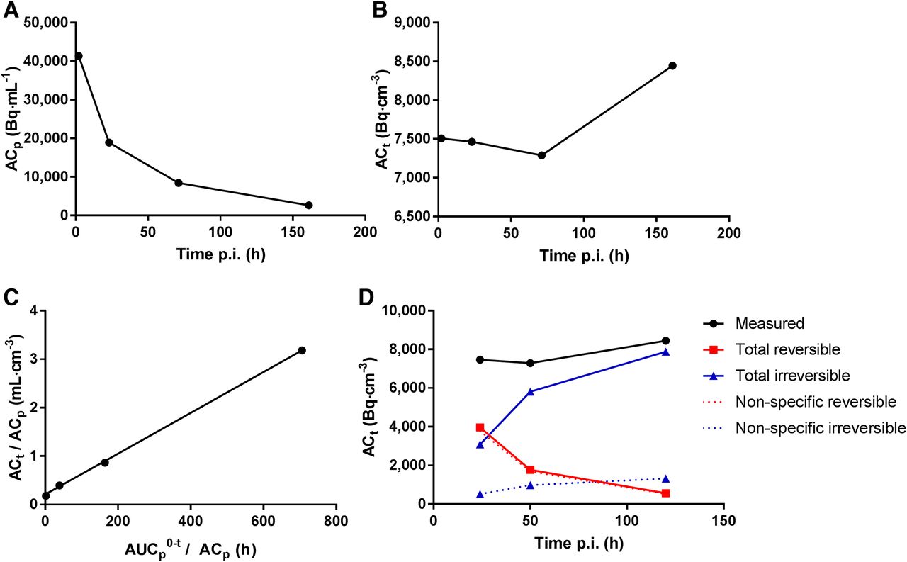
  • FIGURE 3.
    • Download figure
    • Open in new tab
    • Download powerpoint
    FIGURE 3.

    Transfer constants for 89Zr-anti-PSMA in kidney. (A and B) Example for 1 patient in 89Zr-anti-PSMA study, with measured activity concentrations in serum (A) and measured activity concentrations in kidney (B). (C) Patlak linearization to determine offset (VT) and slope (Ki) of linear fit to last 3 time points (same data). (D) Total reversible and total irreversible contributions to measured signal. Dashed lines represent estimated values for nonspecific reversible uptake (calculated as baseline Embedded Image) and nonspecific irreversible uptake (calculated as baseline Embedded Image). Nonspecific uptake accounts for 66%, 34%, and 22% of total measured signal at 1, 3, and 7 d after injection, respectively. Difference between total irreversible uptake and estimated nonspecific irreversible uptake indicates target-mediated uptake. p.i. = after injection.

Tables

  • Figures
  • Additional Files
    • View popup
    TABLE 1

    Data Characteristics

    Characteristic89Zr-anti-CD2089Zr-anti-EGFR89Zr-anti-PSMA89Zr-anti-HER2
    mAbObinutuzumabCetuximabHu-J591Trastuzumab
    TypeHumanizedChimericHumanizedHumanized
    IgG subclassIgG1IgG1IgG1IgG1
    Target expression (20)
     KidneyAbsentAbsentPresentAbsent
     LiverAbsentPresentPresentPresent
     LungAbsentAbsentAbsentPresent
     SpleenPresentAbsentAbsent*Absent
    CenterAmsterdam UMC (n = 4); CHU Lille (n = 5)Amsterdam UMCMemorial Sloan Kettering Cancer CenterMemorial Sloan Kettering Cancer Center
    Patient categoryNon-Hodgkin lymphomaColorectal carcinomaProstate cancerGastric cancer
    Number of patients971010
    Injected activity/mAb dose37 MBq/10 mg; 1,000 mg unlabeled mAb37 MBq/10 mg; 500 mg/m2 unlabeled mAb185 MBq/1.7 mg; total mass of 25 mg mAb185 MBq/3 mg; total mass of 50 mg mAb
    Administration†PredosePredoseCoinjectionCoinjection
    PET scan time points1 h, 72 h, 144 h after injection1 h, 72 h, 144 h after injection2–4 h, 24 h, 48–120 h, 144–168 h after injection4 h, 24 h, 48–96 h, 120–192 h after injection
    Blood sample‡PlasmaPlasmaSerumSerum
    Blood sampling time points5, 30, 60, 120 min; 24¶, 72, 144 h after injection1–2, 24, 48, 72, 144 h5, 30, 60, 120–240 min; 24, 48–120, 144–168 h after injection5, 15, 30, 60 min; 2, 24,48–96, 120–192 h after injection
    ReferenceJauw et al. (16)Menke et al. (17)Pandit-Taskar et al. (18)O’Donoghue et al. (19)
    • ↵* Expression of prostate-specific membrane antigen in spleen has been reported (24).

    • ↵† Predose = 89Zr-mAb within 2 h after administration of unlabeled mAb.

    • ↵‡ Blood samples consisted of plasma or serum samples, assuming no practical difference between these assays for our purposes because mAb binding does not occur to coagulation factors (difference between plasma and serum).

    • ↵¶ Blood samples obtained at 24 h after injection at CHU Lille (n = 5); no 24-h sample obtained at Amsterdam UMC.

    • Coinjection = unlabeled mAb infused intravenously over 5 min followed immediately by 1 min infusion of radiolabeled mAb.

    • View popup
    TABLE 2

    Vt

    Site89Zr-anti-CD2089Zr-anti-EGFR89Zr-anti-PSMA89Zr-anti-HER2Baseline
    Kidney0.18 (0.15–020)0.25 (0.23–0.29)0.28 (0.21–0.32)0.19 (0.15–0.25)0.20 (0.16–0.25)
    Liver0.24 (0.21–0.28)0.64 (0.54–0.91)0.29 (0.23–0.43)0.24 (0.22–0.29)0.24 (0.21–0.28)
    Lung0.08 (0.08–0.10)0.11 (0.09–0.13)0.07 (0.06–0.09)0.08 (0.05–0.11)0.09 (0.07–0.10)
    Spleen0.20 (0.18–0.21)0.23 (0.20–0.27)0.22 (0.20–0.28)0.24 (0.18–0.27)0.24 (0.20–0.27)
    • Vt (mL⋅cm−3) is presented as median followed by IQ in parentheses.

    • View popup
    TABLE 3

    Ki

    Site89Zr-anti-CD2089Zr-anti-EGFR89Zr-anti-PSMA89Zr-anti-HER2Baseline
    Kidney0.4 (0.2–0.6)0.7 (0.4–1.2)2.8 (2.4–3.1)1.5 (0.9–1.8)0.7 (0.4–1.3)
    Liver1.1 (0.8–2.1)3.8 (1.9–5.8)5.7 (4.9–8.4)1.7 (1.4–2.0)1.1 (0.8–2.1)
    Lung0.2 (0.1–0.3)0.4 (0.2–0.6)0.1 (0.0–0.2)0.2 (0.0–0.5)0.2 (0.1–0.3)
    Spleen0.6 (0.5–0.8)0.5 (0.3–0.5)1.5 (1.2–1.7)0.7 (0.4–0.8)0.5 (0.3–0.7)
    • Ki (μL⋅g−1⋅h−1) is presented as median followed by IQ in parentheses.

Additional Files

  • Figures
  • Tables
  • Supplemental Data

    Files in this Data Supplement:

    • Supplemental Data
PreviousNext
Back to top

In this issue

Journal of Nuclear Medicine: 60 (12)
Journal of Nuclear Medicine
Vol. 60, Issue 12
December 1, 2019
  • Table of Contents
  • Table of Contents (PDF)
  • About the Cover
  • Index by author
Print
Download PDF
Article Alerts
Sign In to Email Alerts with your Email Address
Email Article

Thank you for your interest in spreading the word on Journal of Nuclear Medicine.

NOTE: We only request your email address so that the person you are recommending the page to knows that you wanted them to see it, and that it is not junk mail. We do not capture any email address.

Enter multiple addresses on separate lines or separate them with commas.
89Zr-Immuno-PET: Toward a Noninvasive Clinical Tool to Measure Target Engagement of Therapeutic Antibodies In Vivo
(Your Name) has sent you a message from Journal of Nuclear Medicine
(Your Name) thought you would like to see the Journal of Nuclear Medicine web site.
Citation Tools
89Zr-Immuno-PET: Toward a Noninvasive Clinical Tool to Measure Target Engagement of Therapeutic Antibodies In Vivo
Yvonne W.S. Jauw, Joseph A. O’Donoghue, Josée M. Zijlstra, Otto S. Hoekstra, C. Willemien Menke-van der Houven van Oordt, Franck Morschhauser, Jorge A. Carrasquillo, Sonja Zweegman, Neeta Pandit-Taskar, Adriaan A. Lammertsma, Guus. A.M.S. van Dongen, Ronald Boellaard, Wolfgang A. Weber, Marc C. Huisman
Journal of Nuclear Medicine Dec 2019, 60 (12) 1825-1832; DOI: 10.2967/jnumed.118.224568

Citation Manager Formats

  • BibTeX
  • Bookends
  • EasyBib
  • EndNote (tagged)
  • EndNote 8 (xml)
  • Medlars
  • Mendeley
  • Papers
  • RefWorks Tagged
  • Ref Manager
  • RIS
  • Zotero
Share
89Zr-Immuno-PET: Toward a Noninvasive Clinical Tool to Measure Target Engagement of Therapeutic Antibodies In Vivo
Yvonne W.S. Jauw, Joseph A. O’Donoghue, Josée M. Zijlstra, Otto S. Hoekstra, C. Willemien Menke-van der Houven van Oordt, Franck Morschhauser, Jorge A. Carrasquillo, Sonja Zweegman, Neeta Pandit-Taskar, Adriaan A. Lammertsma, Guus. A.M.S. van Dongen, Ronald Boellaard, Wolfgang A. Weber, Marc C. Huisman
Journal of Nuclear Medicine Dec 2019, 60 (12) 1825-1832; DOI: 10.2967/jnumed.118.224568
Twitter logo Facebook logo LinkedIn logo Mendeley logo
  • Tweet Widget
  • Facebook Like
  • Google Plus One
Bookmark this article

Jump to section

  • Article
    • Abstract
    • MATERIALS AND METHODS
    • RESULTS
    • DISCUSSION
    • CONCLUSION
    • DISCLOSURE
    • KEY POINTS
    • Acknowledgments
    • Footnotes
    • REFERENCES
  • Figures & Data
  • Supplemental
  • Info & Metrics
  • PDF

Related Articles

  • This Month in JNM
  • PubMed
  • Google Scholar

Cited By...

  • Immuno-PET and Targeted {alpha}-Therapy Using Anti-Glypican-1 Antibody Labeled with 89Zr or 211At: A Theranostic Approach for Pancreatic Ductal Adenocarcinoma
  • Local and distant response to intratumoral immunotherapy assessed by immunoPET in mice
  • Specific and Nonspecific Uptake in Quantitative 89Zr-Immuno-PET
  • The Role of 89Zr-Immuno-PET in Navigating and Derisking the Development of Biopharmaceuticals
  • Google Scholar

More in this TOC Section

Physics And Instrumentation

  • Performance Evaluation of the uMI Panorama PET/CT System in Accordance with the National Electrical Manufacturers Association NU 2-2018 Standard
  • A Multicenter Study on Observed Discrepancies Between Vendor-Stated and PET-Measured 90Y Activities for Both Glass and Resin Microsphere Devices
  • Ultra-Fast List-Mode Reconstruction of Short PET Frames and Example Applications
Show more Physics And Instrumentation

Translational

  • Quantitative Total-Body Imaging of Blood Flow with High-Temporal-Resolution Early Dynamic 18F-FDG PET Kinetic Modeling
  • [99mTc]Tc-MY6349 Probe for Trop2-Targeted SPECT Imaging: From Preclinical to Pilot Clinical Study
  • Imaging Diverse Pathogenic Bacteria In Vivo with 18F-Fluoromannitol PET
Show more Translational

Similar Articles

Keywords

  • immuno-PET
  • Monoclonal Antibodies
  • 89Zr
  • molecular imaging
  • positron emission tomography
SNMMI

© 2025 SNMMI

Powered by HighWire