


Article Figures & Data
Figures
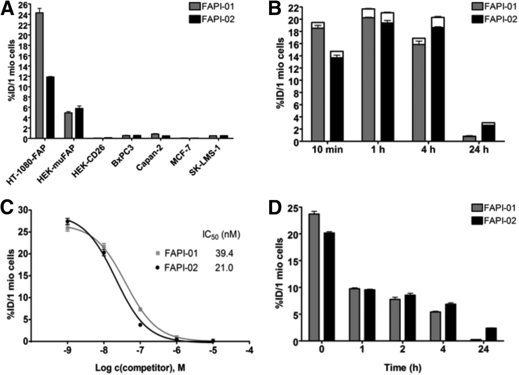
- FIGURE 1.
(A) Binding of radiolabeled FAPI-01 and FAPI-02 to the 4 human cancer cell lines and to the HT-1080-FAP, HEK-muFAP, and HEK-CD26 cell lines after 60 min of incubation. (B) Internalization of radiolabeled FAPI-01 and FAPI-02 into HT-1080-FAP cells after incubation for 10 min to 24 h. Internalized fraction is gray or black, and extracellularly bound fraction is white. (C) Competitive binding of radiolabeled FAPI-01 and FAPI-02 to HT-1080-FAP cells after adding increasing concentrations of unlabeled FAPI-01 and Lu-FAPI-02. (D) Efflux kinetics of FAPI-01 and FAPI-02 after 1 h of incubation of HT-1080-FAP cells with radiolabeled compounds, followed by incubation with compound-free medium for 1–24 h. All data are %ID normalized to 1 million (mio) cells.
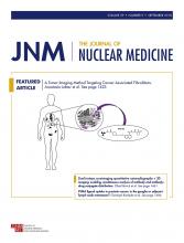
- FIGURE 2.
Internalization of FAPI-02 into HT-1080-FAP cells, HEK-muFAP cells, and HEK-CD26 cells after incubation for 2 h. Blue indicates 4′,6-diamidino-2-phenylindole, and green indicates FAPI-02-Atto488.
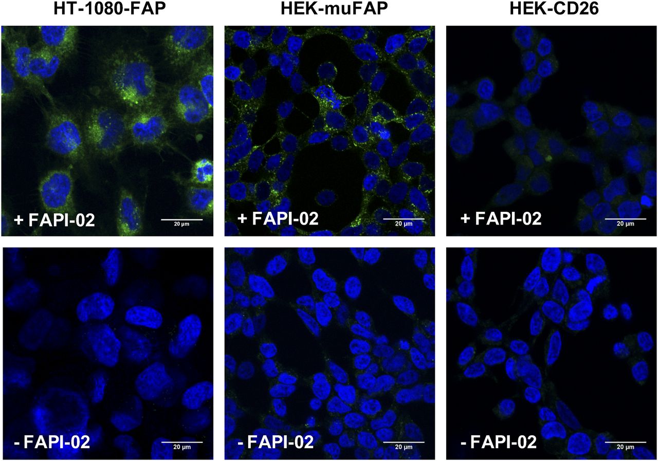
- FIGURE 3.
68Ga-FAPI-02 PET in mice bearing FAP-negative (Capan-2) (A) and human FAP-expressing (HT-1080-FAP) (B) xenografts. Images were obtained at the indicated times after injection and show rapid uptake in tumor (arrows), no accumulation in noncancerous tissue, and rapid elimination via kidneys and bladder. Quantification of PET images shows solid clearance from cardiovascular system and constant uptake into tumor.
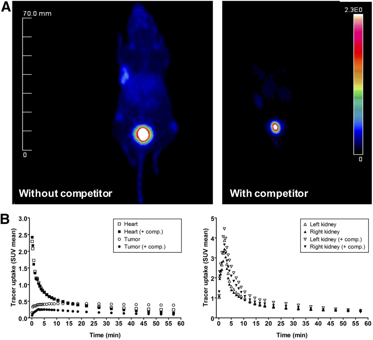
- FIGURE 4.
(A) Blocking of 68Ga-FAPI-02 tumor accumulation by coadministration of 30 nmol of unlabeled FAPI-02 in mice bearing HT-1080-FAP tumors. (B) Time–activity curves for 68Ga-FAPI-02 in selected organs after administration with and without unlabeled FAPI-02 as competitor.
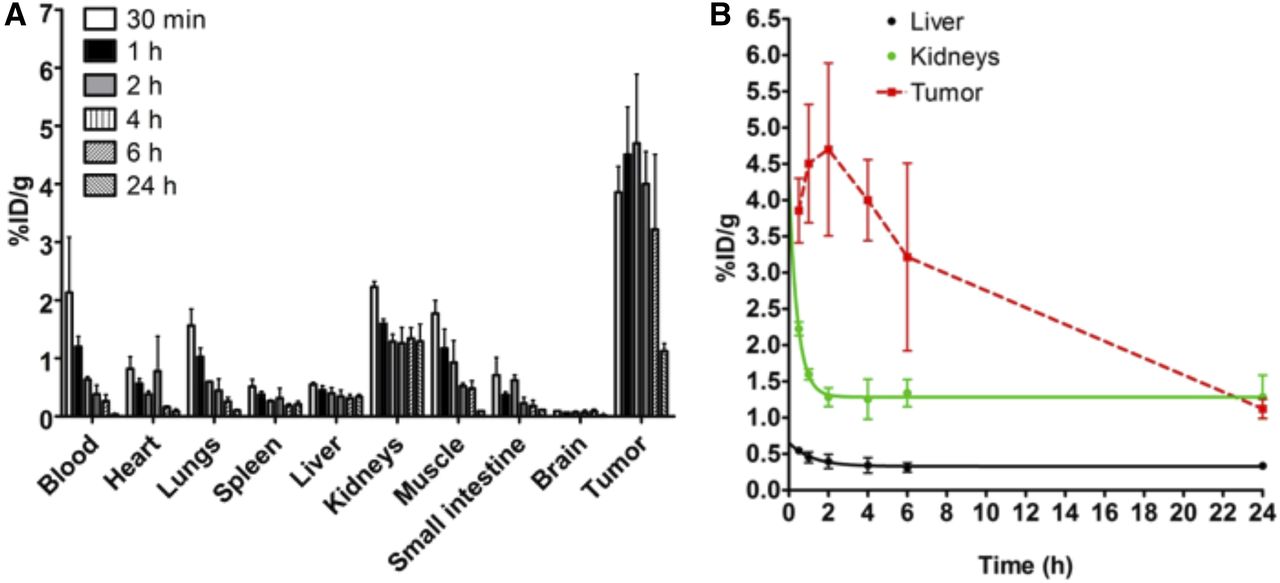
- FIGURE 5.
(A) Ex vivo biodistribution of 177Lu-FAPI-02 after administration to mice bearing HT-1080-FAP tumors. Tumor uptake is highest after 2 h (4.7 %ID/g). (B) Pharmacokinetic profile of 177Lu-FAPI-02 up to 24 h after administration.
- FIGURE 6.
PET/CT maximum-intensity projections of patient with metastasized pancreatic cancer (A) and patient with breast cancer (C). (B) Maximum uptake of 68Ga-FAPI-02 at 10 min, 1 h, and 3 h after administration to breast cancer patient. Me = metastases.
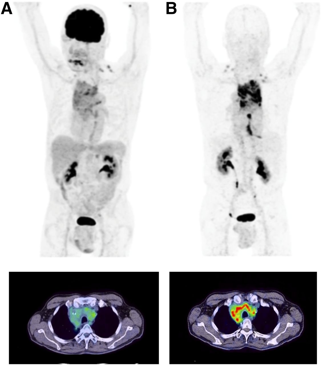
- FIGURE 7.
PET/CT maximum-intensity projections (top) and transaxial views (bottom) 1 h after administration of 18F-FDG (A) and 68Ga-FAPI-02 (B) to patient with locally advanced lung adenocarcinoma. 68Ga-FAPI-02 is seen to selectively accumulate in FAP-expressing tissue and to be significantly higher than 18F-FDG in malignant lesions. Unlike 18F-FDG, 68Ga-FAPI-02 shows no uptake in brain, spleen, or liver.


Additional Files
Supplemental Data
Files in this Data Supplement:


Jump to section
Related Articles
Cited By...
- Exploring molecular imaging to investigate immune checkpoint inhibitor-related toxicity
- Feasibility, Tolerability, and Preliminary Clinical Response of Fractionated Radiopharmaceutical Therapy with 213Bi-FAPI-46: Pilot Experience in Patients with End-Stage, Progressive Metastatic Tumors
- Evaluation of Fibroblast Activation Protein Expression Using 68Ga-FAPI46 PET in Hypertension-Induced Tissue Changes
- Fibroblast Activation Protein-Directed Imaging Outperforms 18F-FDG PET/CT in Malignant Mesothelioma: A Prospective, Single-Center, Observational Trial
- 68Ga-FAP-2286 PET of Solid Tumors: Biodistribution, Dosimetry, and Comparison with 18F-FDG
- Diagnostic Potential of Supplemental Static and Dynamic 68Ga-FAPI-46 PET for Primary 18F-FDG-Negative Pulmonary Lesions
- Fibroblast Activation Protein Inhibitor Tracers and Their Preclinical, Translational, and Clinical Status in China
- 68Ga-FAPI PET/CT as an Alternative to 18F-FDG PET/CT in the Imaging of Invasive Lobular Breast Carcinoma
- Expression of fibroblast activation protein-{alpha} in human deep venous thrombus
- Diagnostic Accuracy of 68Ga-FAPI Versus 18F-FDG PET in Patients with Various Malignancies
- Immunohistochemical FAP Expression Reflects 68Ga-FAPI PET Imaging Properties of Low- and High-Grade Intraductal Papillary Mucinous Neoplasms and Pancreatic Ductal Adenocarcinoma
- Impact of 68Ga-FAPI PET/CT on Staging and Oncologic Management in a Cohort of 226 Patients with Various Cancers
- [68Ga]Ga-FAPI-46 PET for Visualization of Postinfarction Renal Fibrosis
- PET imaging of fibroblast activation protein alpha (FAP) detects incipient cardiotoxicity due to anthracycline chemotherapy
- Performance of 68Ga-Labeled Fibroblast Activation Protein Inhibitor PET/CT in Evaluation of Erdheim-Chester Disease: A Comparison with 18F-FDG PET/CT
- Evaluation of the Diagnostic Accuracy of FAPI PET/CT in Oncologic Studies: Systematic Review and Metaanalysis
- Clinical Evaluation of 68Ga-FAPI-RGD for Imaging of Fibroblast Activation Protein and Integrin {alpha}v{beta}3 in Various Cancer Types
- [68Ga]Ga-FAPI-46 PET for Visualization of Postinfarction Renal Fibrosis
- Evaluation of the Diagnostic Accuracy of FAPI PET/CT in Oncologic Studies: Systematic Review and Metaanalysis
- Fibroblast Activation Protein Inhibitor-Based Radionuclide Therapies: Current Status and Future Directions
- Clinical Evaluation of 68Ga-FAPI-RGD for Imaging of Fibroblast Activation Protein and Integrin {alpha}v{beta}3 in Various Cancer Types
- Fibroblast-Activation Protein PET and Histopathology in a Single-Center Database of 324 Patients and 21 Tumor Entities
- Static and Dynamic 68Ga-FAPI PET/CT for the Detection of Malignant Transformation of Intraductal Papillary Mucinous Neoplasia of the Pancreas
- Staging Liver Fibrosis by Fibroblast Activation Protein Inhibitor PET in a Human-Sized Swine Model
- Fibroblast Activation Protein Inhibitor Imaging in Nonmalignant Diseases: A New Perspective for Molecular Imaging
- Head-to-Head Comparison of 68Ga-FAPI-46 and 18F-FDG PET/CT for Evaluation of Head and Neck Squamous Cell Carcinoma: A Single-Center Exploratory Study
- FAPI PET Opens a New Window to Understanding Immune-Mediated Inflammatory Diseases
- Correlation of 68Ga-FAPi-46 PET Biodistribution with FAP Expression by Immunohistochemistry in Patients with Solid Cancers: Interim Analysis of a Prospective Translational Exploratory Study
- Synthesis, Preclinical Evaluation, and a Pilot Clinical PET Imaging Study of 68Ga-Labeled FAPI Dimer
- Albumin Binder-Conjugated Fibroblast Activation Protein Inhibitor Radiopharmaceuticals for Cancer Therapy
- Feasibility of In Vivo Imaging of Fibroblast Activation Protein in Human Arterial Walls
- 68Ga-FAPI as a Diagnostic Tool in Sarcoma: Data from the 68Ga-FAPI PET Prospective Observational Trial
- Fibroblast Activation Protein-Specific PET/CT Imaging in Fibrotic Interstitial Lung Diseases and Lung Cancer: A Translational Exploratory Study
- Building the Bridge: Molecular Imaging Biomarkers for 21st Century Cancer Therapies
- The Annual Journal Impact Factor Saga
- Impact of 68Ga-FAPI PET/CT Imaging on the Therapeutic Management of Primary and Recurrent Pancreatic Ductal Adenocarcinomas
- FAPI PET/CT: Will It End the Hegemony of 18F-FDG in Oncology?
- The Latest Developments in Imaging of Fibroblast Activation Protein
- Fibroblast Activation Protein-Targeted PET/CT with 68Ga-FAPI for Imaging IgG4-Related Disease: Comparison to 18F-FDG PET/CT
- FAPI-74 PET/CT Using Either 18F-AlF or Cold-Kit 68Ga Labeling: Biodistribution, Radiation Dosimetry, and Tumor Delineation in Lung Cancer Patients
- Targeting Fibroblast Activation Protein: Radiosynthesis and Preclinical Evaluation of an 18F-Labeled FAP Inhibitor
- Design and Development of 99mTc-Labeled FAPI Tracers for SPECT Imaging and 188Re Therapy
- Imaging Fibroblast Activation Protein Alpha Improves Diagnosis of Metastatic Prostate Cancer with Positron Emission Tomography
- The Role of 68Ga-FAPI PET/CT for Patients with Malignancies of the Lower Gastrointestinal Tract: First Clinical Experience
- Radiation Dosimetry and Biodistribution of 68Ga-FAPI-46 PET Imaging in Cancer Patients
- Theranostics Targeting Fibroblast Activation Protein in the Tumor Stroma: 64Cu- and 225Ac-Labeled FAPI-04 in Pancreatic Cancer Xenograft Mouse Models
- FAP: The Next Billion Dollar Nuclear Theranostics Target?
- Molecular Imaging of Fibroblast Activity After Myocardial Infarction Using a 68Ga-Labeled Fibroblast Activation Protein Inhibitor, FAPI-04
- A Conversation Between Uwe Haberkorn and Johannes Czernin
- Development of Fibroblast Activation Protein-Targeted Radiotracers with Improved Tumor Retention
- Future of Theranostics: An Outlook on Precision Oncology in Nuclear Medicine
- The Future of Nuclear Medicine as an Independent Specialty
- Toward Independent Nuclear Medicine, Molecular Imaging, and Theranostic Programs
- 68Ga-FAPI PET/CT: Tracer Uptake in 28 Different Kinds of Cancer
- 68Ga-FAPI PET/CT: Biodistribution and Preliminary Dosimetry Estimate of 2 DOTA-Containing FAP-Targeting Agents in Patients with Various Cancers
- Visualisation of interstitial lung disease by molecular imaging of integrin {alpha}v{beta}3 and somatostatin receptor 2