


Article Figures & Data
Figures
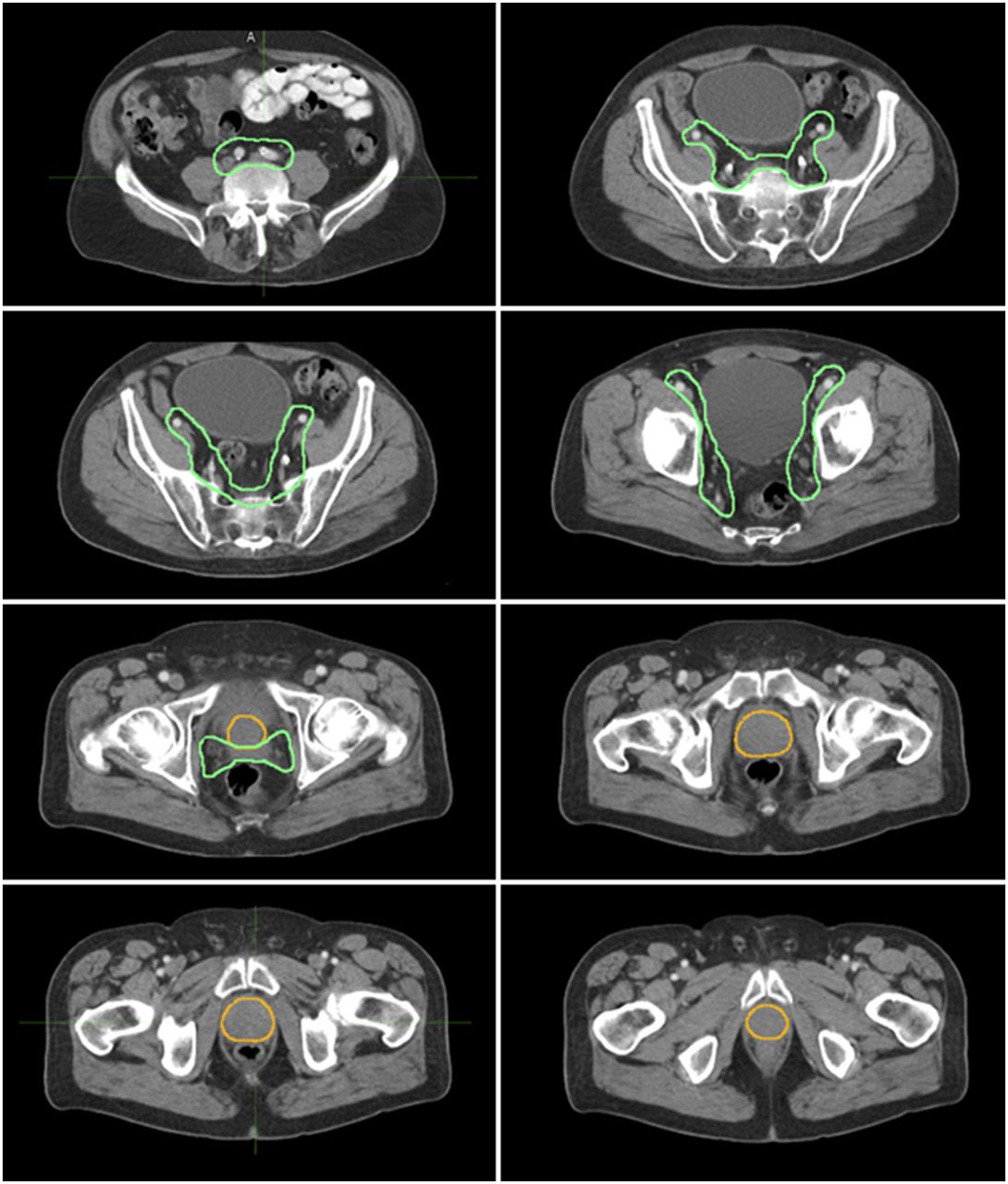
- FIGURE 1.
Axial CT views of prostate CTV (yellow) and of pelvic LN and seminal vesicle CTV (green). CTVs were contoured on CT dataset of PET/CT for all 73 patients by experienced radiation oncologist who was masked to 68Ga-PSMA-11 PET findings. Pelvic LN CTV included presacral, distal common iliac, internal iliac, external iliac, and obturator LNs (upper limit, L4/L5).
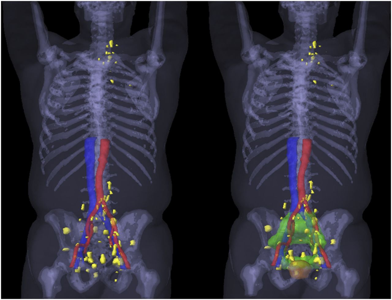
- FIGURE 2.
(Left) Three-dimensional rendering of all 68Ga-PSMA-11–positive lesions (yellow) in patients with extraprostatic metastasis: 20 N1M0 lesions (5 with out-of-field positive lesions), 3 N1M1a lesions, 2 N0M1b lesions, 1 N1M1aM1b lesion, and 1 N1M1bM1c lesion. (Right) Three-dimensional rendering of targeted volumes for prostate (yellow) and for pelvic LN plus seminal vesicles (green).
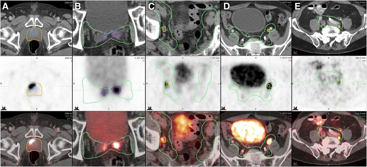
- FIGURE 3.
Examples of 68Ga-PSMA-11–positive disease within radiation fields on axial CT (top), PET (middle), and PET/CT (bottom). Once positive lesions were identified on PET, contours of prostate CTV (yellow) and pelvic LN CTV (green) were drawn on the basis of CT. (A) Primary prostate tumor (MTV, 4 cm3; SUVmax, 34.6). (B) Invaded seminal vesicles (SUVmax, 18.0). (C) Right obturator LN (short axis, 6 mm; SUVmax, 4.6). (D) Left external iliac LN (short axis, 7 mm; SUVmax, 22.3). (E) Left common iliac LN (short axis, 5 mm; SUVmax, 4.1).
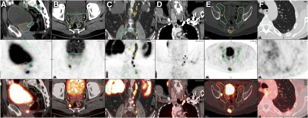
- FIGURE 4.
Examples of 68Ga-PSMA-11–positive disease outside radiation fields on CT (top), PET (middle), and PET/CT (bottom). Once positive lesions were identified on PET, contours of prostate CTV (yellow) and pelvic LN CTV (green) were drawn on the basis of CT. (A) Primary prostate tumor (MTV, 3 cm3; SUVmax, 12) without CT correlate, located more than 1 cm below CTV. (B) Right perirectal LN (short axis, 8 mm; SUVmax, 6.1). (C) Multiple abdominal LNs (short axis, 4–7 mm; SUVmax, 4.7–17.2). (D) Multiple left subclavicular LNs (short axis, 3–4 mm; SUVmax, 3.0–9.1). (E) Sacral bone metastasis without CT correlate (SUVmax, 8.4). (F) Left lung nodule (short axis, 7 mm; SUVmax, 1.5).

Tables
Characteristic Data Age at PET/CT (y) Median 66 Range 45–91 PSA before surgery Median (ng/mL) 13.9 Range (ng/mL) 0.22–909 10 ng/mL (n) 28 (38.5%) ≥10 to <20 ng/mL (n) 18 (24.5%) ≥20 ng/mL (n) 27 (37%) Gleason score (n) ≤6 2 (2.5%) 7 27 (37%) ≥8 44 (60%) Initial tumor stage* (n) T1–T2a 14 (19%) T2b–T2c 8 (11%) T3a 20 (27.5%) T3b–T4 8 (11%) N1 6 (8%) Unknown 17 (23%) NCCN risk group (n) Intermediate 11 (15%) High 33 (45%) Very high 22 (30%) N1 7 (9.5%) Prior ADT (n) 7 (9.5%) ↵* Clinical examination and CT/MRI.
NCCN (National Comprehensive Cancer Network) risk groups: intermediate (T2b–T2c, or Gleason score 3 + 4 = 7 [grade group 2], or Gleason score 4 + 3 = 7 [grade group 3], or PSA = 10–20 ng/mL); high (T3a, or Gleason score 8 [grade group 4], or Gleason score 9–10 [grade group 5], or PSA > 20 ng/mL); very high (T3b–T4, or primary Gleason pattern 5 [grade group 5], or >4 cores with Gleason score 8–10 [grade group 4 or 5]).
Parameter Total population (n = 73) Patients without radiographic N1 disease (n = 66) PSMA-positive findings* N1 25 (34%) 19 (29%) M1 7 (9.5%) 7 (10.5%) M1a 4 (5.5%) 4 (6%) M1b 4 (5.5%) 4 (6%) M1c 1 (1.5%) 1 (1.5%) PSMA patterns N0M0 46 (63%) 45 (68%) N1M0 20 (27.5%) 14 (21%) N1M1a 3 (4%) 3 (4.5%) N0M1b 2 (2.5%) 2 (3%) N1M1aM1b 1 (1.5%) 1 (1.5%) N1M1bM1c 1 (1.5%) 1 (1.5%) ↵* Percentages do not add up to 100 because multiple disease locations per patient were possible.
Data are number of patients.
- TABLE 3
Anatomic Repartition and Radiation Field Coverage of 68Ga-PSMA-11 PET/CT–Positive Findings, per Patient and per Lesion
Patients (n) Lesions (n) Volume (cm3) or size (mm) SUVmax Lesion site PSMA-positive Outside CTV PSMA-positive Outside CTV Median Range Median Range Prostate gland (T+) 73 (100%) 4 (5.5%) 107 4 7.46 cm3 1–65 cm3 11.2 3–53 Pelvic LNs (N+) 25 (34%) 5 (7%) 73 11 6.0 mm 3.0–24.0 mm 4.6 1.7–58.2 External iliac 15 (20.5%) 1 (1.5%) 27 1 8.0 mm 4.0–24.0 mm 5.8 1.7–31.5 Common iliac 10 (13.5%) 3 (4%) 15 5 5.0 mm 3.5–12.0 mm 3.9 2.0–25.6 Internal iliac 9 (12.5%) 0 (0%) 10 0 5.0 mm 4.0–12.0 mm 4.9 1.7–11.4 Obturator 9 (12.5%) 0 (0%) 14 0 6.0 mm 4.0–11.0 mm 4.8 2.9–16.5 Perirectal 3 (4%) 3 (4%) 4 4 9 mm 5–17.0 mm 11.4 2.1–58.2 Presacral 1 (1.5%) 1 (1.5%) 3 1 7.0 mm 3.0–7.0 mm 8.9 3.5–16.2 Extrapelvic LNs (M1a) 4 (5.5%) 4 (5.5%) 27 27 4.0 mm 3.0–7.0 mm 4.3 1.7–17.2 Abdominal 3 (4%) 3 (4%) 13 13 4.0 mm 3.0–7.0 mm 4.3 1.7–17.2 Upper diaphragm 3 (4%) 3 (4%) 14 14 4.5 mm 3.5–7.0 mm 3.4 2.7–9.1 Bone (M1b) 4 (5.5%) 4 (5.5%) 6 6 NA 4.0 3.0–8.0 Lung (M1c) 1 (1.5%) 1 (1.5%) 1 1 7.0 mm 1.50 1.50 + = positive; NA = not applicable.
Percentages do not add up to 100 because multiple disease locations per patient were possible.
- TABLE 4
Potential Impact of 68Ga-PSMA-11 PET/CT on RT Planning Based on CTVs Treating Prostate and Seminal Vesicles With or Without Pelvic LNs
Parameter n Out-of-field PSMA-positive findings PSMA pattern RT to prostate and seminal vesicles with pelvic LNs 73 Major impact on RT planning outside CTV 12 (16.5%) Extension of prostate CTV 1 (1.5%) 1 T out N0M0 Extension of consensus pelvic LN CTV 2 (2.5%) 2 N out N1M0 Extension of both prostate CTV and consensus pelvic LN CTV 2 (2.5%) 2 T out + N out N1M0 Oligometastasis-directed SBRT (≤5 M1a or M1b) 3 (4%) 2 M1b N0M1b 1 M1a + M1b N1M1aM1b Oligometastasis-directed SBRT + extension of prostate CTV 1 (1.5%) 1 T out + M1b N1M1a RT futile because of polymetastatic or visceral disease 3 (4%) 1 M1a N1M1a 1 N out + M1a N1M1a 1 M1b + M1c N1M1bM1c RT to prostate and seminal vesicles without pelvic LNs 66 Major impact on RT planning outside CTV 21 (32%) Addition of whole pelvic LN CTV 13 (19.5%) 13 N out N1M0 Extension of prostate CTV 1 (1.5%) 1 T out N0M0 Extension of both prostate and consensus pelvic LN CTV 1 (1.5%) 1 T out + N out N1M0 Oligometastasis-directed SBRT (≤5 M1a or M1b) 3 (4.5%) 2 M1b N0M1b 1 M1a + M1b N1M1aM1b Oligometastasis-directed SBRT + extension of prostate CTV 1 (1.5%) 1 T out + M1a N1M1a RT futile because of polymetastatic or visceral disease 3 (4.5%) 1 M1a N1M1a 1 N out + M1a N1M1a 1 M1b + M1c N1M1bM1c SBRT = stereotactic body RT.


Jump to section
Related Articles
Cited By...
- Randomized Trial of Prostate-Specific Membrane Antigen PET/CT Before Definitive Radiotherapy for Unfavorable Intermediate- and High-Risk Prostate Cancer (PSMA-dRT Trial)
- aPROMISE: A Novel Automated PROMISE Platform to Standardize Evaluation of Tumor Burden in 18F-DCFPyL Images of Veterans with Prostate Cancer
- Intraprostatic Tumor Segmentation on PSMA PET Images in Patients with Primary Prostate Cancer with a Convolutional Neural Network
- Mars Shot for Nuclear Medicine, Molecular Imaging, and Molecularly Targeted Radiopharmaceutical Therapy
- 3-Year Freedom from Progression After 68Ga-PSMA PET/CT-Triaged Management in Men with Biochemical Recurrence After Radical Prostatectomy: Results of a Prospective Multicenter Trial
- Total-Body 68Ga-PSMA-11 PET/CT for Bone Metastasis Detection in Prostate Cancer Patients: Potential Impact on Bone Scan Guidelines
- Impact of 68Ga-PSMA PET/CT on the Radiotherapeutic Approach to Prostate Cancer in Comparison to CT: A Retrospective Analysis
- Prospective, Multisite, International Comparison of 18F-Fluoromethylcholine PET/CT, Multiparametric MRI, and 68Ga-HBED-CC PSMA-11 PET/CT in Men with High-Risk Features and Biochemical Failure After Radical Prostatectomy: Clinical Performance and Patient Outcomes