


Article Figures & Data
Figures
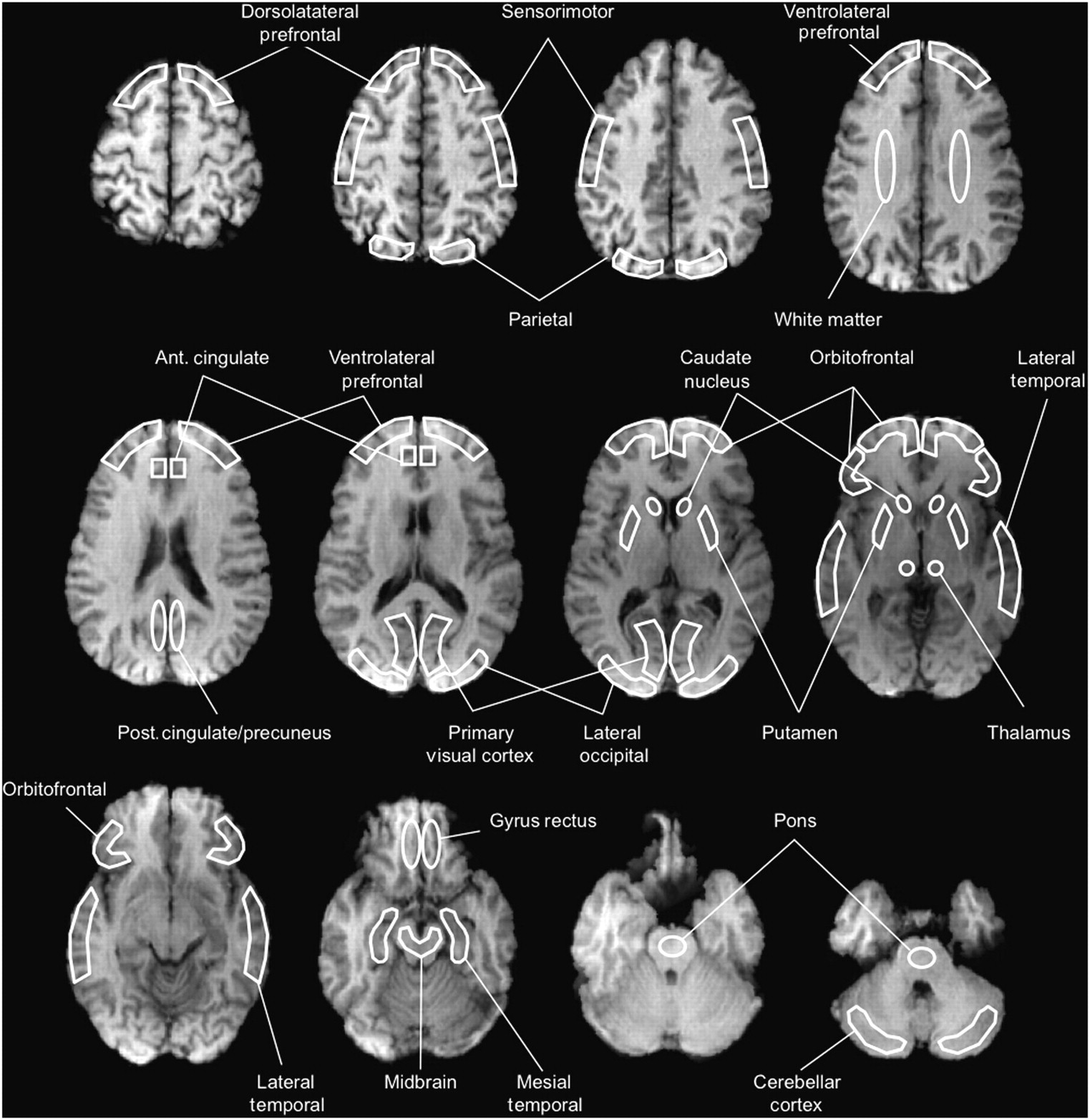
- FIGURE 1.
Schematic representation of ROI template defined on MR image. In white, with their respective labels, are enhanced examples of cortical and subcortical ROIs examined in this study. Also shown is the cerebellar cortex ROI used as reference region. Ant. = anterior; post. = posterior.
- FIGURE 2.
18F-florbetaben imaging with PET. Representative 18F-florbetaben PET transaxial images overlaid on individual coregistered MR images of 68-y-old control (MMSE, 29), 73-y-old patient with PD (MMSE, 29), 73-y-old patient with DLB (MMSE, 10), 70-y-old subject with MCI (MMSE, 26), 80-y-old AD patient (MMSE, 25), 62-y-old patient with FTLD (MMSE, 25), and 79-y-old patient with VaD (MMSE, 26). PET images show clear differences when comparing cortical 18F-florbetaben binding in controls, PD, VaD, and FTLD with MCI, DLB, or AD patients. Only nonspecific 18F-florbetaben binding in white matter is observed in controls, PD, VaD, and FTLD, compared with 18F-florbetaben binding in cortical areas of AD, MCI, and DLB patients. All images are scaled to same SUVR maximum. HC = controls.
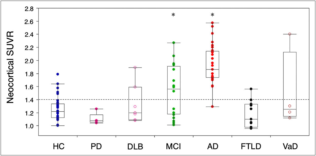
- FIGURE 3.
In vivo quantification of neocortical Aβ burden with 18F-florbetaben.Box and whiskers plots of Aβ burden by clinical classification. Aβ burden in AD and MCI groups was significantly higher (*) than in controls. About 60% of MCI subjects showed high florbetaben retention. Two of 7 DLB, 1 of 11 FTLD, and 1 of 4 VaD patients presented with high florbetaben retention although lower than that observed in AD. PD patients showed no cortical florbetaben retention. Dotted line = threshold (SUVR 1.4) between high and low florbetaben binding. HC = controls. *Significantly different from controls (P < 0.05).


Tables
Parameter Controls (n = 32) PD (n = 5) DLB (n = 7) MCI (n = 20) AD (n = 30) FTLD (n = 11) VaD (n = 4) Age (y) 70.7 ± 6.3 72.6 ± 6.5 71.7 ± 5.7 73.4 ± 6.7 72.0 ± 9.2 63.5 ± 7.0* 73.0 ± 11.0 Sex M 19 5* 7* 12 14 7 0* F 13 0* 0* 8 16 4 4* MMSE 29.6 ± 0.7 27.4 ± 2.7 24.0 ± 6.6* 27.4 ± 1.9 22.8 ± 3.7* 24.5 ± 2.9* 27.8 ± 2.1 Clinical dementia rating 0.0 0.3 ± 0.3* 0.8 ± 0.3* 0.5 ± 0.2* 1.0 ± 0.0* 1.0 ± 0.4* 0.6 ± 0.3* Injected activity (MBq) 282 ± 47 294 ± 17 271 ± 31 295 ± 14 268 ± 41 301 ± 29 263 ± 39 Injected mass (μg) 1.2 ± 0.7 2.1 ± 1.0 2.2 ± 1.5 1.7 ± 0.7 1.5 ± 1.6 1.7 ± 1.5 2.4 ± 1.1 ↵* Significantly different from controls (P < 0.05).
Region Controls (n = 32) PD (n = 5) DLB (n = 7) MCI (n = 20) AD (n = 30) FTLD (n = 11) VaD (n = 4) Dorsolateral prefrontal 1.19 ± 0.2 1.09 ± 0.1 1.24 ± 0.3 1.36 ± 0.4 1.82 ± 0.3* 1.10 ± 0.2 1.43 ± 0.6 Ventrolateral prefrontal 1.26 ± 0.2 1.08 ± 0.1 1.37 ± 0.4 1.54 ± 0.5* 2.02 ± 0.3* 1.18 ± 0.3 1.54 ± 0.6 Orbitofrontal 1.31 ± 0.2 1.11 ± 0.1 1.38 ± 0.3 1.56 ± 0.4* 2.00 ± 0.4* 1.21 ± 0.2 1.59 ± 0.8 Posterior cingulate 1.24 ± 0.2 1.03 ± 0.0 1.37 ± 0.5 1.60 ± 0.5* 2.05 ± 0.4* 1.21 ± 0.3 1.52 ± 0.7 Anterior cingulate 1.20 ± 0.2 0.90 ± 0.1 1.19 ± 0.3 1.45 ± 0.5 1.95 ± 0.4* 1.20 ± 0.3 1.43 ± 1.0 Parietal cortex 1.18 ± 0.2 1.12 ± 0.1 1.34 ± 0.4 1.42 ± 0.4* 1.82 ± 0.3* 1.07 ± 0.2 1.35 ± 0.3 Occipital cortex 1.41 ± 0.2 1.41 ± 0.2 1.45 ± 0.2 1.61 ± 0.3 1.83 ± 0.2* 1.27 ± 0.1 1.59 ± 0.3 Lateral temporal cortex 1.29 ± 0.2 1.15 ± 0.1 1.32 ± 0.2 1.56 ± 0.4* 1.96 ± 0.3* 1.19 ± 0.2 1.61 ± 0.6 Mesial temporal cortex 1.23 ± 0.1 1.14 ± 0.1 1.21 ± 0.2 1.26 ± 0.2 1.40 ± 0.2* 1.17 ± 0.1 1.29 ± 0.2 Caudate nuclei 1.32 ± 0.2 1.24 ± 0.1 1.49 ± 0.4 1.63 ± 0.5* 2.00 ± 0.4* 1.24 ± 0.2 1.48 ± 0.6 Putamen 1.30 ± 0.1 1.24 ± 0.1 1.45 ± 0.4 1.50 ± 0.3* 1.81 ± 0.3* 1.27 ± 0.2 1.40 ± 0.4 Thalamus 1.30 ± 0.2 1.21 ± 0.2 1.31 ± 0.2 1.34 ± 0.2 1.51 ± 0.3* 1.27 ± 0.2 1.30 ± 0.2 Mid brain 1.75 ± 0.2 1.88 ± 0.3 1.89 ± 0.2 1.74 ± 0.1 1.81 ± 0.3 1.65 ± 0.3 1.97 ± 0.3 Pons 1.97 ± 0.2 2.06 ± 0.3 1.93 ± 0.1 1.96 ± 0.2 1.93 ± 0.4 1.80 ± 0.2 2.10 ± 0.4 White matter 1.97 ± 0.2 1.82 ± 0.2 1.86 ± 0.1 1.95 ± 0.2 1.93 ± 0.4 1.80 ± 0.3 1.95 ± 0.2 Neocortex 1.26 ± 0.2 1.14 ± 0.1 1.38 ± 0.3 1.52 ± 0.4* 1.93 ± 0.3* 1.18 ± 0.1 1.51 ± 0.6 Effect size (d) 0.9 0.8 0.8 2.8 0.4 0.6 ↵* Significantly different from controls (P < 0.05).


Jump to section
Related Articles
Cited By...
- Quantitative PET imaging and modeling of molecular blood-brain barrier permeability
- Impact of PET Reconstruction on Amyloid-{beta} Quantitation in Cross-Sectional and Longitudinal Analyses
- Traumatic brain injury and Alzheimers Disease biomarkers: A systematic review of findings from amyloid and tau positron emission tomography (PET)
- Inter-scanner A{beta}-amyloid PET harmonization using barrel phantom spatial resolution matching
- Impact of PET reconstruction on A{beta}-amyloid quantitation in cross-sectional and longitudinal analyses
- Synthesizing Images of Tau Pathology from Cross-modal Neuroimaging using Deep Learning
- p-tau/A{beta}42 Ratio Associates with Cognitive Decline in Alzheimers disease, Mild Cognitive Impairment, and Cognitively Unimpaired Older Adults
- A Fully Automatic Technique for Precise Localization and Quantification of Amyloid-{beta} PET Scans
- Progressive Tau Accumulation in Alzheimer Disease: 2-Year Follow-up Study
- 3D Mapping Reveals Network-specific Amyloid Progression and Subcortical Susceptibility
- Spatial Normalization of 18F-Flutemetamol PET Images Using an Adaptive Principal-Component Template
- Test-Retest Reproducibility for the Tau PET Imaging Agent Flortaucipir F 18
- {beta}-Amyloid accumulation in the human brain after one night of sleep deprivation
- 18F-AV-1451 binds to motor-related subcortical gray and white matter in corticobasal syndrome
- Cardiac Amyloid Imaging with 18F-Florbetaben PET: A Pilot Study
- Visualization and Quantification of 3-Dimensional Stereotactic Surface Projections for 18F-Flutemetamol PET Using Variable Depth
- Performance of 11C-Pittsburgh Compound B PET Binding Potential Images in the Detection of Amyloid Deposits on Equivocal Static Images
- Optimal Target Region for Subject Classification on the Basis of Amyloid PET Images
- A{beta} imaging with 18F-florbetaben in prodromal Alzheimer's disease: a prospective outcome study
- Improved Power for Characterizing Longitudinal Amyloid-{beta} PET Changes and Evaluating Amyloid-Modifying Treatments with a Cerebral White Matter Reference Region
- 18F-Florbetapir PET in Patients with Frontotemporal Dementia and Alzheimer Disease
- Automated Quantification of 18F-Flutemetamol PET Activity for Categorizing Scans as Negative or Positive for Brain Amyloid: Concordance with Visual Image Reads
- Systematic Comparison of the Performance of Integrated Whole-Body PET/MR Imaging to Conventional PET/CT for 18F-FDG Brain Imaging in Patients Examined for Suspected Dementia
- Molecular Imaging of Alzheimer Disease Pathology
- Implementation and Validation of an Adaptive Template Registration Method for 18F-Flutemetamol Imaging Data
- Head-to-Head Comparison of 11C-PiB and 18F-AZD4694 (NAV4694) for {beta}-Amyloid Imaging in Aging and Dementia
- PET Quantification of 18F-Florbetaben Binding to {beta}-Amyloid Deposits in Human Brains
- Brain {beta}-amyloid load approaches a plateau
- The history of cerebral PET scanning: From physiology to cutting-edge technology
- Brain Amyloid Imaging
- Preclinical Characterization of a Novel Class of 18F-Labeled PET Tracers for Amyloid-{beta}
- A{beta}-amyloid deposition in patients with Parkinson disease at risk for development of dementia
- Brain Amyloid Imaging