


Article Figures & Data
Figures
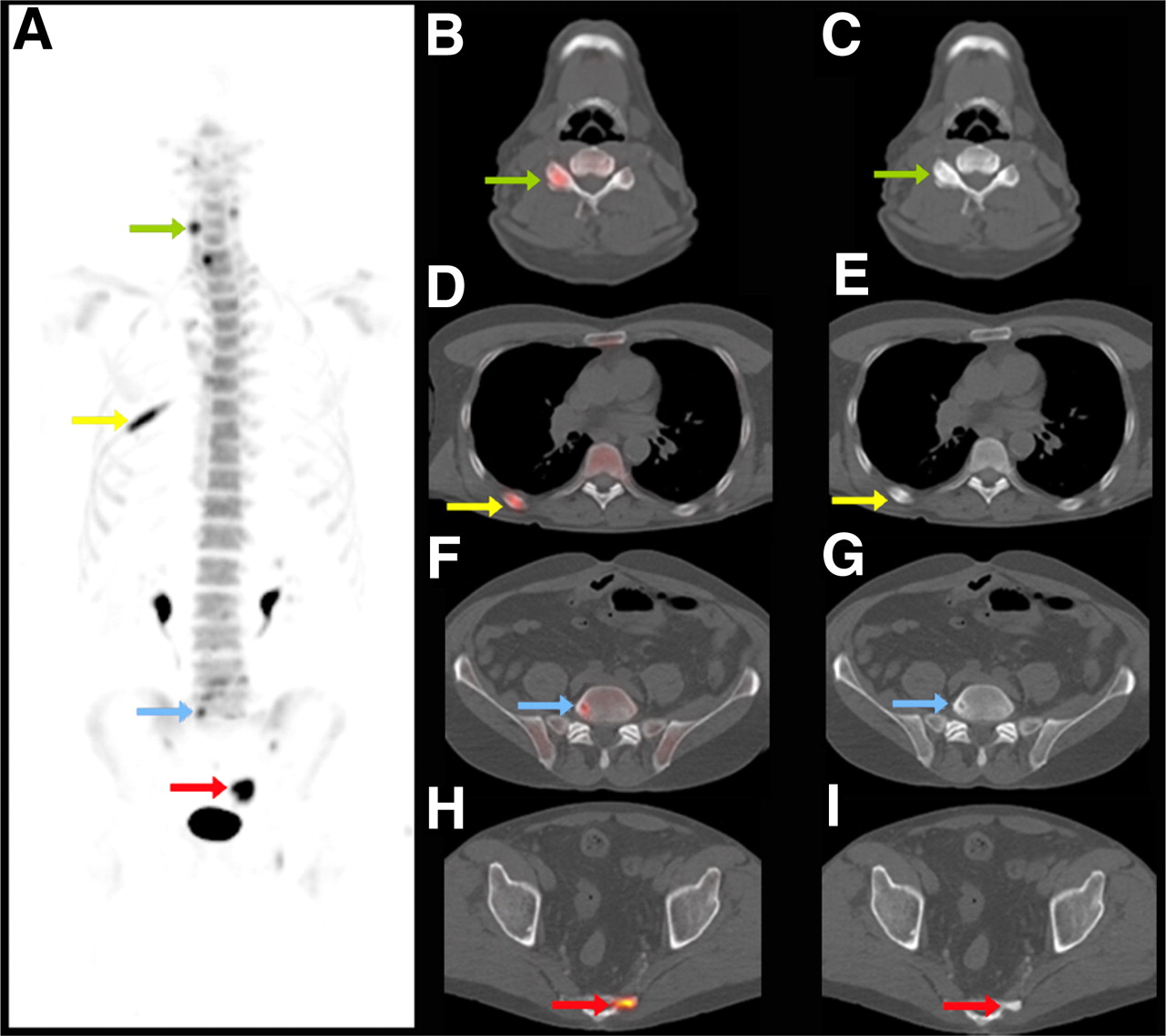
- FIGURE 1.
Maximum-intensity-projection PET image (A) and selected axial PET/CT (B, D, F, H) and CT (C, E, G, I) images of 18F-NaF PET/CT scan of 63-y-old man with prostate cancer. Increased 18F-NaF uptake can be seen in benign changes of right cervical facet joint (green arrow in A, B, and C), healing rib fracture (yellow arrow in A, D, and E), benign lumbar vertebral bone cyst (blue arrow in A, F, and G), and blastic metastasis in sacrum (red arrow in A, H, and I). Additional value of CT to characterize focally increased tracer uptake is evident.
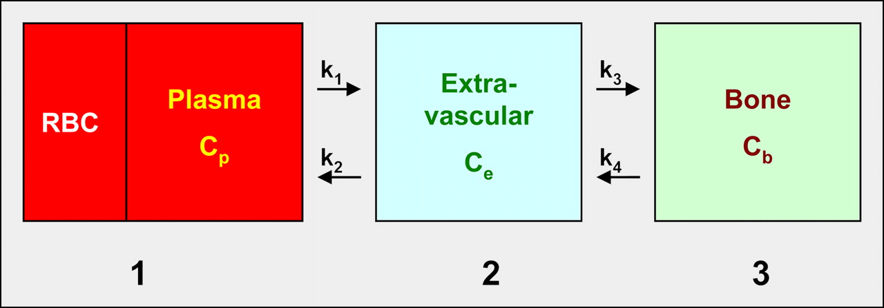
- FIGURE 2.
18F-NaF kinetic model with 3 compartments: vascular (1), extravascular (2), and bone (3). Plasma clearance of 18F-NaF is measured in left ventricle or aorta with PET scanner or from arterial blood draw; k1–k4 are rate constants; k1 and k2 represent forward and reverse transport from plasma, and k3 and k4 represent uptake and release from bone. If extraction fraction equals 1, then k1 represents local bone blood flow. Influx rate Ki = k1 · k3 · (k2 + k3)−1 is related to Ca2+ influx and bone apposition rate and, presumably, represents bone remodeling rate. Ki is determined by both bone blood flow and bone turnover. Therefore, measurements have to be interpreted in context of each individual study. Respective concentrations of fluoride are denoted as Cp (in plasma), Ce (in extravascular space), and Cb (in bone compartment). RBC = red blood cells
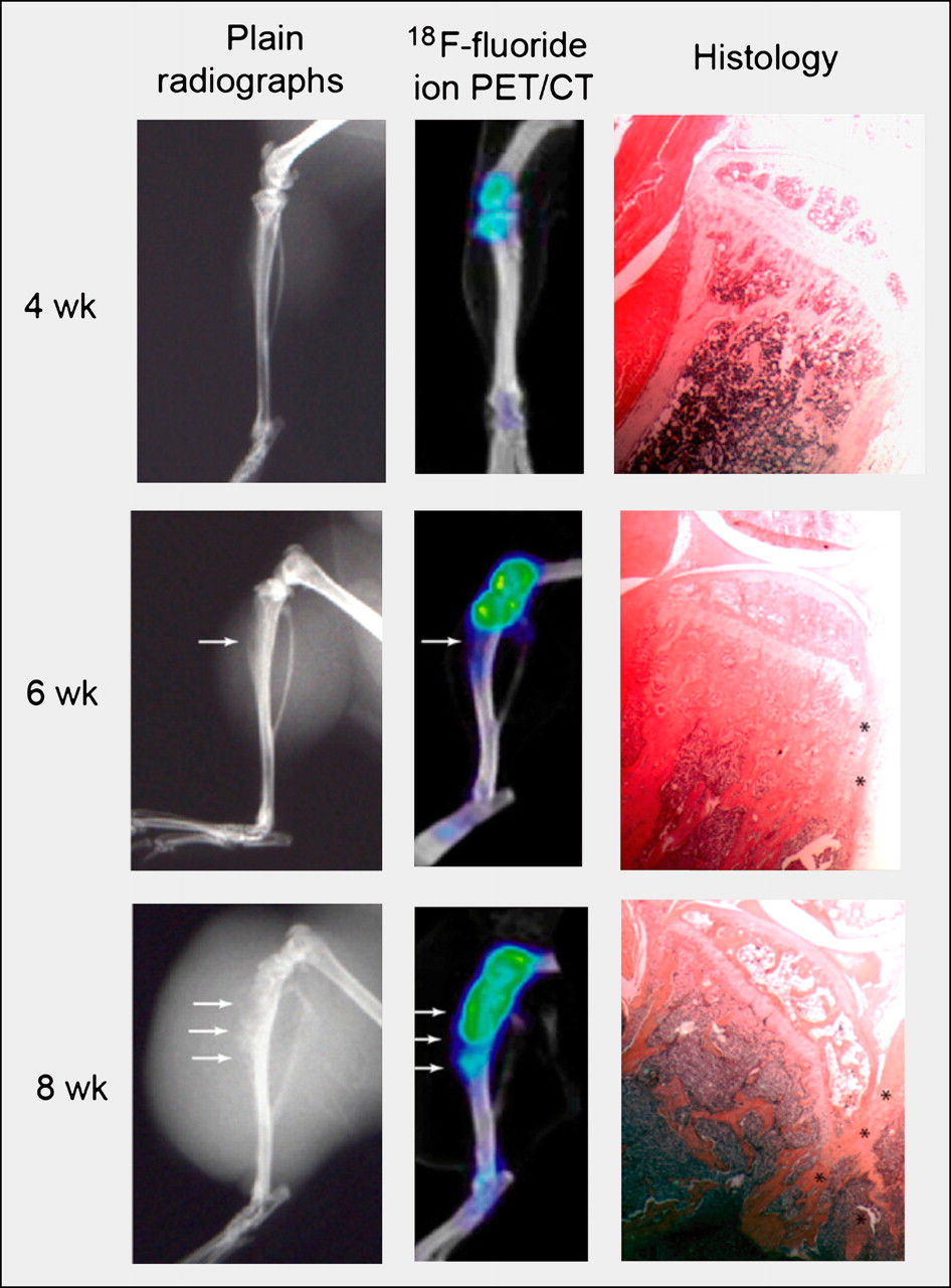
- FIGURE 3.
Radiographs (left), 18F-NaF PET/CT scans (middle), and photomicrographs of histologic specimen (right). PET/CT images reveal osteoblastic lesion earlier (4 wk) than radiography (arrows denote bone lesions). Increasing 18F-NaF uptake over time corresponds to increased bone formation seen on histology (asterisks). (Reprinted from (36).)


Jump to section
Related Articles
Cited By...
- Multimodality Imaging of Aortic Valve Calcification and Function in a Murine Model of Calcific Aortic Valve Disease and Bicuspid Aortic Valve
- Multimodality Imaging of Aortic Valve Calcification and Function in a Murine Model of Calcific Aortic Valve Disease and Bicuspid Aortic Valve
- 68Ga-Bisphosphonates for the Imaging of Extraosseous Calcification by Positron Emission Tomography
- Reducing Radiation Exposure from PET Patients
- 18F-NaF PET/CT of Obese Patients on a Lutetium-Yttrium Oxyorthosilicate PET/CT System: Patient Dosimetry, Optimization of Injected Activity, and Acquisition Time
- HAP-multitag, a PET and positive MRI contrast nanotracer for the longitudinal characterization of vascular calcifications in atherosclerosis
- Observer Agreement and Accuracy of 18F-Sodium Fluoride PET/CT in the Diagnosis of Bone Metastases in Prostate Cancer
- 18F-Sodium Fluoride PET: History, Technical Feasibility, Mechanism of Action, Normal Biodistribution, and Diagnostic Performance in Bone Metastasis Detection Compared with Other Imaging Modalities
- Three-Hour Delayed Imaging Improves Assessment of Coronary 18F-Sodium Fluoride PET
- Characterization of Bone Lesions in Myeloma Before and During Anticancer Therapy Using 18F-FDG-PET/CT and 18F-NaF-PET/CT
- Assessment of Physiologic Intracranial Calcification in Healthy Adults Using 18F-NaF PET/CT
- A Prospective Study Comparing 99mTc-Hydroxyethylene-Diphosphonate Planar Bone Scintigraphy and Whole-Body SPECT/CT with 18F-Fluoride PET/CT and 18F-Fluoride PET/MRI for Diagnosing Bone Metastases
- Repeatability of Quantitative 18F-NaF PET: A Multicenter Study
- The Role of 18F-Sodium Fluoride PET/CT Bone Scans in the Diagnosis of Metastatic Bone Disease from Breast and Prostate Cancer
- Bone-Targeted Imaging and Radionuclide Therapy in Prostate Cancer
- 18F-Fluoride PET in the Assessment of Malignant Bone Disease
- Impact of Personal Characteristics and Technical Factors on Quantification of Sodium 18F-Fluoride Uptake in Human Arteries: Prospective Evaluation of Healthy Subjects
- AEG-1 Promoter-Mediated Imaging of Prostate Cancer
- A Compartmental Model of Mouse Thrombopoiesis and Erythropoiesis to Predict Bone Marrow Toxicity After Internal Irradiation
- An Approach to Breast Cancer Diagnosis via PET Imaging of Microcalcifications Using 18F-NaF
- Mammary Cancer Bone Metastasis Follow-up Using Multimodal Small-Animal MR and PET Imaging
- Dynamic Bone Imaging with 99mTc-Labeled Diphosphonates and 18F-NaF: Mechanisms and Applications
- Clinical utility of fluoride-18 positron emission tomography/CT in temporomandibular disorder with osteoarthritis: comparisons with 99mTc-MDP bone scan