
Main menu

  • Home
  • Content
    • Current
    • Ahead of print
    • Past Issues
    • JNM Supplement
    • SNMMI Annual Meeting Abstracts
    • Continuing Education
    • JNM Podcasts
  • Subscriptions
    • Subscribers
    • Institutional and Non-member
    • Rates
    • Journal Claims
    • Corporate & Special Sales
  • Authors
    • Submit to JNM
    • Information for Authors
    • Assignment of Copyright
    • AQARA requirements
  • Info
    • Reviewers
    • Permissions
    • Advertisers
  • About
    • About Us
    • Editorial Board
    • Contact Information
  • More
    • Alerts
    • Feedback
    • Help
    • SNMMI Journals
  • SNMMI
    • JNM
    • JNMT
    • SNMMI Journals
    • SNMMI

User menu

  • Subscribe
  • My alerts
  • Log in
  • My Cart

Search

  • Advanced search
Journal of Nuclear Medicine
  • SNMMI
    • JNM
    • JNMT
    • SNMMI Journals
    • SNMMI
  • Subscribe
  • My alerts
  • Log in
  • My Cart
Journal of Nuclear Medicine

Advanced Search

  • Home
  • Content
    • Current
    • Ahead of print
    • Past Issues
    • JNM Supplement
    • SNMMI Annual Meeting Abstracts
    • Continuing Education
    • JNM Podcasts
  • Subscriptions
    • Subscribers
    • Institutional and Non-member
    • Rates
    • Journal Claims
    • Corporate & Special Sales
  • Authors
    • Submit to JNM
    • Information for Authors
    • Assignment of Copyright
    • AQARA requirements
  • Info
    • Reviewers
    • Permissions
    • Advertisers
  • About
    • About Us
    • Editorial Board
    • Contact Information
  • More
    • Alerts
    • Feedback
    • Help
    • SNMMI Journals
  • View or Listen to JNM Podcast
  • Visit JNM on Facebook
  • Join JNM on LinkedIn
  • Follow JNM on Twitter
  • Subscribe to our RSS feeds
Research ArticleCLINICAL INVESTIGATIONS

Characterization of PiB Binding to White Matter in Alzheimer Disease and Other Dementias

Michelle T. Fodero-Tavoletti, Christopher C. Rowe, Catriona A. McLean, Laura Leone, Qiao-Xin Li, Colin L. Masters, Roberto Cappai and Victor L. Villemagne
Journal of Nuclear Medicine February 2009, 50 (2) 198-204; DOI: https://doi.org/10.2967/jnumed.108.057984
Michelle T. Fodero-Tavoletti
  • Find this author on Google Scholar
  • Find this author on PubMed
  • Search for this author on this site
Christopher C. Rowe
  • Find this author on Google Scholar
  • Find this author on PubMed
  • Search for this author on this site
Catriona A. McLean
  • Find this author on Google Scholar
  • Find this author on PubMed
  • Search for this author on this site
Laura Leone
  • Find this author on Google Scholar
  • Find this author on PubMed
  • Search for this author on this site
Qiao-Xin Li
  • Find this author on Google Scholar
  • Find this author on PubMed
  • Search for this author on this site
Colin L. Masters
  • Find this author on Google Scholar
  • Find this author on PubMed
  • Search for this author on this site
Roberto Cappai
  • Find this author on Google Scholar
  • Find this author on PubMed
  • Search for this author on this site
Victor L. Villemagne
  • Find this author on Google Scholar
  • Find this author on PubMed
  • Search for this author on this site
  • Article
  • Figures & Data
  • Info & Metrics
  • PDF
Loading

Article Figures & Data

Figures

  • Tables
  • FIGURE 1. 
    • Download figure
    • Open in new tab
    • Download powerpoint
    FIGURE 1. 

    In vitro binding studies demonstrate that 3H-PiB fails to bind to white matter brain homogenates. Scatchard plots of 3H-PiB binding to gray matter (AD patients [A] and age-matched HCs [B]) and white matter (AD patients [C] and age-matched HCs [D]) brain homogenates. Scatchard analysis indicated that 3H-PiB binds to AD gray matter (dissociation constant, 3.77 nM; maximum number of binding sites, 9,254 pmol 3H-PiB/g tissue) brain homogenates. No significant binding of 3H-PiB to HC gray matter or white matter homogenates was observed. Consequently, no binding parameters could be calculated. Binding data were analyzed using software (version 1.0; GraphPad). Figure is representative of at least 3 independent experiments.

  • FIGURE 2. 
    • Download figure
    • Open in new tab
    • Download powerpoint
    FIGURE 2. 

    IHC and IF analysis indicates PiB staining does not bind to white matter. Microscopy images of 2 serial sections (7 μm) from frontal cortex of AD patient (A) and centrum semiovale from age-matched HC (B) and AD patient (C). First section was immunostained with antibody to Aβ (1E8) to identify Aβ plaques; second serial section was stained with 100 μM PiB. Presence of Aβ plaques was detected in AD frontal cortex sections (black arrowheads) and colocalized with positive PiB staining (white arrowheads). Centrum semiovale sections exhibited no immunoreactivity with 1E8, indicating absence of plaques (B and C). At higher magnification, PiB staining was observed to highlight blood vessels. (D) Serial sections (5 μm) isolated from gray matter (frontal cortex; top) and white matter (centrum semiovale; bottom) of patient with DLB who underwent 11C-PiB PET 23 mo before his death. Sections (from left to right) were stained with antibodies to Aβ (1E8) to Aβ plaques, PiB, α-syn (97/8), and tau-protein (Dako) and stained with PiB (100 μM). Presence of Aβ plaques was detected in DLB frontal cortex sections and colocalized with positive PiB staining. Centrum semiovale sections exhibited no immunoreactivity with any reagent used, indicating absence of Aβ plaques, Lewy bodies, or neurofibrillary tangles. Images were taken on Leica microscope. Scale bars, 50 μm. α-syn = α-synuclein.
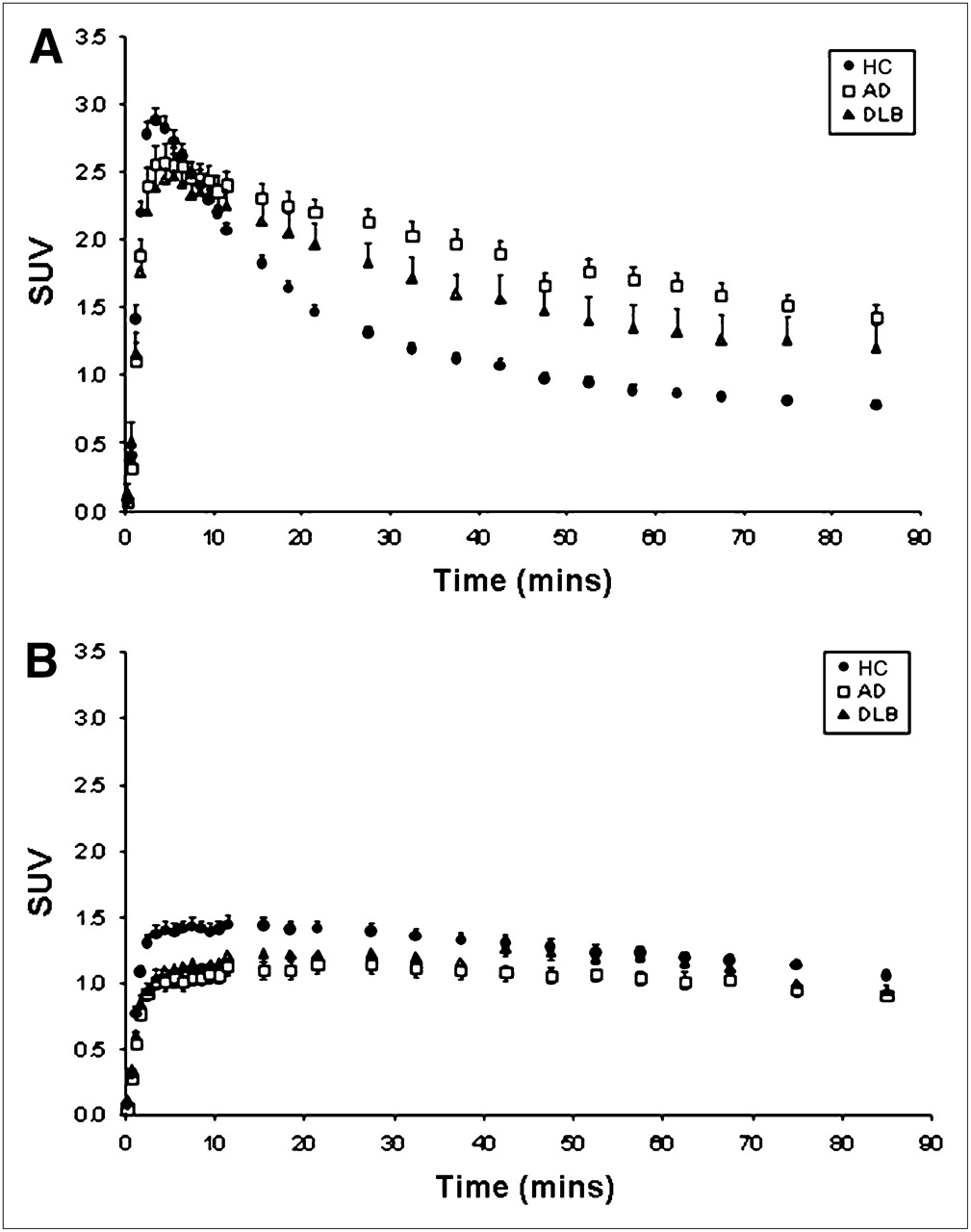
  • FIGURE 3. 
    • Download figure
    • Open in new tab
    • Download powerpoint
    FIGURE 3. 

    Time–activity curves demonstrate no difference in 11C-PiB clearance rates in white matter of all patients analyzed. Time–activity curves demonstrate uptake and clearance of 11C-PiB in frontal cortex (A) and white matter (B). There is greater retention of PiB in frontal cortex of AD patients and to lesser extent in DLB patients than in HCs. In contrast, there was no significant difference in rate of 11C-PiB clearance in all groups analyzed. SUV = standardized uptake value.

Tables

  • Figures
    • View popup
    TABLE 1

    ELISA Analysis of Aβ in Human Brain Tissue Homogenates

    TissueAβ1–40 (pmol/g protein)Aβ1–42 (pmol/g protein)Total Aβ (pmol/g protein)
    White matter (centrum semiovale)
     AD (n = 3)2.98 ± 0.1313.37 ± 2.8716.35 ± 3.0
     HC (n = 3)0.77 ± 0.058.12 ± 5.468.89 ± 5.5
    Gray matter (frontal cortex)
     AD (n = 3)128.0 ± 39.01,082.0 ± 337.01,210.0 ± 376.0
     HC (n = 3)4.61 ± 0.134.20 ± 0.618.81 ± 0.74
    • View popup
    TABLE 2

    Group Mean Demographic Data and PiB DVRs and Clearance Values in Aging HCs and Patients with Dementia

    PiB DVR and clearance value
    Demographic dataHCs (n = 27)AD patients (n = 17)DLB patients (n = 10)
    Age (y)72.6 ± 6.9 (range, 59–84)74.0 ± 11.3 (range, 56–91)72.0 ± 5.4 (range, 63–81)
    Female:male13:149:82:8
    Education (y)12.5 ± 3.512.3 ± 4.89.3 ± 0.9
    MMSE29.2 ± 0.922.1 ± 5.5*20.8 ± 7.2*
    Clinical dementia rating0.0 ± 0.01.2 ± 0.7*1.6 ± 0.8*
    ApoE ε4/non ε48/1910/77/3
    Neocortical† DVR1.22 ± 0.211.99 ± 0.23*1.67 ± 0.32*‡
    White matter DVR1.39 ± 0.19§1.36 ± 0.24§1.33 ± 0.36
    Neocortical clearance t1/2 (min)68.7 ± 11.6112.5 ± 28.3*94.6 ± 25.7*
    White matter clearance t1/2 (min)224.4 ± 89.9§268.7 ± 98.7§229.1 ± 107.0§
    • ↵* Statistically significant results, compared with controls (P < 0.05).

    • ↵† Neocortex comprises average DVR values for frontal, parietal, cingulate, occipital, and lateral temporal cortices.

    • ↵‡ Statistically significant results for DLB patients, compared with AD patients (P < 0.05).

    • ↵§ Statistically significant results for white matter, compared with gray matter (P < 0.05).

    • ApoE = apolipoprotein E.

PreviousNext
Back to top

In this issue

Journal of Nuclear Medicine: 50 (2)
Journal of Nuclear Medicine
Vol. 50, Issue 2
February 2009
  • Table of Contents
  • Table of Contents (PDF)
  • About the Cover
  • Index by author
Print
Download PDF
Article Alerts
Sign In to Email Alerts with your Email Address
Email Article

Thank you for your interest in spreading the word on Journal of Nuclear Medicine.

NOTE: We only request your email address so that the person you are recommending the page to knows that you wanted them to see it, and that it is not junk mail. We do not capture any email address.

Enter multiple addresses on separate lines or separate them with commas.
Characterization of PiB Binding to White Matter in Alzheimer Disease and Other Dementias
(Your Name) has sent you a message from Journal of Nuclear Medicine
(Your Name) thought you would like to see the Journal of Nuclear Medicine web site.
Citation Tools
Characterization of PiB Binding to White Matter in Alzheimer Disease and Other Dementias
Michelle T. Fodero-Tavoletti, Christopher C. Rowe, Catriona A. McLean, Laura Leone, Qiao-Xin Li, Colin L. Masters, Roberto Cappai, Victor L. Villemagne
Journal of Nuclear Medicine Feb 2009, 50 (2) 198-204; DOI: 10.2967/jnumed.108.057984

Citation Manager Formats

  • BibTeX
  • Bookends
  • EasyBib
  • EndNote (tagged)
  • EndNote 8 (xml)
  • Medlars
  • Mendeley
  • Papers
  • RefWorks Tagged
  • Ref Manager
  • RIS
  • Zotero
Share
Characterization of PiB Binding to White Matter in Alzheimer Disease and Other Dementias
Michelle T. Fodero-Tavoletti, Christopher C. Rowe, Catriona A. McLean, Laura Leone, Qiao-Xin Li, Colin L. Masters, Roberto Cappai, Victor L. Villemagne
Journal of Nuclear Medicine Feb 2009, 50 (2) 198-204; DOI: 10.2967/jnumed.108.057984
Twitter logo Facebook logo LinkedIn logo Mendeley logo
  • Tweet Widget
  • Facebook Like
  • Google Plus One
Bookmark this article

Jump to section

  • Article
    • Abstract
    • MATERIALS AND METHODS
    • RESULTS
    • DISCUSSION
    • CONCLUSION
    • Acknowledgments
    • Footnotes
    • References
  • Figures & Data
  • Info & Metrics
  • PDF

Related Articles

  • This Month in JNM
  • PubMed
  • Google Scholar

Cited By...

  • Development of a 213Bi-Labeled Pyridyl Benzofuran for Targeted {alpha}-Therapy of Amyloid-{beta} Aggregates
  • Amyloid-PET of the white matter: relationship to free water, fiber integrity, and cognition in patients with dementia and small vessel disease
  • Comparison of 11C-Pittsburgh Compound B and 18F-Flutemetamol White Matter Binding in PET
  • A Fully Automatic Technique for Precise Localization and Quantification of Amyloid-{beta} PET Scans
  • White Matter Reference Region in PET Studies of 11C-Pittsburgh Compound B Uptake: Effects of Age and Amyloid-{beta} Deposition
  • Optimizing Longitudinal Amyloid-{beta} PET Measurement: The Challenges of Intensity Normalization
  • Performance of 11C-Pittsburgh Compound B PET Binding Potential Images in the Detection of Amyloid Deposits on Equivocal Static Images
  • Voxelwise Relationships Between Distribution Volume Ratio and Cerebral Blood Flow: Implications for Analysis of {beta}-Amyloid Images
  • 18F-Florbetapir PET in Patients with Frontotemporal Dementia and Alzheimer Disease
  • Quantitative Analysis of Amyloid Deposition in Alzheimer Disease Using PET and the Radiotracer 11C-AZD2184
  • In vivo amyloid imaging in autopsy-confirmed Parkinson disease with dementia
  • Phase 1 Study of the Pittsburgh Compound B Derivative 18F-Flutemetamol in Healthy Volunteers and Patients with Probable Alzheimer Disease
  • Google Scholar

More in this TOC Section

  • Cardiac Presynaptic Sympathetic Nervous Function Evaluated by Cardiac PET in Patients with Chronotropic Incompetence Without Heart Failure
  • Validation and Evaluation of a Vendor-Provided Head Motion Correction Algorithm on the uMI Panorama PET/CT System
  • Efficacy and Safety of 124I-MIBG Dosimetry-Guided High-Activity 131I-MIBG Therapy of Advanced Pheochromocytoma or Neuroblastoma
Show more Clinical Investigations

Similar Articles

SNMMI

© 2025 SNMMI

Powered by HighWire