


Article Figures & Data
Figures
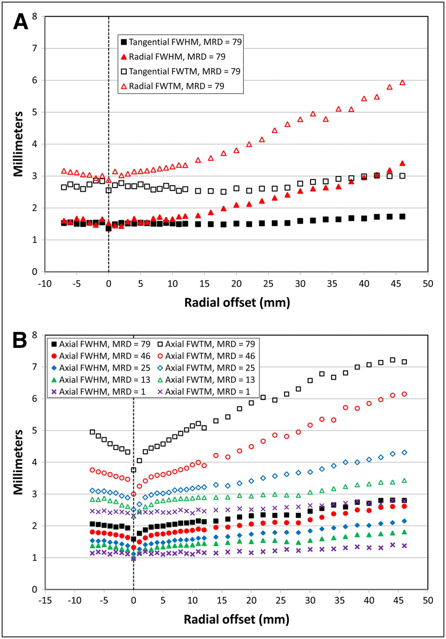
- FIGURE 1.
(A) Transaxial spatial resolutions (FWHM and FWTM) obtained with FORE and FBP, using MRD of 79 as function of radial distance from CFOV. Other settings for MRD yielded highly similar results, which have been left out for clarity. (B) Axial spatial resolutions (FWHM and FWTM) obtained with FORE and FBP as function of radial distance from CFOV for different MRD settings.
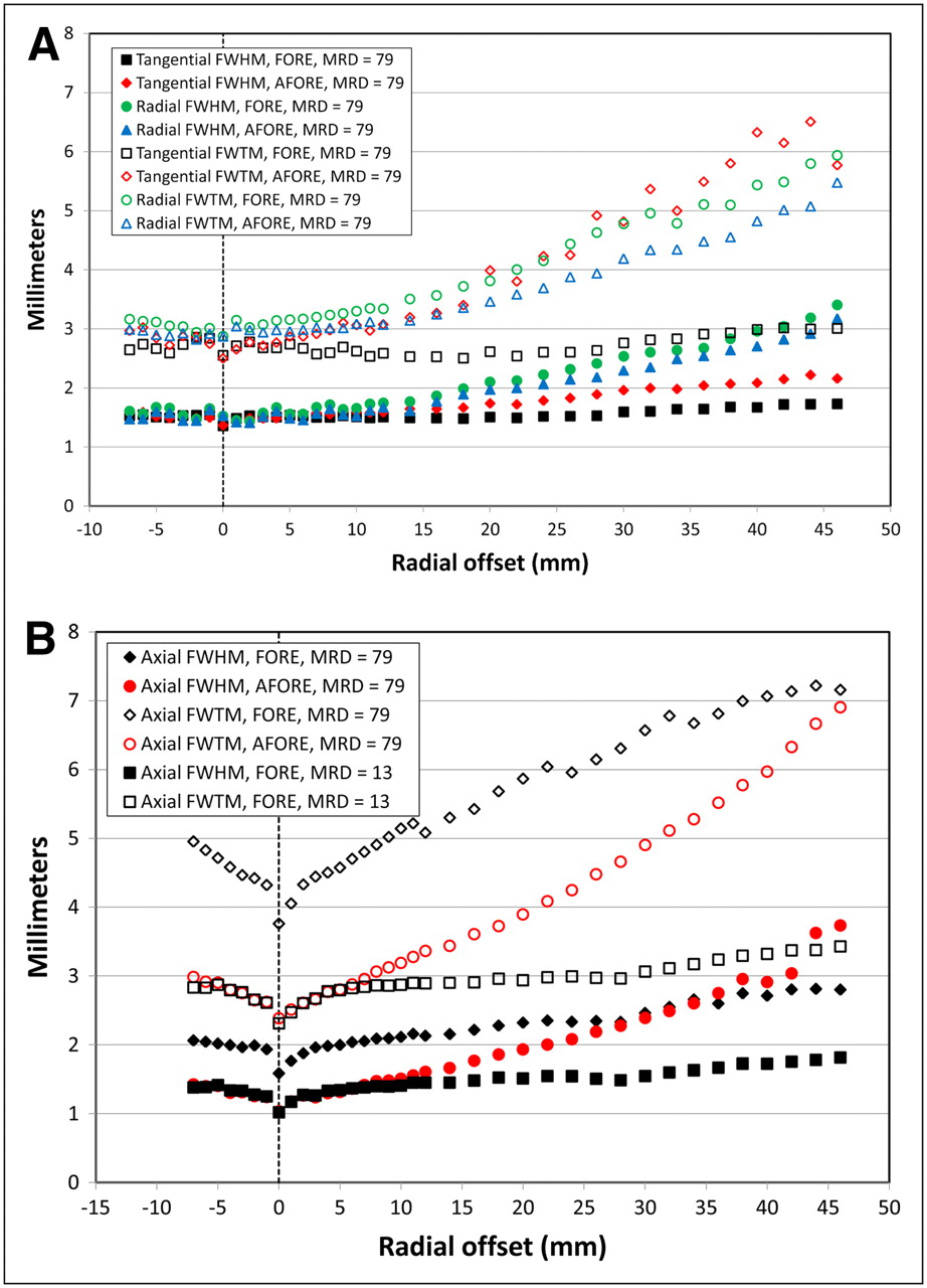
- FIGURE 2.
(A) Comparison of transaxial spatial resolutions using FORE and AFORE, with FBP as function of radial distance from CFOV. Only results for MRD of 79 have been plotted, because both algorithms yielded negligible differences upon variation of MRD. (B) Comparison of axial spatial resolutions using FORE and AFORE as function of radial distance from CFOV. For AFORE, only results for MRD of 79 have been plotted, because differences upon variation of MRD were negligible. FORE results depended strongly on MRD. As example for small MRDs, FORE results with MRD of 13 are shown.
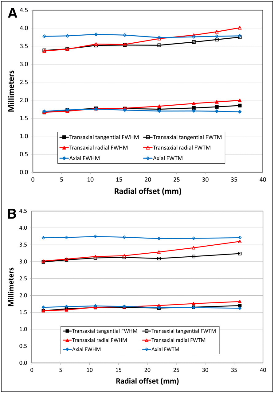
- FIGURE 3.
Spatial resolution (FWHM and FWTM) in all directions as function of radial distance from CFOV for OSEM3D/MAP with β of 1.5 (A) and β of 0.5 (B).
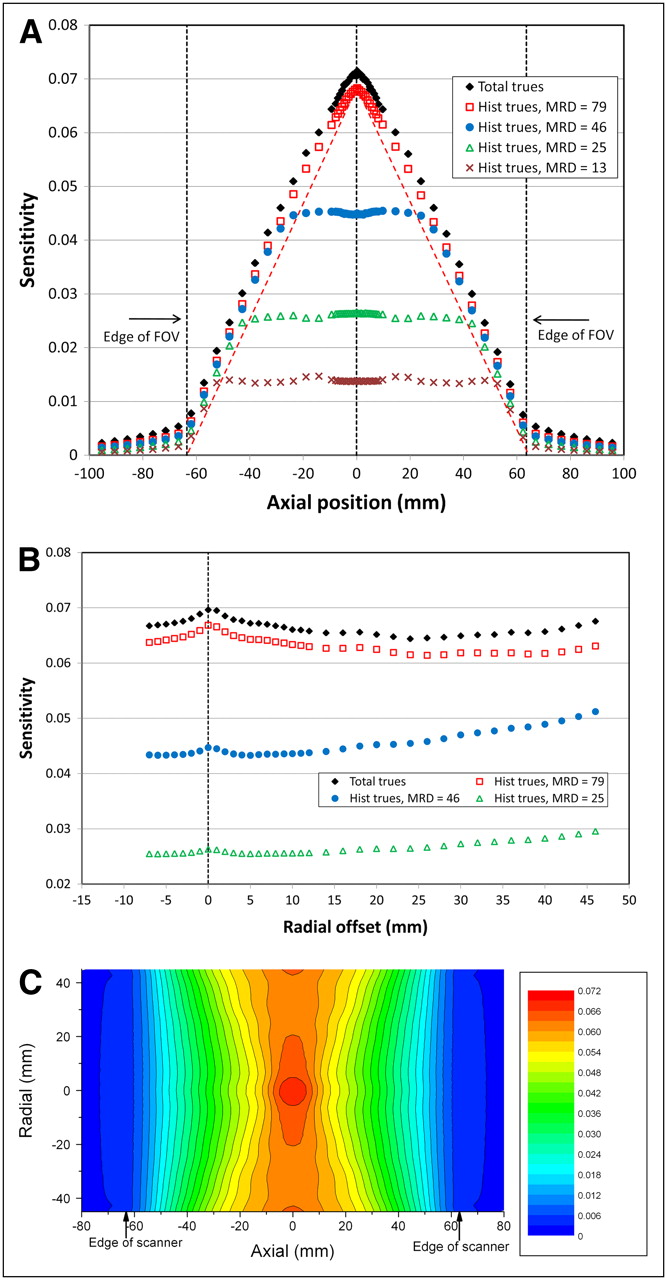
- FIGURE 4.
(A) Axial sensitivity profiles at radial center for different MRDs. Dashed red line indicates sensitivity as would be obtained for MRD of 79, with linear decrease down to axial edge of FOV of scanner. (B) Radial sensitivity profiles for middle plane for different MRDs. (C) Sensitivity for complete FOV for MRD of 79. All sensitivities correspond to default ΔE and Δt and are based on histogrammed (Hist) trues rates, except for ♦, which were calculated using total trues rate.
- FIGURE 5.
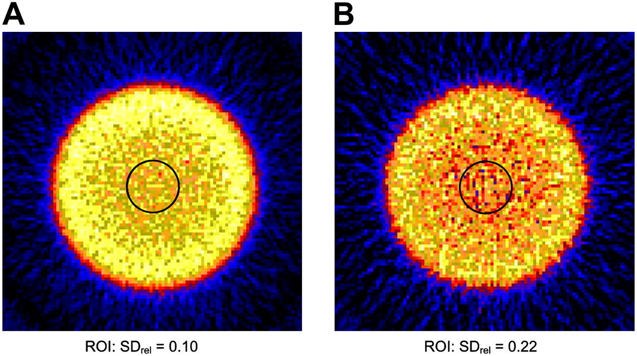
Comparison of reconstructed images of uniform 68Ge cylinder with 6-cm diameter using FORE (A) and AFORE (B) for central transaxial plane. Reconstruction algorithm for both images was FBP with matrix size of 128 × 128. Color was scaled to maximum pixel value in both images. SDrel was calculated for circular regions of interest. ROI = region of interest.


Tables
- TABLE 1
Geometric Properties of Several Commercial, Crystal-Based Small-Animal PET Scanners
PET scanner Crystal element size (mm3) No. of crystal rings Detector material Axial length (mm) Crystal ring diameter (mm) Aspect ratio* Largest LOR acceptance angle† (degrees) Inveon 1.5 × 1.5 × 10 80 LSO 127 161 0.79 38.3 F120 (12) 1.5 × 1.5 × 10 48 LSO 76 147 0.52 27.3 Mosaic (13) 2 × 2 × 10 52 GSO 119 197 0.60 28.0‡ Vista (14) 1.45 × 1.45 × (8 + 7) 26 LYSO/GSO phoswich 48 118 0.41 22.1 ClearPET (15,16) 2 × 2× (10 + 10) 32 LYSO/LuYAP phoswich 110 135 0.81 39.2 ↵* Aspect ratio is crystal ring diameter divided by axial length.
↵† LOR acceptance angle is angle between LOR and transaxial planes.
↵‡ This LOR angle is determined by software; LOR angle from aspect ratio would be somewhat larger.
GSO = gadolinium oxyorthosilicate; LYSO = lutetium yttrium orthosilicate; LuYAP = lutetium yttrium aluminum perovskite.
CFOV* Radial EFOV† Method of measurement Tangential Radial Axial Tangential Radial Axial FORE, FBP MRD = 79 1.52 ± 0.02 1.57 ± 0.09 1.98 ± 0.04 1.70 ± 0.04 3.02 ± 0.26 2.75 ± 0.08 MRD = 46 1.52 ± 0.02 1.56 ± 0.09 1.72 ± 0.05 1.74 ± 0.04 2.99 ± 0.23 2.53 ± 0.09 MRD = 25 1.52 ± 0.02 1.54 ± 0.09 1.47 ± 0.04 1.74 ± 0.04 2.95 ± 0.17 2.06 ± 0.06 MRD = 13 1.51 ± 0.02 1.52 ± 0.08 1.32 ± 0.05 1.78 ± 0.04 2.95 ± 0.21 1.75 ± 0.05 MRD = 1 1.47 ± 0.02 1.51 ± 0.08 1.14 ± 0.03 1.83 ± 0.07 2.94 ± 0.20 1.51 ± 0.07 AFORE, FBP MRD = 79 1.50 ± 0.02 1.51 ± 0.08 1.30 ± 0.05 2.12 ± 0.07 2.80 ± 0.23 3.17 ± 0.40 OSEM3D/MAP β = 1.5 mm 1.69 ± 0.04 1.68 ± 0.02 1.71 ± 0.03 1.85 1.99 1.68 β = 0.5 mm 1.57 ± 0.04 1.56 ± 0.02 1.66 ± 0.02 1.70 1.82 1.62 ↵* Values for CFOV for 2D reconstructions were obtained by averaging over 8 point source positions (2−5 mm from radial center). For OSEM3D/MAP, averaging was done for 2 positions (2 and 6 mm from radial center).
↵† Values for radial EFOV for 2D reconstructions were obtained by averaging over 6 point source positions (36–46 mm from radial center). For OSEM3D/MAP, value at 36 mm from radial center was taken.
Errors are SD belonging to averaging as indicated.
Δt (ns) ΔE (keV) 2.8 3.4 4.1 4.7 350–650 0.068 (0.071) 0.068 (0.072) 0.068 (0.072) 0.068 (0.072) 250–750 0.099 (0.107) 0.100 (0.109) 0.101 (0.110) 0.101 (0.111) Values belong to CFOV and are based on histogrammed trues rate. Numbers in parentheses are based on total trues rate.


Jump to section
Related Articles
Cited By...
- Multimodality Imaging of Aortic Valve Calcification and Function in a Murine Model of Calcific Aortic Valve Disease and Bicuspid Aortic Valve
- Multimodality Imaging of Aortic Valve Calcification and Function in a Murine Model of Calcific Aortic Valve Disease and Bicuspid Aortic Valve
- Asymmetry of Fibrillar Plaque Burden in Amyloid Mouse Models
- Linking imaging to omics utilizing image-guided tissue extraction
- A Promising Future: Comparable Imaging Capability of MRI-Compatible Silicon Photomultiplier and Conventional Photosensor Preclinical PET Systems
- Immuno-PET and Immuno-SPECT of Rheumatoid Arthritis with Radiolabeled Anti-Fibroblast Activation Protein Antibody Correlates with Severity of Arthritis
- Early Response Monitoring with 18F-FDG PET and Cetuximab-F(ab')2-SPECT After Radiotherapy of Human Head and Neck Squamous Cell Carcinomas in a Mouse Model
- Small-Animal PET Imaging of Isolated Perfused Rat Heart
- Quantitative ImmunoPET of Prostate Cancer Xenografts with 89Zr- and 124I-Labeled Anti-PSCA A11 Minibody
- N-Acetylcysteine- and MK-801-Induced Changes in Glutamate Levels Do Not Affect In Vivo Binding of Metabotropic Glutamate 5 Receptor Radioligand 11C-ABP688 in Rat Brain
- Performance Evaluation of the Small-Animal nanoScan PET/MRI System
- Bone Marrow Stromal Cell Transplantation Enhances Recovery of Local Glucose Metabolism After Cerebral Infarction in Rats: A Serial 18F-FDG PET Study
- NEMA NU 4-2008 Comparison of Preclinical PET Imaging Systems
- PET of Tumors Expressing Gastrin-Releasing Peptide Receptor with an 18F-Labeled Bombesin Analog
- Imaging of Human Epidermal Growth Factor Receptor Type 2 Expression with 18F-Labeled Affibody Molecule ZHER2:2395 in a Mouse Model for Ovarian Cancer
- National Electrical Manufacturers Association NU-4 Performance Evaluation of the PET Component of the NanoPET/CT Preclinical PET/CT Scanner
- Detection of Macrophages in Aortic Aneurysms by Nanoparticle Positron Emission Tomography-Computed Tomography
- ImmunoSPECT and ImmunoPET of IGF-1R Expression with the Radiolabeled Antibody R1507 in a Triple-Negative Breast Cancer Model
- Performance Evaluation of the FLEX Triumph X-PET Scanner Using the National Electrical Manufacturers Association NU-4 Standards
- PET of Hypoxia with 89Zr-Labeled cG250-F(ab')2 in Head and Neck Tumors
- Image-Quality Assessment for Several Positron Emitters Using the NEMA NU 4-2008 Standards in the Siemens Inveon Small-Animal PET Scanner
- Pretargeted Immuno-Positron Emission Tomography Imaging of Carcinoembryonic Antigen-Expressing Tumors with a Bispecific Antibody and a 68Ga- and 18F-Labeled Hapten Peptide in Mice with Human Tumor Xenografts
- A Novel Facile Method of Labeling Octreotide with 18F-Fluorine
- NEMA NU4-2008 Image Quality Performance Report for the microPET Focus 120 and for Various Transmission and Reconstruction Methods