
Main menu

  • Home
  • Content
    • Current
    • Ahead of print
    • Past Issues
    • JNM Supplement
    • SNMMI Annual Meeting Abstracts
    • Continuing Education
    • JNM Podcasts
  • Subscriptions
    • Subscribers
    • Institutional and Non-member
    • Rates
    • Journal Claims
    • Corporate & Special Sales
  • Authors
    • Submit to JNM
    • Information for Authors
    • Assignment of Copyright
    • AQARA requirements
  • Info
    • Reviewers
    • Permissions
    • Advertisers
  • About
    • About Us
    • Editorial Board
    • Contact Information
  • More
    • Alerts
    • Feedback
    • Help
    • SNMMI Journals
  • SNMMI
    • JNM
    • JNMT
    • SNMMI Journals
    • SNMMI

User menu

  • Subscribe
  • My alerts
  • Log in
  • Log out
  • My Cart

Search

  • Advanced search
Journal of Nuclear Medicine
  • SNMMI
    • JNM
    • JNMT
    • SNMMI Journals
    • SNMMI
  • Subscribe
  • My alerts
  • Log in
  • Log out
  • My Cart
Journal of Nuclear Medicine

Advanced Search

  • Home
  • Content
    • Current
    • Ahead of print
    • Past Issues
    • JNM Supplement
    • SNMMI Annual Meeting Abstracts
    • Continuing Education
    • JNM Podcasts
  • Subscriptions
    • Subscribers
    • Institutional and Non-member
    • Rates
    • Journal Claims
    • Corporate & Special Sales
  • Authors
    • Submit to JNM
    • Information for Authors
    • Assignment of Copyright
    • AQARA requirements
  • Info
    • Reviewers
    • Permissions
    • Advertisers
  • About
    • About Us
    • Editorial Board
    • Contact Information
  • More
    • Alerts
    • Feedback
    • Help
    • SNMMI Journals
  • View or Listen to JNM Podcast
  • Visit JNM on Facebook
  • Join JNM on LinkedIn
  • Follow JNM on Twitter
  • Subscribe to our RSS feeds
OtherBASIC SCIENCE INVESTIGATIONS

MRI-Based Attenuation Correction for PET/MRI: A Novel Approach Combining Pattern Recognition and Atlas Registration

Matthias Hofmann, Florian Steinke, Verena Scheel, Guillaume Charpiat, Jason Farquhar, Philip Aschoff, Michael Brady, Bernhard Schölkopf and Bernd J. Pichler
Journal of Nuclear Medicine November 2008, 49 (11) 1875-1883; DOI: https://doi.org/10.2967/jnumed.107.049353
Matthias Hofmann
  • Find this author on Google Scholar
  • Find this author on PubMed
  • Search for this author on this site
Florian Steinke
  • Find this author on Google Scholar
  • Find this author on PubMed
  • Search for this author on this site
Verena Scheel
  • Find this author on Google Scholar
  • Find this author on PubMed
  • Search for this author on this site
Guillaume Charpiat
  • Find this author on Google Scholar
  • Find this author on PubMed
  • Search for this author on this site
Jason Farquhar
  • Find this author on Google Scholar
  • Find this author on PubMed
  • Search for this author on this site
Philip Aschoff
  • Find this author on Google Scholar
  • Find this author on PubMed
  • Search for this author on this site
Michael Brady
  • Find this author on Google Scholar
  • Find this author on PubMed
  • Search for this author on this site
Bernhard Schölkopf
  • Find this author on Google Scholar
  • Find this author on PubMed
  • Search for this author on this site
Bernd J. Pichler
  • Find this author on Google Scholar
  • Find this author on PubMed
  • Search for this author on this site
  • Article
  • Figures & Data
  • Info & Metrics
  • PDF
Loading

Article Figures & Data

Figures

  • Tables
  • FIGURE 1. 
    • Download figure
    • Open in new tab
    • Download powerpoint
    FIGURE 1. 

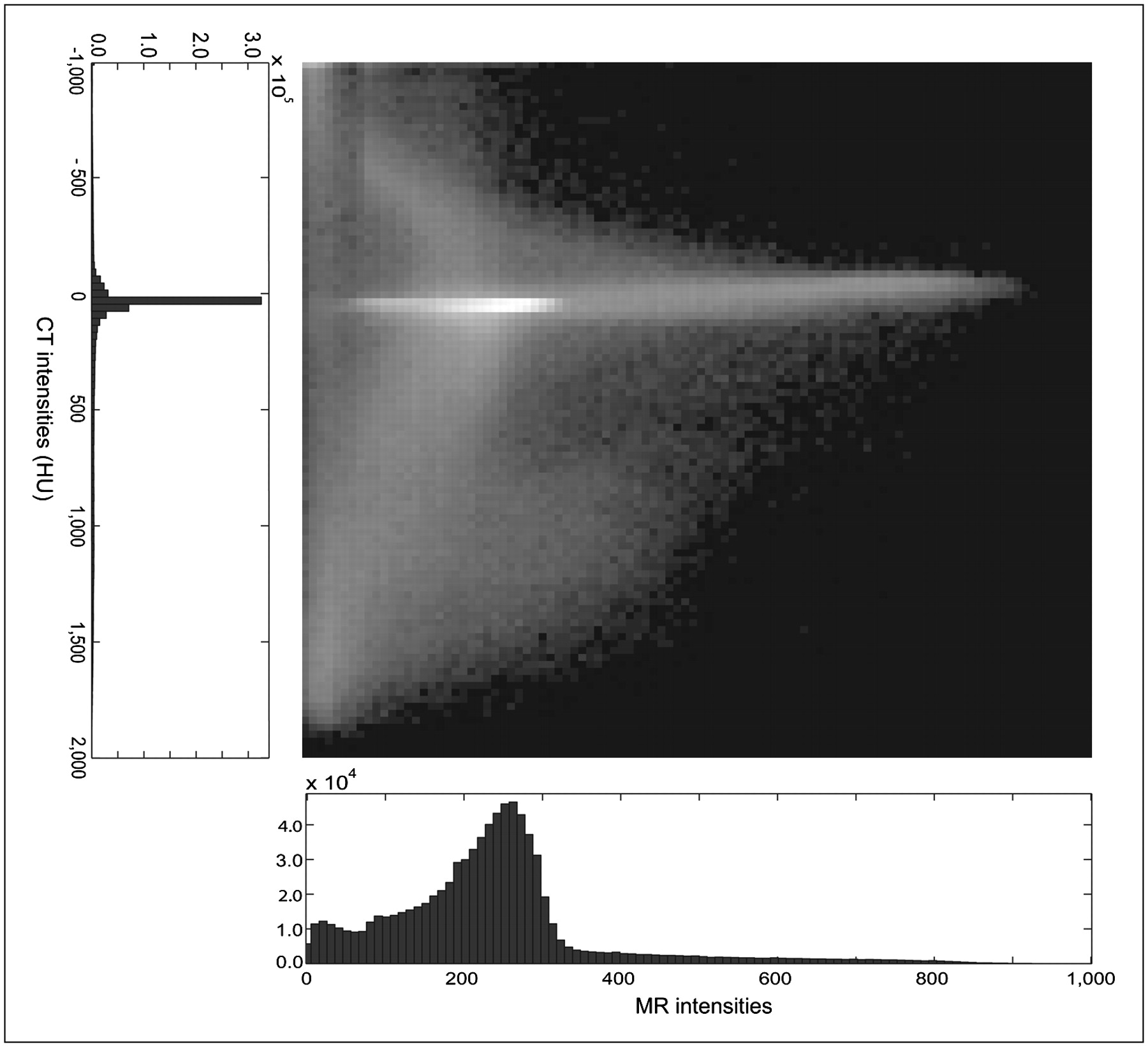
    2-dimensional histogram of MRI and CT intensities in T1-weighted head scan.
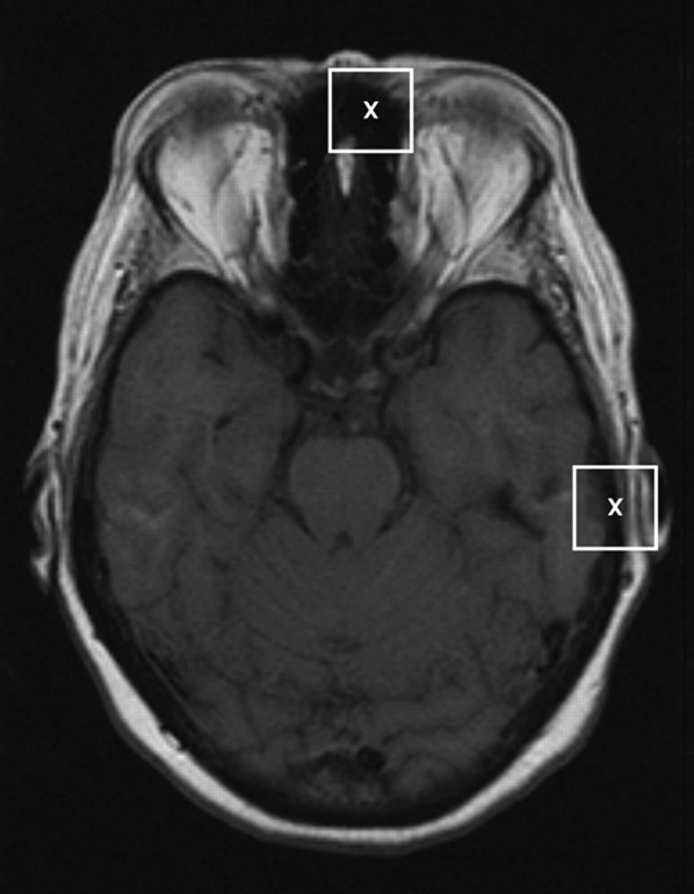
  • FIGURE 2. 
    • Download figure
    • Open in new tab
    • Download powerpoint
    FIGURE 2. 

    Xs indicate 2 voxels in MR image whose tissue classes (air and bone) cannot be distinguished on basis of their intensity alone. If we include surrounding patch (rectangle), we see patterns that are typical for either bone or air, and different attenuation values can be assigned.

  • FIGURE 3. 
    • Download figure
    • Open in new tab
    • Download powerpoint
    FIGURE 3. 

    Overview of steps involved in our method for obtaining attenuation-corrected PET image, based on PET detector sinogram and MR image. Resampling of MRI, PET, and CT to required resolution is performed wherever necessary.

  • FIGURE 4. 
    • Download figure
    • Open in new tab
    • Download powerpoint
    FIGURE 4. 

    Images from patient's T1-weighted spin-echo MRI (left), pseudo-CT (as predicted using our method; middle), and real CT (right) scans. MR T1SE = T1-weighted spin-echo MRI.
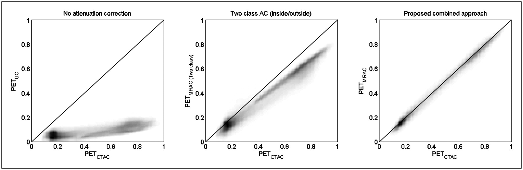
  • FIGURE 5. 
    • Download figure
    • Open in new tab
    • Download powerpoint
    FIGURE 5. 

    Joint histograms showing no AC (A), simple AC using attenuation map with only 2 attenuation values (B), and AC using our MRI-based predicted attenuation map (C). No significant systematic over- or underestimation of activity is demonstrated.
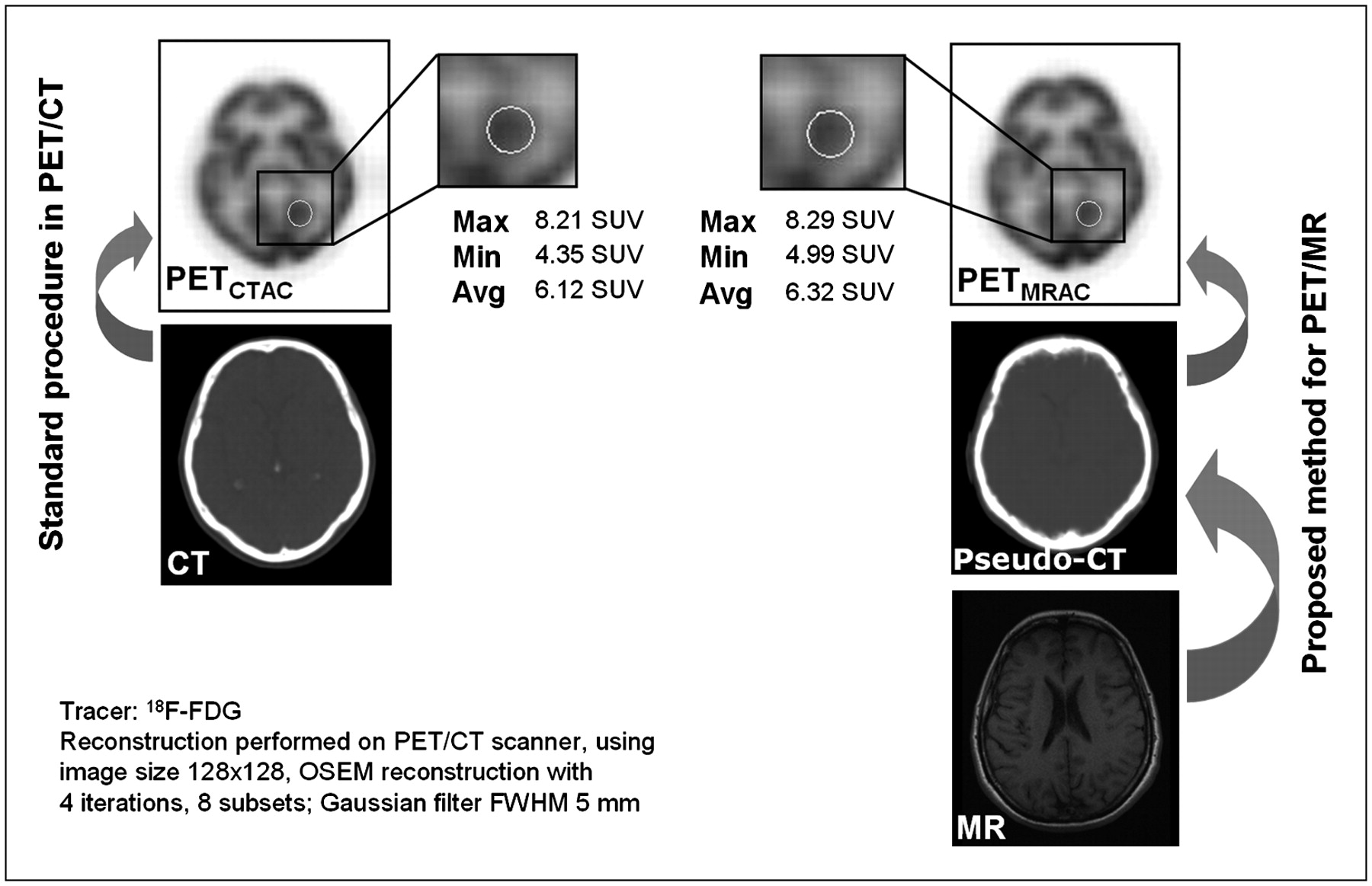
  • FIGURE 6. 
    • Download figure
    • Open in new tab
    • Download powerpoint
    FIGURE 6. 

    Direct comparison of CT attenuation-corrected PET image and PET image that was obtained with AC based on pseudo-CT that was calculated from MR image only. Maximum (Max), minimum (Min), and average (Avg) standardized uptake values (SUV) are given for shown ROI. OSEM = ordered-subset expectation maximization; FWHM = full width at half maximum.
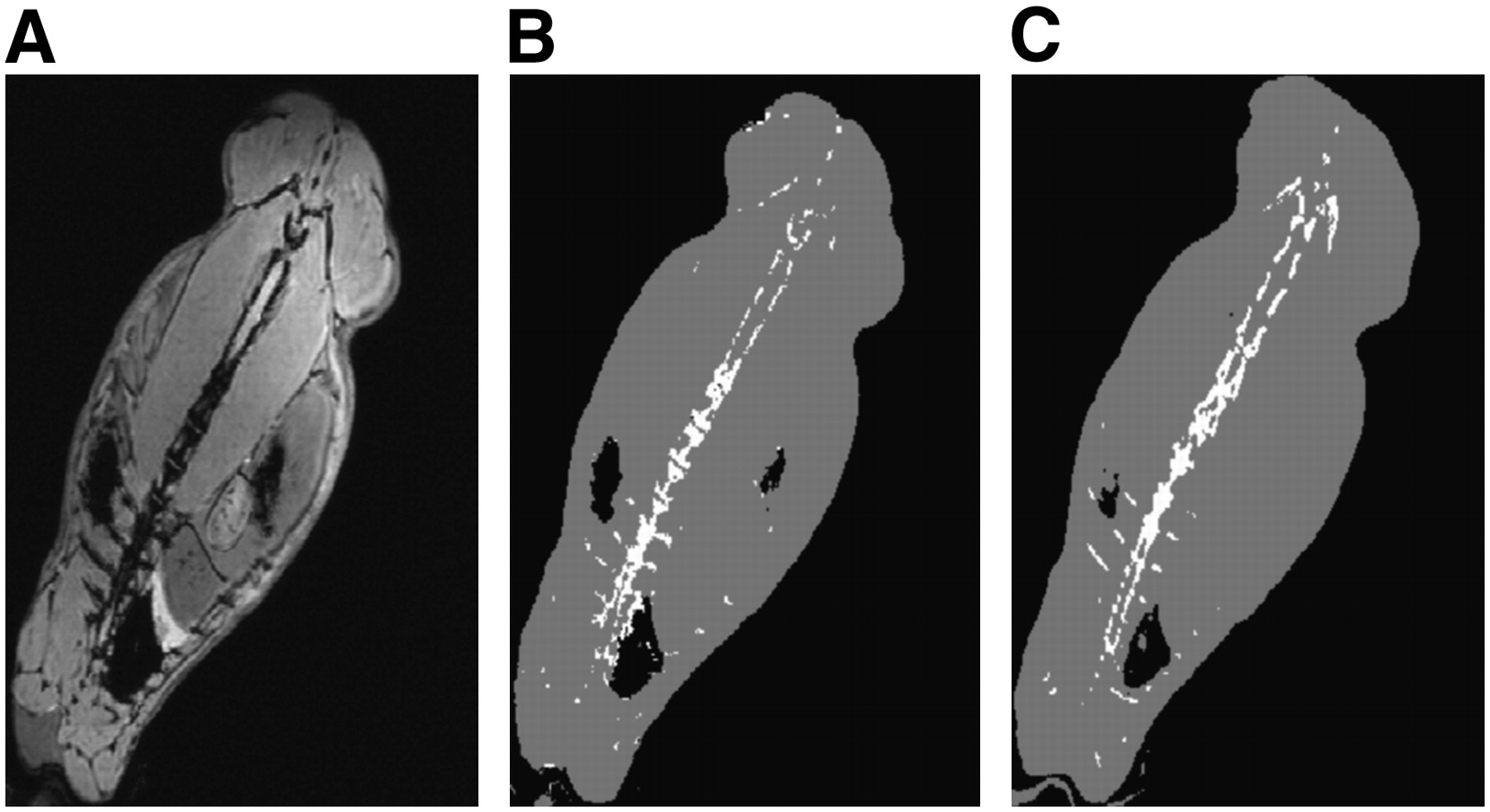
  • FIGURE 7. 
    • Download figure
    • Open in new tab
    • Download powerpoint
    FIGURE 7. 

    (A) MRI T2-weighted multi-echo data-imaging combination of rabbit. (B) Three-class labels as predicted using simple variation of our method for 3-class classification. (C) Three-class labels obtained by thresholding of CT image of rabbit. Not all differences between B and C are because of false predictions. Despite best efforts to physically fixate rabbit between MRI and CT scans, there was still some slight movement between scans, which explains misalignment between A and C.

Tables

  • Figures
    • View popup
    TABLE 1

    Regression Coefficients and R2 Values

    R2 value for…
    CoefficientProposed methodUncorrected imagesTwo-class AC images
    α−0.006 ± 0.0070.025 ± 0.0040.014 ± 0.009
    β0.990 ± 0.0260.138 ± 0.0190.78 ± 0.030
    R20.968 ± 0.0110.476 ± 0.0760.884 ± 0.044
PreviousNext
Back to top

In this issue

Journal of Nuclear Medicine: 49 (11)
Journal of Nuclear Medicine
Vol. 49, Issue 11
November 2008
  • Table of Contents
  • Table of Contents (PDF)
  • About the Cover
  • Index by author
Print
Download PDF
Article Alerts
Sign In to Email Alerts with your Email Address
Email Article

Thank you for your interest in spreading the word on Journal of Nuclear Medicine.

NOTE: We only request your email address so that the person you are recommending the page to knows that you wanted them to see it, and that it is not junk mail. We do not capture any email address.

Enter multiple addresses on separate lines or separate them with commas.
MRI-Based Attenuation Correction for PET/MRI: A Novel Approach Combining Pattern Recognition and Atlas Registration
(Your Name) has sent you a message from Journal of Nuclear Medicine
(Your Name) thought you would like to see the Journal of Nuclear Medicine web site.
Citation Tools
MRI-Based Attenuation Correction for PET/MRI: A Novel Approach Combining Pattern Recognition and Atlas Registration
Matthias Hofmann, Florian Steinke, Verena Scheel, Guillaume Charpiat, Jason Farquhar, Philip Aschoff, Michael Brady, Bernhard Schölkopf, Bernd J. Pichler
Journal of Nuclear Medicine Nov 2008, 49 (11) 1875-1883; DOI: 10.2967/jnumed.107.049353

Citation Manager Formats

  • BibTeX
  • Bookends
  • EasyBib
  • EndNote (tagged)
  • EndNote 8 (xml)
  • Medlars
  • Mendeley
  • Papers
  • RefWorks Tagged
  • Ref Manager
  • RIS
  • Zotero
Share
MRI-Based Attenuation Correction for PET/MRI: A Novel Approach Combining Pattern Recognition and Atlas Registration
Matthias Hofmann, Florian Steinke, Verena Scheel, Guillaume Charpiat, Jason Farquhar, Philip Aschoff, Michael Brady, Bernhard Schölkopf, Bernd J. Pichler
Journal of Nuclear Medicine Nov 2008, 49 (11) 1875-1883; DOI: 10.2967/jnumed.107.049353
Twitter logo Facebook logo LinkedIn logo Mendeley logo
  • Tweet Widget
  • Facebook Like
  • Google Plus One
Bookmark this article

Jump to section

  • Article
    • Abstract
    • MATERIALS AND METHODS
    • RESULTS
    • DISCUSSION
    • CONCLUSION
    • Acknowledgments
    • Footnotes
    • References
  • Figures & Data
  • Info & Metrics
  • PDF

Related Articles

  • This Month in JNM
  • PubMed
  • Google Scholar

Cited By...

  • An Efficient Approach to Perform MR-Assisted PET Data Optimization in Simultaneous PET/MR Neuroimaging Studies
  • PET/MRI in the Presence of Metal Implants: Completion of the Attenuation Map from PET Emission Data
  • 4-Dimensional MRI and Attenuation Map Generation in PET/MRI with 4-Dimensional PET-Derived Deformation Matrices: Study of Feasibility for Lung Cancer Applications
  • Prediction of CT Substitutes from MR Images Based on Local Diffeomorphic Mapping for Brain PET Attenuation Correction
  • Multi-Atlas-Based Attenuation Correction for Brain 18F-FDG PET Imaging Using a Time-of-Flight PET/MR Scanner: Comparison with Clinical Single-Atlas- and CT-Based Attenuation Correction
  • Dixon Sequence with Superimposed Model-Based Bone Compartment Provides Highly Accurate PET/MR Attenuation Correction of the Brain
  • Evaluation of Atlas-Based Attenuation Correction for Integrated PET/MR in Human Brain: Application of a Head Atlas and Comparison to True CT-Based Attenuation Correction
  • Fast Patch-Based Pseudo-CT Synthesis from T1-Weighted MR Images for PET/MR Attenuation Correction in Brain Studies
  • Molecular Imaging to Plan Radiotherapy and Evaluate Its Efficacy
  • Whole-Body PET/MR Imaging: Quantitative Evaluation of a Novel Model-Based MR Attenuation Correction Method Including Bone
  • Clinical Assessment of Emission- and Segmentation-Based MR-Guided Attenuation Correction in Whole-Body Time-of-Flight PET/MR Imaging
  • Clinical Evaluation of Zero-Echo-Time MR Imaging for the Segmentation of the Skull
  • PET Attenuation Correction Using Synthetic CT from Ultrashort Echo-Time MR Imaging
  • An SPM8-Based Approach for Attenuation Correction Combining Segmentation and Nonrigid Template Formation: Application to Simultaneous PET/MR Brain Imaging
  • Qualitative and Quantitative Performance of 18F-FDG-PET/MRI versus 18F-FDG-PET/CT in Patients with Head and Neck Cancer
  • Principles of PET/MR Imaging
  • Systematic Comparison of the Performance of Integrated Whole-Body PET/MR Imaging to Conventional PET/CT for 18F-FDG Brain Imaging in Patients Examined for Suspected Dementia
  • Anatomic Evaluation of 3-Dimensional Ultrashort-Echo-Time Bone Maps for PET/MR Attenuation Correction
  • PET/MR Imaging in the Detection and Characterization of Pulmonary Lesions: Technical and Diagnostic Evaluation in Comparison to PET/CT
  • Improvement of Attenuation Correction in Time-of-Flight PET/MR Imaging with a Positron-Emitting Source
  • MR-Based Attenuation Correction Methods for Improved PET Quantification in Lesions Within Bone and Susceptibility Artifact Regions
  • PET and MR Imaging: The Odd Couple or a Match Made in Heaven?
  • Comparison of Segmentation-Based Attenuation Correction Methods for PET/MRI: Evaluation of Bone and Liver Standardized Uptake Value with Oncologic PET/CT Data
  • Variable Lung Density Consideration in Attenuation Correction of Whole-Body PET/MRI
  • First Clinical Experience with Integrated Whole-Body PET/MR: Comparison to PET/CT in Patients with Oncologic Diagnoses
  • MRI-Based Attenuation Correction for Hybrid PET/MRI Systems: A 4-Class Tissue Segmentation Technique Using a Combined Ultrashort-Echo-Time/Dixon MRI Sequence
  • MRI-Based Attenuation Correction for Whole-Body PET/MRI: Quantitative Evaluation of Segmentation- and Atlas-Based Methods
  • Attenuation Correction Methods Suitable for Brain Imaging with a PET/MRI Scanner: A Comparison of Tissue Atlas and Template Attenuation Map Approaches
  • Toward Implementing an MRI-Based PET Attenuation-Correction Method for Neurologic Studies on the MR-PET Brain Prototype
  • Hybrid PET/MRI of Intracranial Masses: Initial Experiences and Comparison to PET/CT
  • MRI-Based Attenuation Correction for PET/MRI Using Ultrashort Echo Time Sequences
  • Switching on the Lights for Real-Time Multimodality Tumor Neuroimaging: The Integrated Positron-Emission Tomography/MR Imaging System
  • PET/MRI: Paving the Way for the Next Generation of Clinical Multimodality Imaging Applications
  • Tissue Classification as a Potential Approach for Attenuation Correction in Whole-Body PET/MRI: Evaluation with PET/CT Data
  • Google Scholar

More in this TOC Section

  • 11C-Methionine PET of Myocardial Inflammation in a Rat Model of Experimental Autoimmune Myocarditis
  • Counting Rate Characteristics and Image Distortion in Preclinical PET Imaging During Radiopharmaceutical Therapy
  • Design and Fabrication of Kidney Phantoms for Internal Radiation Dosimetry Using 3D Printing Technology
Show more Basic Science Investigations

Similar Articles

SNMMI

© 2025 SNMMI

Powered by HighWire