


Article Figures & Data
Figures
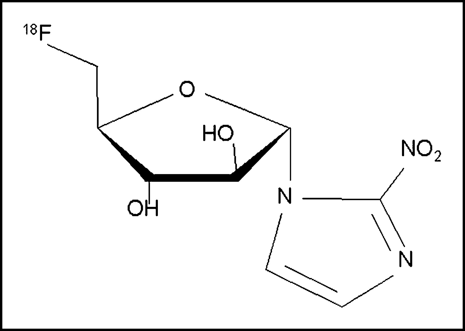
- FIGURE 1.
Chemical structure of 18F-fluoroazomycin arabinoside (18F-FAZA). Nucleoside analog contains nitroimidazole ring in α-position of arabinose ring.
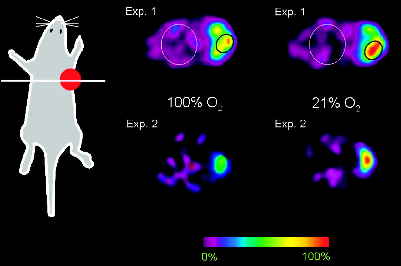
- FIGURE 2.
Typical serial transaxial small animal 18F-FAZA PET scans of 2 Swiss nude mice bearing subcutaneous A431 tumors at right side of thorax. Note decreased tracer uptake after breathing 100% O2 for 8 h. ROI analyses, as shown in experiment 1 (Exp. 1), revealed that the mean T/Bk ratio decreased from 4.7:1 (breathing room air) to 3.7:1 (breathing 100% O2) in experiment 1 and from 11.3:1 to 5.4:1 in experiment 2 (Exp. 2), respectively.
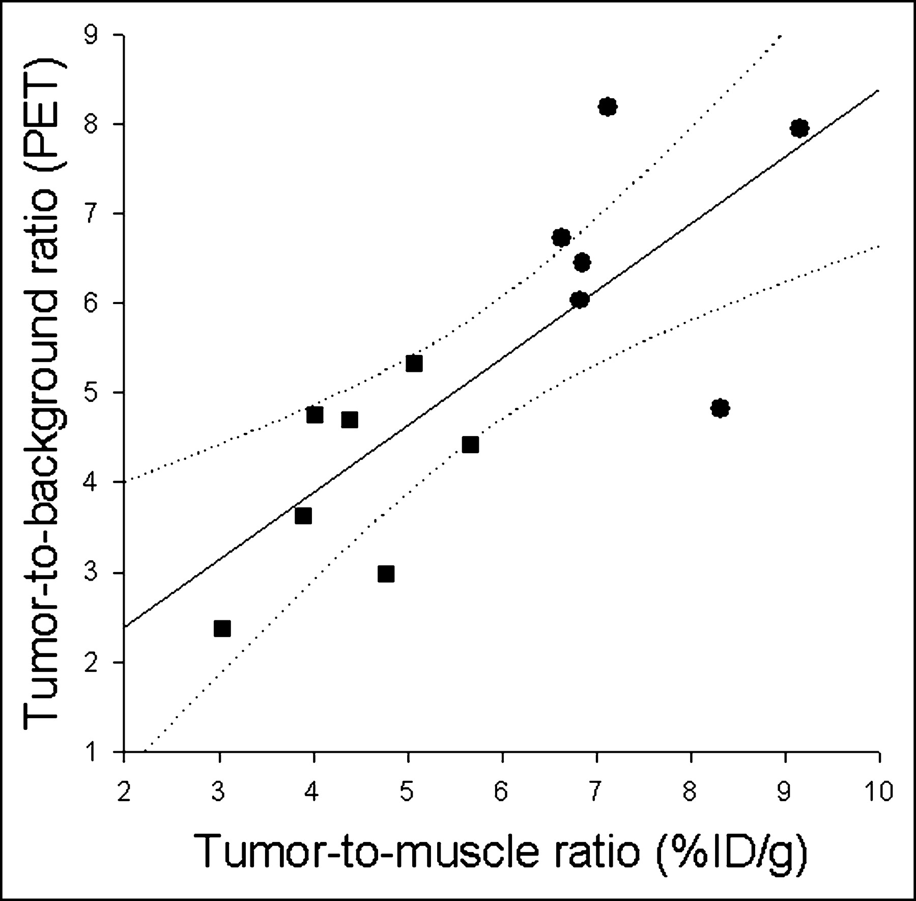
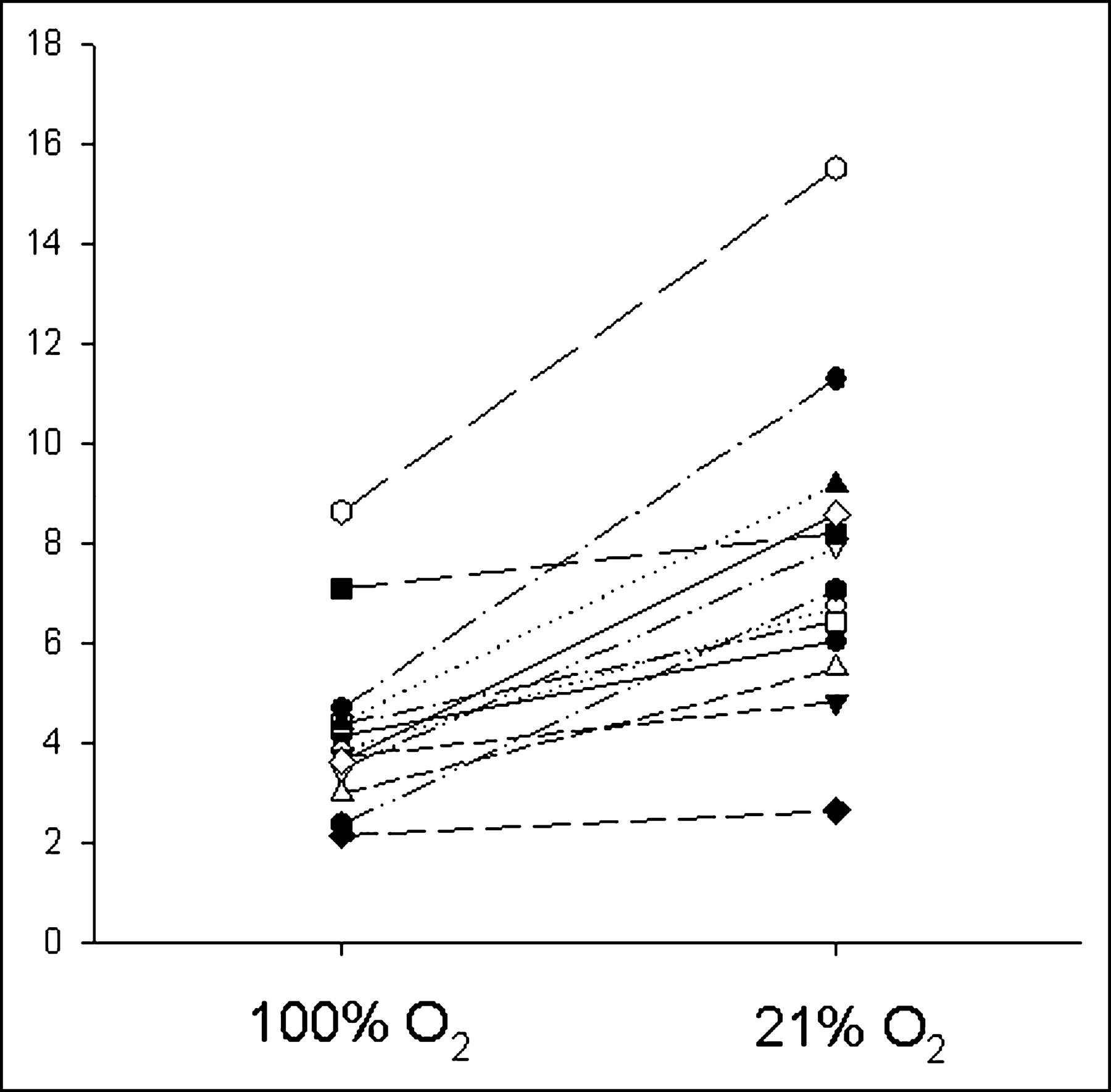
- FIGURE 3.
T/Bk ratio of 18F-FAZA (y-axis) in 13 Swiss nude mice bearing A431 tumor using serial MADPET studies with and without oxygen breathing. Animals breathing oxygen on the first day are displayed with open symbols, whereas animals breathing room air on the first day are shown with filled symbols.
- FIGURE 4.
Relationship of T/M ratio derived from biodistribution studies and T/Bk ratio of small animal PET studies. PET measurements were performed 150–180 min after injection of ∼10 MBq 18F-FAZA into Swiss nude mice bearing A431 tumor. During scanning, animals breathed either room air (circles) or 100% oxygen (squares). Regression analysis revealed a significant linear correlation between both measurements (regression line y = 0.89 + 0.75x; adjusted r2 = 0.57; P < 0.01; dotted lines, 95% confidence intervals).
- FIGURE 5.
Digital autoradiographic image of radioactivity 3 h after 18F-FAZA administration (20-μm slice thickness).


Tables
- TABLE 1
Biodistribution (%ID/g) at 180 Minutes of 18F-FMISO and at 10, 60, and 180 Minutes of 18F-FAZA in Swiss Nude Mice Bearing AR42J Tumor
Tissue 18F-FMISO 18F-FAZA 180 min (n = 5) 10 min (n = 6) 60 min (n = 6) 180 min (n = 8) Blood 0.69 ± 0.19 3.22 ± 0.66 0.90 ± 0.13 0.23 ± 0.26 Heart 1.05 ± 0.19 3.75 ± 0.89 1.14 ± 0.18 0.44 ± 0.37 Lung 1.32 ± 0.29 3.48 ± 0.72 1.05 ± 0.21 0.46 ± 0.21 Liver 4.07 ± 1.43 5.58 ± 1.06 2.47 ± 0.46 0.92 ± 0.71 Spleen 0.71 ± 0.21 3.57 ± 0.82 0.94 ± 0.07 0.48 ± 0.17 Kidney 1.95 ± 0.44 7.08 ± 2.02 2.38 ± 0.35 1.05 ± 0.42 Brain 0.62 ± 0.16 0.39 ± 0.05 0.60 ± 0.11 0.27 ± 0.32 Muscle 0.79 ± 0.14 3.26 ± 0.67 2.03 ± 1.08 0.31 ± 0.29 Bone 1.31 ± 0.22 2.27 ± 0.35 0.74 ± 0.04 0.42 ± 0.27 Skin 1.02 ± 0.26 3.00 ± 0.60 2.71 ± 2.61 0.87 ± 0.21 Stomach 1.25 ± 0.25 3.17 ± 0.51 1.19 ± 0.13 0.87 ± 0.43 Small intestine 3.61 ± 1.16 4.30 ± 0.67 1.88 ± 0.46 1.22 ± 0.61 Large intestine 8.26 ± 4.75 3.84 ± 0.85 1.59 ± 0.09 1.19 ± 0.90 Tumor 2.27 ± 0.39 2.30 ± 1.17 2.87 ± 1.30 1.35 ± 0.89* T/Bl ratio 3.39 ± 0.52 0.73 ± 0.39 3.27 ± 1.66 9.06 ± 4.07† T/M ratio 2.92 ± 0.66 0.72 ± 0.39 1.69 ± 1.02 5.49 ± 2.26‡ - TABLE 2
Biodistribution (%ID/g) at 180 Minutes of 18F-FMISO and 18F-FAZA in BALB/c Mice Bearing EMT6 Tumor
Tissue 18F-FMISO (n = 5) 18F-FAZA (n = 8) Blood 1.44 ± 0.26 0.19 ± 0.19 Heart 1.42 ± 0.35 0.42 ± 0.43 Lung 1.50 ± 0.38 0.39 ± 0.23 Liver 4.23 ± 0.82 0.61 ± 0.62 Spleen 1.15 ± 0.27 0.34 ± 0.13 Kidney 1.84 ± 0.49 0.61 ± 0.28 Brain 0.99 ± 0.27 0.24 ± 0.24 Muscle 1.38 ± 0.32 0.26 ± 0.24 Bone 1.52 ± 0.27 0.50 ± 0.50 Skin 0.96 ± 0.22 0.49 ± 0.48 Stomach 2.56 ± 0.46 0.62 ± 0.25 Small intestine 4.00 ± 1.99 0.76 ± 0.48 Large intestine 5.53 ± 2.67 0.90 ± 0.65 Tumor 4.32 ± 0.72 1.38 ± 0.62* T/Bl ratio 3.03 ± 0.30 9.82 ± 3.94* T/M ratio 3.22 ± 0.68 7.10 ± 2.91* ↵* P < 0.01 18F-FMISO vs. 18F-FAZA.
Data are presented as mean ± SD.
- TABLE 3
Biodistribution (%ID/g) at 180 Minutes of 18F-FMISO and 18F-FAZA in Swiss Nude Mice Bearing A431 Tumor
Tissue 18F-FMISO (n = 7) 18F-FAZA (n = 11) Blood 0.74 ± 0.16 0.31 ± 0.12 Heart 0.85 ± 0.46 0.61 ± 0.25 Lung 1.01 ± 0.56 0.38 ± 0.21 Liver 3.26 ± 2.00 1.23 ± 0.63 Spleen 0.66 ± 0.26 0.31 ± 0.14 Kidney 1.54 ± 0.99 0.65 ± 0.37 Brain 0.51 ± 0.21 0.56 ± 0.22 Muscle 1.05 ± 0.48 0.38 ± 0.16 Bone 1.77 ± 1.77 0.50 ± 0.17 Skin 1.00 ± 0.52 0.71 ± 0.85 Stomach 1.40 ± 0.72 0.64 ± 0.31 Small intestine 1.43 ± 0.77 0.90 ± 0.52 Large intestine 7.73 ± 4.00 1.63 ± 1.07 Tumor 3.67 ± 1.00 2.96 ± 1.27* T/Bl ratio 4.92 ± 0.77 9.62 ± 1.44† T/M ratio 3.95 ± 1.34 7.81 ± 0.94†
In this issue


Jump to section
Related Articles
Cited By...
- Concomitant [18F]F-FAZA and [18F]F-FDG Imaging of Gynecological Cancer Xenografts: Insight into Tumor Hypoxia
- GdDO3NI allows imaging of hypoxia after brain injury
- First Evaluation of PET-Based Human Biodistribution and Dosimetry of 18F-FAZA, a Tracer for Imaging Tumor Hypoxia
- In Vivo Hypoxia PET Imaging Quantifies the Severity of Arthritic Joint Inflammation in Line with Overexpression of Hypoxia-Inducible Factor and Enhanced Reactive Oxygen Species Generation
- 18F-FAZA PET Imaging Response Tracks the Reoxygenation of Tumors in Mice upon Treatment with the Mitochondrial Complex I Inhibitor BAY 87-2243
- Comparison of 18F-Fluoroazomycin-Arabinofuranoside and 64Cu-Diacetyl-Bis(N4-Methylthiosemicarbazone) in Preclinical Models of Cancer
- Prognostic and Predictive Significance of Plasma HGF and IL-8 in a Phase III Trial of Chemoradiation with or without Tirapazamine in Locoregionally Advanced Head and Neck Cancer
- Preclinical evaluation and validation of [18F]HX4, a promising hypoxia marker for PET imaging
- Location, Location, Location-Makes All the Difference for Hypoxia in Lung Tumors
- Hypoxia in Models of Lung Cancer: Implications for Targeted Therapeutics
- Quantitative Assessment of Hypoxia Kinetic Models by a Cross-Study of Dynamic 18F-FAZA and 15O-H2O in Patients with Head and Neck Tumors
- Pharmacologically Increased Tumor Hypoxia Can Be Measured by 18F-Fluoroazomycin Arabinoside Positron Emission Tomography and Enhances Tumor Response to Hypoxic Cytotoxin PR-104
- Radiopharmaceuticals in Preclinical and Clinical Development for Monitoring of Therapy with PET
- Reply: Intratumoral Spatial Distribution of Hypoxia and Angiogenesis Assessed by 18F-FAZA and 125I-Gluco-RGD Autoradiography
- Molecular Imaging of Hypoxia
- Molecular Imaging of Metastatic Potential
- Applications of Nitroimidazole In Vivo Hypoxia Imaging in Ischemic Stroke
- Correlative Imaging of Hypoxia and Angiogenesis in Oncology
- Intratumoral Spatial Distribution of Hypoxia and Angiogenesis Assessed by 18F-FAZA and 125I-Gluco-RGD Autoradiography
- Nuclear Imaging Probes: from Bench to Bedside
- Pretreatment 18F-FAZA PET Predicts Success of Hypoxia-Directed Radiochemotherapy Using Tirapazamine
- Experience of PET for target localisation in radiation oncology
- Modulation of intratumoral hypoxia by the epidermal growth factor receptor inhibitor gefitinib detected using small animal PET imaging