


Hypoxia plays a critical role in tumor development and aggressiveness and is an important prognostic factor for resistance to antineoplastic treatments. Hypoxic cells in tumors are about 3-fold more resistant to radiation therapy than their well-oxygenated counterparts (1). In the early stage of development, tumors remain dormant in an avascular phase, with active cellular proliferation balanced by cell apoptosis. The phenomenon, in which a tumor progresses from a nonangiogenic to angiogenic phenotype, has been termed the angiogenic switch (2). Hypoxia is a key player in the angiogenic See page 597
switch during tumor development. A key regulator of hypoxia-induced angiogenesis is the hypoxia-inducible factor 1 (HIF-1). Multiple HIF-1 target genes have been shown to modulate angiogenesis by promoting the mitogenic and migratory activities of endothelial cells (3). Accumulating evidence, however, indicates that HIF-1–independent pathways can also control angiogenesis (4). Hypoxia-induced angiogenesis has become an important area in basic, translational, and clinical cancer research and an attractive target for cancer therapy (5). The mechanisms involved during this process and how best to target it for cancer therapy are still under investigation. Oxygenation therapy, hypoxia-directed intensity-modulated radiotherapy, and antiangiogenic therapy in combination with cytotoxic chemotherapy are some of the current approaches to improve the therapeutic success in various tumor entities (2,5,6). Consequently, there is a growing demand for imaging modalities that allow for pretherapeutic stratification and response assessment of patients receiving targeted therapies.
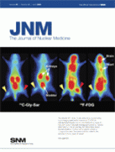
In this issue of The Journal of Nuclear Medicine, Picchio et al. (7) report on the spatial distribution of hypoxia and angiogenesis in rat tumor xenografts using double-tracer autoradiography. The authors applied the second-generation nitroimidazole-based hypoxia tracer 18F-fluoroazomycin arabinoside (FAZA) and 125I-gluco-RGD, which binds with high affinity to the αvβ3-integrin receptors that are highly expressed on activated endothelial cells during angiogenesis. A corresponding PET tracer, that is, 18F-galacto-RGD, is currently under clinical evaluation (8,9).
The comparison of 18F-FAZA distribution with immunohistochemical staining for HIF-1 as a gold standard of tissue hypoxia demonstrated that HIF-1 staining underestimated the extent of hypoxia as determined by 18F-FAZA. The authors put the role of HIF-1 as a surrogate marker for hypoxia into question, as is supported by several other publications (7). Roughly 60% of the tumor surface displayed the expected coupling between hypoxia and angiogenesis, that is, both low 18F-FAZA and 125I-gluco-RGD uptake on the one hand or elevated 18F-FAZA and 125I-gluco-RGD uptake on the other hand. However, approximately 40% of the tumor surface showed a discordant pattern indicating that hypoxia and angiogenesis are not necessarily spatially linked in malignant tumors. The authors speculate that tumor areas with increased 18F-FAZA uptake and low 125I-gluco-RGD uptake may indicate acute hypoxia when angiogenesis is not yet activated. In contrast, areas with low 18F-FAZA uptake and high 125I-gluco-RGD uptake may indicate tumor areas that have escaped hypoxia because of activated angiogenesis.
Certainly, this concept needs further confirmation, especially given the limited specificity of αvβ3-integrin ligands for imaging of tumor angiogenesis and the fact that the results concerning angiogenesis were not confirmed by a standard immunohistochemical method. Nevertheless, the study by Picchio et al. highlights the possible contribution of combined in vivo imaging of hypoxia and angiogenesis before and during therapy to the optimization of targeted chemotherapeutic and antiangiogenic therapy approaches in individual patients. With the rapid development and spread of integrated PET and CT scanners featuring a capability for full planning of radiation therapy, and specific PET tracers that are able to target hypoxia and angiogenesis in vivo, the required tools to integrate these parameters into clinical research and practice are becoming more and more available.
Although these experimental results are rather encouraging, a short consideration of the tracers that are currently under clinical evaluation for in vivo imaging of hypoxia and angiogenesis with PET is needed. At present, most experience concerning noninvasive imaging of tumor hypoxia is available for 18F-labeled fluoromisonidazole (10). This tracer has successfully been applied in numerous studies and turned out to be useful to predict the outcome of radiotherapy in lung and head and neck cancers and to detect tumor reoxygenation during radiotherapy (10–12). Recently, the evaluation of 18F-fluoromisonidazole kinetics was found to be helpful to characterize the hypoxia-perfusion pattern of head and neck tumors—a pattern that correlated with the outcome of radiotherapy (12,13). Nevertheless, the tracer is not optimal for clinical application because of slow accumulation in hypoxic tumors, a low tumor-to-background (T/B) contrast, and a significant amount of radioactive metabolites (14). The second-generation hypoxia tracer 18F-FAZA exhibits a faster clearance than 18F-fluoromisonidazole and higher T/B contrast in animal experiments (15,16). A first study on patients with head and neck cancer, however, showed only slightly increased T/B ratios at 2 h after injection, compared with 18F-MISO (14,17). In many tumors, the T/B ratios of 18F-FAZA uptake decreased between 2 and 4 h after injection, indicating that 18F-FAZA binding to hypoxic tumor tissue is not irreversible. Although 18F-FAZA shows clinical potential similar to that of 18F-fluoromisonidazole (18,19), there are only minor advantages, and further efforts for the development of an optimal hypoxia tracer with higher T/B contrast are needed.
The field of angiogenesis is a rapidly growing biomedical discipline, and great efforts are being undertaken to develop antiangiogenesis drugs for the treatment of cancer. One target structure is the αvβ3 integrin receptor, which is highly expressed on activated endothelial cells during angiogenesis. Various ligands based on the tripeptide RGD, which binds with high affinity to the αvβ3-integrin receptor, have been developed for SPECT and PET (20). The glycosylated cyclic pentapeptide 18F-galacto-RGD resulted from tracer optimization based on the first-generation peptide 125I-3-iodo-d-Tyr4-cyclo(-Arg-Gly-Asp-d-Tyr-Val-) (21). Initial clinical studies using 18F-galacto-RGD and PET demonstrated that tracer accumulation in the tumor correlates with αvβ3 expression but also revealed high inter- and intraindividual variance in tracer accumulation in tumors, indicating great diversity in receptor expression (8,9). In a comparative study, no correlation between 18F-FDG and 18F-galacto-RGD tracer uptake in the tumors was found, and the sensitivity of 18F-galacto-RGD for tumor detection was clearly inferior to that of 18F-FDG (9). Another limitation is that αvβ3 can be expressed on tumor cells as well as on endothelial cells, making it difficult to attribute the resulting signal exclusively to molecular processes during tumor-induced angiogenesis. Furthermore, the exact role of αvβ3 expression in the context of angiogenesis is still a matter of discussion. Experiments on knockout mice lacking the integrin αvβ3 led to a reevaluation of the role of αvβ3 with regard to angiogenesis, because the knockout mice showed normal developmental angiogenesis and even excessive tumor angiogenesis (22).
In summary, molecular imaging of hypoxia and angiogenesis using PET is an exciting field that has made considerable progress in recent years and may soon contribute significantly to individually targeted therapies. The tracers currently available already have a high potential to approach these goals, but further efforts in the development of new ligands with higher T/B contrast and specificity are desirable.
Footnotes
-
COPYRIGHT © 2008 by the Society of Nuclear Medicine, Inc.
References
- Received for publication February 6, 2008.
- Accepted for publication February 20, 2008.


Jump to section
Related Articles
Cited By...
- No citing articles found.