


Article Figures & Data
Figures
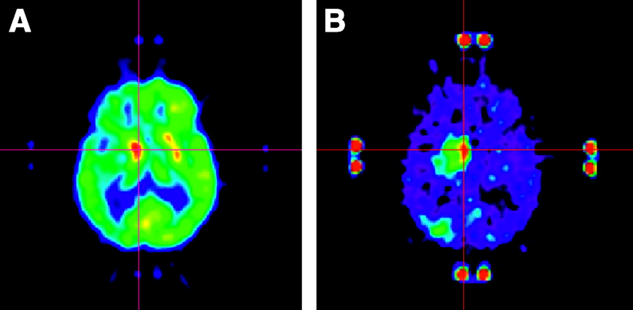
- FIGURE 1.
PET performed with 18F-FDG (A) and Met (B) on a 32-y-old man with an AA in the left frontorolandic cortical area. Uptake of 18F-FDG in tumor was equivalent to that in cortical gray matter. Uptake of Met was higher in tumor than in cortex, allowing definition of a target for biopsy. When PET images obtained with the 2 tracers were coregistered, the focus of highest Met uptake corresponded to the unique focus of 18F-FDG uptake within the tumor (crosses).
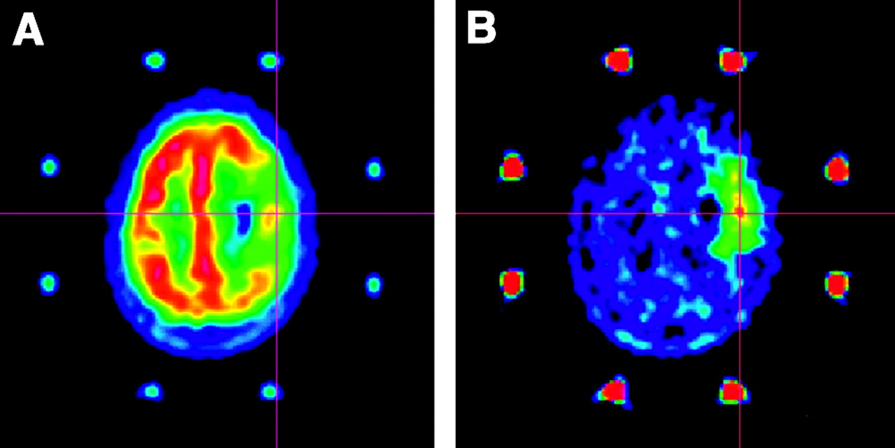
- FIGURE 2.
PET performed with 18F-FDG (A) and Met (B) on a 62-y-old woman with a GB in the right prerolandic cortical area. Uptake of 18F-FDG was reduced in the tumor area except for 1 spot of uptake equivalent to that in the surrounding gray matter. Uptake of Met was higher in tumor than in cortex, allowing definition of a target for biopsy. When PET images obtained with the 2 tracers were coregistered, the highest focus of Met uptake corresponded to the hot spot of 18F-FDG (crosses).
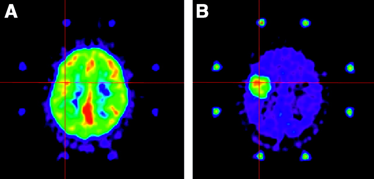
- FIGURE 3.
PET performed with 18F-FDG (A) and Met (B) on a 74-y-old man with a GB in the right basal ganglia. Uptake of 18F-FDG was higher in tumor than in surrounding gray matter and allowed definition of a target for biopsy. Uptake of Met was also higher in tumor than in surrounding gray matter, and zones of highest uptake of both tracers corresponded on coregistered images (crosses).
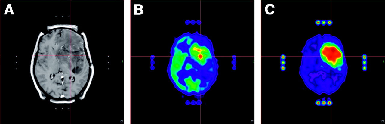
- FIGURE 4.
MRI (A) and PET performed with 18F-FDG (B) and Met (C) on a 57-y-old woman with an infiltrating AA in the left basal ganglia and subcortical frontal region. Uptake of 18F-FDG was higher in tumor than in surrounding gray matter and allowed definition of a target for biopsy. Uptake of Met was also higher in tumor than in surrounding gray matter. Coregistration of PET images showed that Met uptake was more extended than 18F-FDG uptake but that zones of highest uptake corresponded (crosses).


Tables
Diagnosis 18F-FDG-defined target (FDGt > FDGgm) Met-defined target FDGt = FDGgm FDGt < FDGgm GB 7 3 — AA 6 5 1 LGG 1 5 4 Total 14 13 5 FDGt = 18F-FDG uptake in tumor; FDGgm = 18F-FDG uptake in surrounding gray matter.
Diagnosis Met(+) trajectories Met(−) trajectories (FDGt < FDGgm) FDGt > FDGgm FDGt = FDGgm FDGt < FDGgm GB 11 7 — — AA 10 9 2 — LGG 1 6 15 — Nondiagnostic — — — 9 Total 22 22 17 9 FDGt = 18F-FDG uptake in tumor; FDGgm = 18F-FDG uptake in surrounding gray matter.


Jump to section
Related Articles
Cited By...
- Amino Acid PET in Neurooncology
- Amino Acid PET in Neurooncology
- Extent and Instability of Trimethylation of Histone H3 Lysine Increases With Degree of Malignancy and Methionine Addiction
- Metabolic Phenotypes, Dependencies, and Adaptation in Lung Cancer
- The linkage of methionine addiction, overmethylation of histone H3 lysines and malignancy demonstrated when cancer cells revert to methionine-independence
- Chronic Treatment of an Advanced Prostate-cancer Patient With Oral Methioninase Resulted in Long-term Stabilization of Rapidly Rising PSA Levels
- Lowering and Stabilizing PSA Levels in Advanced-prostate Cancer Patients With Oral Methioninase
- Histone H3K4me3 and H3K9me3 are super over-methylated in soft tissue sarcoma compared to normal muscle in patient-derived xenograft (PDX) mouse models: an indicator of cancer methionine addiction
- Differentiation between Treatment-Induced Necrosis and Recurrent Tumors in Patients with Metastatic Brain Tumors: Comparison among 11C-Methionine-PET, FDG-PET, MR Permeability Imaging, and MRI-ADC--Preliminary Results
- Molecular Imaging to Plan Radiotherapy and Evaluate Its Efficacy
- Imaging Intratumor Heterogeneity: Role in Therapy Response, Resistance, and Clinical Outcome
- Clinical Impact of Amino Acid PET in Gliomas
- Correlation of 18F-FLT Uptake with Tumor Grade and Ki-67 Immunohistochemistry in Patients with Newly Diagnosed and Recurrent Gliomas
- Clinical applications of imaging biomarkers. Part 2. The neurosurgeon's perspective
- Multimodality Assessment of Brain Tumors and Tumor Recurrence
- Voxel-Based Analysis of Dual-Time-Point 18F-FDG PET Images for Brain Tumor Identification and Delineation
- Evaluation of 18F-FDG PET and MRI Associations in Pediatric Diffuse Intrinsic Brain Stem Glioma: A Report from the Pediatric Brain Tumor Consortium
- Quantitative, Preclinical PET of Translocator Protein Expression in Glioma Using 18F-N-Fluoroacetyl-N-(2,5-Dimethoxybenzyl)-2-Phenoxyaniline
- 1-11C-Acetate Versus 18F-FDG PET in Detection of Meningioma and Monitoring the Effect of {gamma}-Knife Radiosurgery
- Evaluation of Focal Cortical Dysplasia and Mixed Neuronal and Glial Tumors in Pediatric Epilepsy Patients Using 18F-FDG and 11C-Methionine PET
- Tumor Cell Metabolism Imaging
- Evaluation of Primary Brain Tumors Using 11C-Methionine PET with Reference to a Normal Methionine Uptake Map
- Clinical Applications of PET in Brain Tumors