


Article Figures & Data
Figures
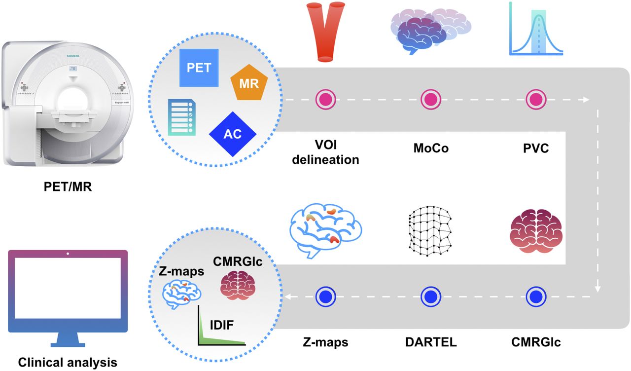
- FIGURE 1.
Noninvasive absolute quantification pipeline consisting of 6 nodes (depicted as circles); pink nodes correspond to IDIF-generating components, and blue nodes correspond to quantification components. Input consists of synergistic data from PET/MRI study along with parameter file, with output yielding IDIF as well as CMRGlc and abnormality maps (Z-maps). MoCo = motion correction; PVC = partial-volume correction; VOI = volume of interest.
- FIGURE 2.
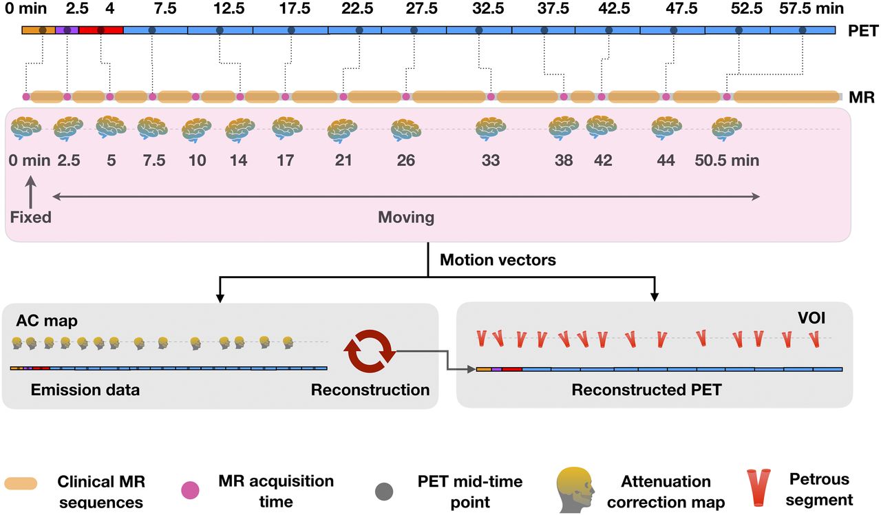
MR-driven motion correction as implemented in developed pipeline. MRI navigators are assigned to each PET frame on basis of smallest temporal difference, and obtained motion vectors are used for aligning both AC map and petrous volume of interest to PET image data. VOI = volume of interest.
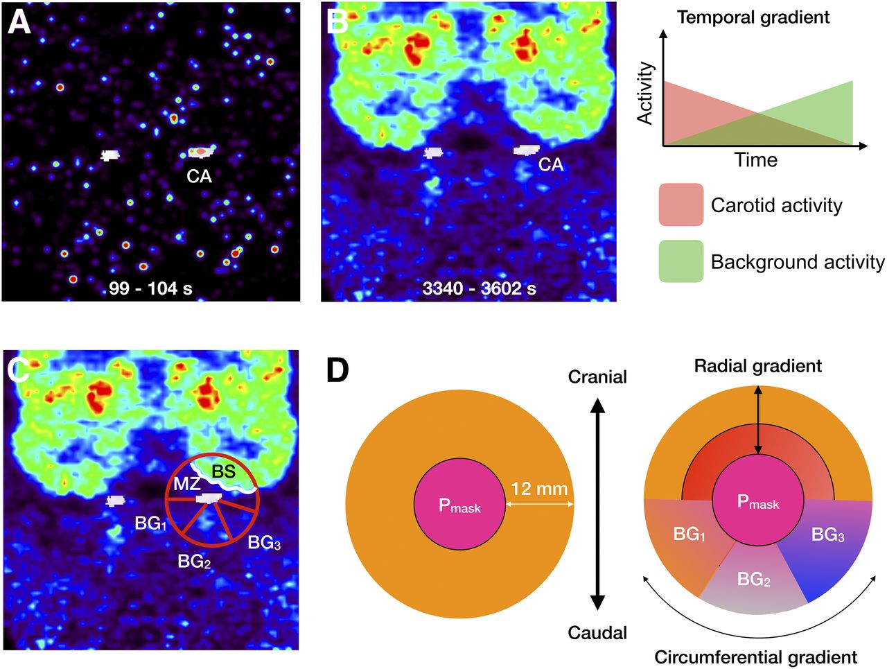
- FIGURE 3.
(A and B) PET frame reconstruction with ICA overlay (white) for early (A) and late (B) times after injection. Temporal and spatial variabilities of ICA background can be clearly deduced from images. (C) Tracer distribution in vicinity of ICA displays both radial and circumferential tracer concentration gradients. (D) Definition of subregions in vicinity of Pmask used to account for partial-volume distortions. BG1, BG2, and BG3 = various background regions with homogeneous tracer concentrations; BS = brain activity; CA = measured activity in ICA; MZ = activity in MZ.
- FIGURE 4.
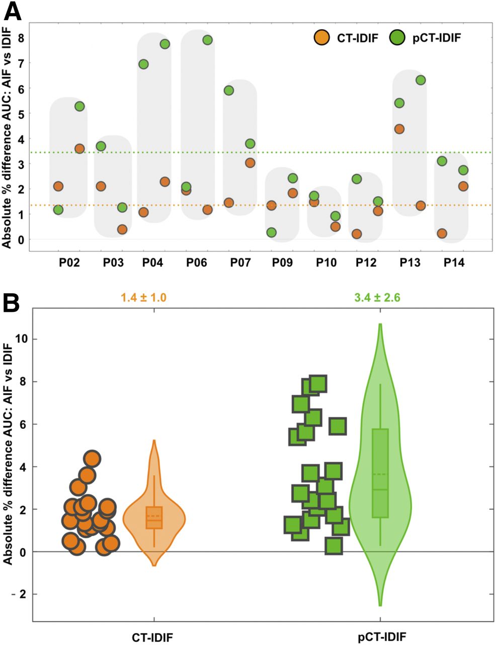
Comparison of IDIFs using AUCs. (A) Individual absolute percentage differences in AUCs for CT-IDIF and pCT-IDIF against AIF. Shaded areas indicate test-retest results for same subject. Broken lines represent mean difference over all scans between AUCs derived using AIF and CT-IDIF (orange) and those derived using AIF and pCT-IDIF (green). (B) Plot of absolute percentage differences in AUCs for AIF and IDIFs (CT-IDIF and pCT-IDIF). Shaded area enclosing box plot indicates probability density distribution for absolute percentage differences. Average absolute percentage difference for both methods was <5% (shown above graph).
- FIGURE 5.
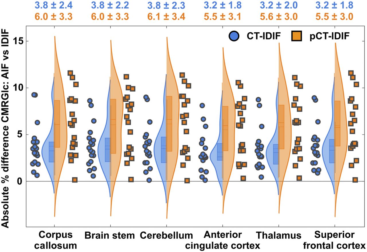
Probability density distribution for absolute percentage differences between CMRGlc values derived using AIF and those derived using IDIFs (CT-IDIF and pCT-IDIF) for 6 different brain regions. Absolute percentage differences in CMRGlc values derived using CT-AC are shown in blue, and those derived using pCT-AC are shown in orange. Mean and SD for each region and 2 AC methods are shown above graph.
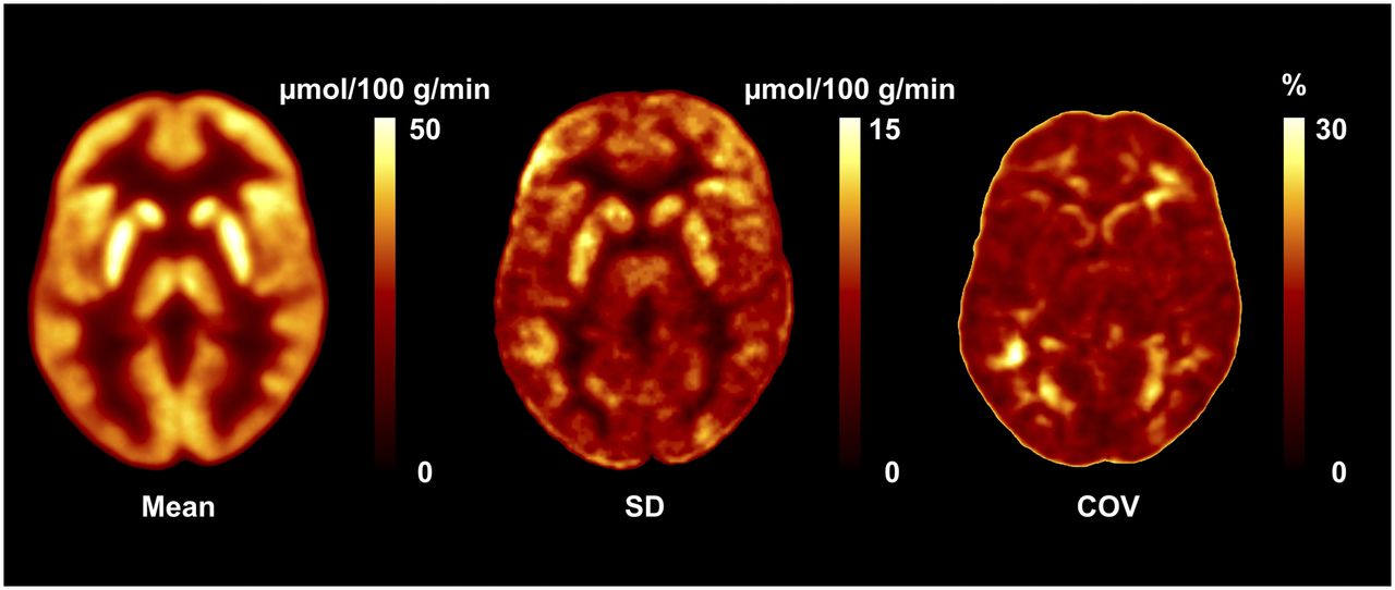
- FIGURE 6.
Database images in Montreal Neurological Institute space representing mean, SD, and COV maps for absolute values of CMRGlc. COV map indicates normal physiologic variability of 15%–25%.


Tables
CMRGlc values* obtained from: AIF CT-IDIF pCT-IDIF Region Mean ± SD COV (%) Mean ± SD COV (%) Mean ± SD COV (%) Corpus callosum 16.1 ± 3.6 22 16.1 ± 3.6 22 15.9 ± 4.0 25 Brain stem 20.0 ± 2.7 14 19.9 ± 2.7 14 19.6 ± 3.1 16 Cerebellum 24.6 ± 3.4 14 24.6 ± 3.5 14 24.3 ± 4.1 17 Anterior cingulate 31.9 ± 6.4 21 31.8 ± 6.3 20 31.4 ± 7.0 22 Thalamus 34.3 ± 5.9 17 34.2 ± 5.8 17 33.7 ± 6.6 20 Superior frontal 34.4 ± 6.6 19 34.2 ± 6.6 19 33.8 ± 7.3 22 ↵* Reported as μmol/100 g/min.
Maximum deviations from AIF standard of CMRGlc obtained from CT-IDIF and fully automated pCT-IDIF were 10% and 12%, respectively.
Supplemental Data
Files in this Data Supplement:


Jump to section
Related Articles
Cited By...
- Fully Automated, Fast Motion Correction of Dynamic Whole-Body and Total-Body PET/CT Imaging Studies
- Fully Automated, Semantic Segmentation of Whole-Body 18F-FDG PET/CT Images Based on Data-Centric Artificial Intelligence
- Conditional Generative Adversarial Networks Aided Motion Correction of Dynamic 18F-FDG PET Brain Studies