


Article Figures & Data
Figures
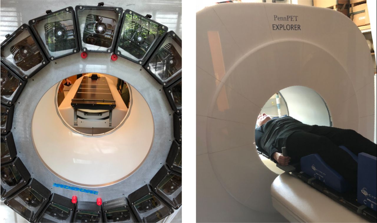
- FIGURE 1.
PennPET Explorer in its prototype configuration with 3 ring segments, housed in dry, cool enclosure. View of back of gantry shows modular detector and electronic bays. Also shown is couch with flat pallet installed for human studies.
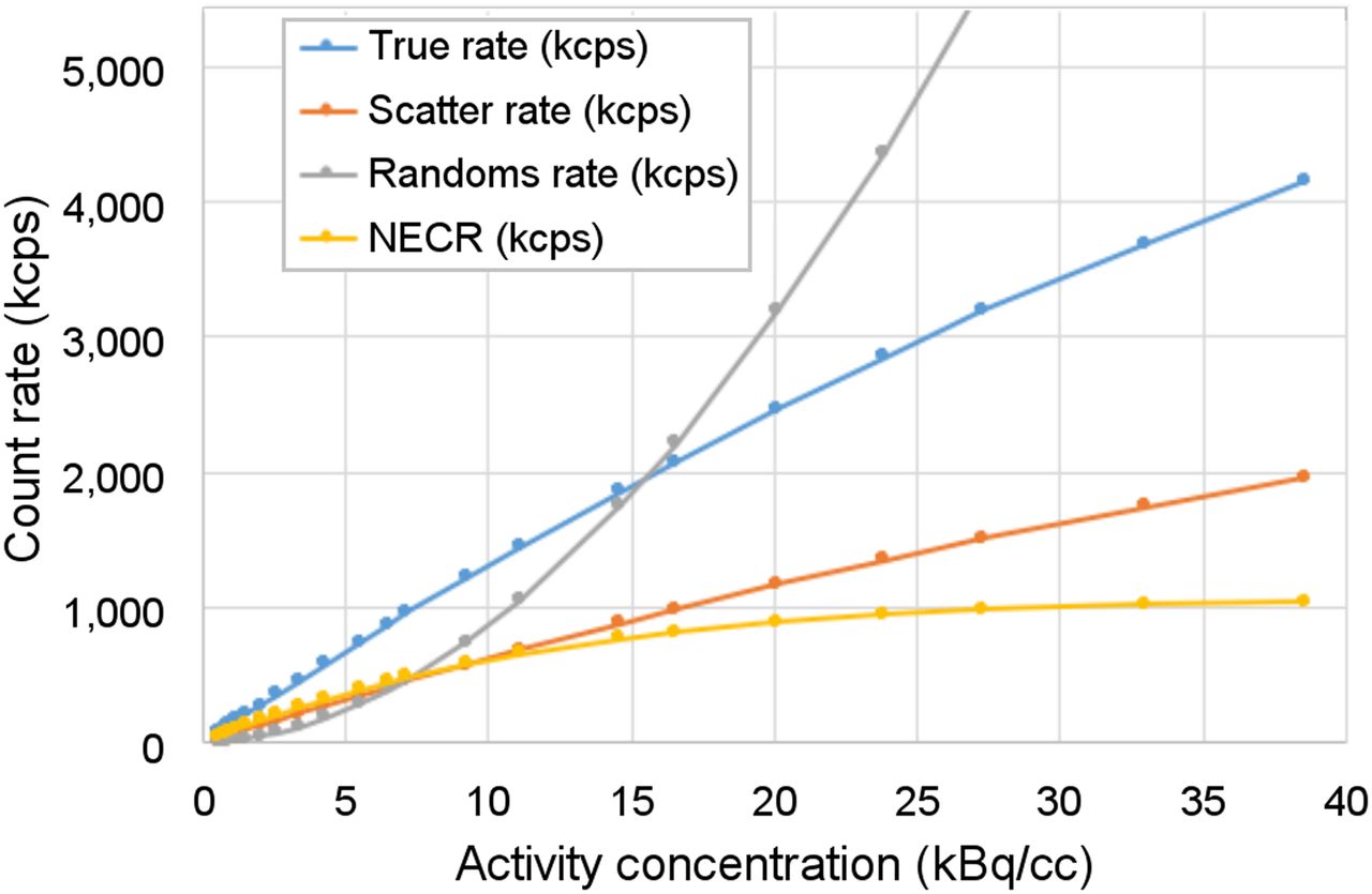
- FIGURE 2.
NEMA NU-2 count-rate performance with 70-cm line source inside 20-cm-diameter polyethylene scatter cylinder. Count-rate results were acquired up to 40 kBq/mL, although clinical 18F-FDG studies are typically performed with activity concentrations of less than 5 kBq/mL.
- FIGURE 3.
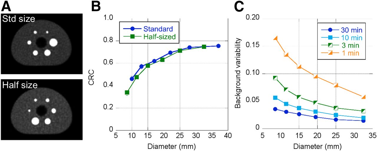
(A) NEMA image-quality phantom shown with standard spheres and half-sized spheres. Body activity concentration is 2 kBq/mL, sphere contrast is 9.7:1, and scan duration is 7.5 min. (B) CRCs as per NEMA guidelines for both standard and half-sized spheres. (C) Background variability as per NEMA guidelines for half-sized spheres.
- FIGURE 4.
(A) Clinical Trials Network torso phantom with activity concentration of 5.9 kBq/mL and lesion contrast of 4.2:1. (B) CRC of representative lesions as function of scan duration. (C) SD of CRC, determined from replicates of data.
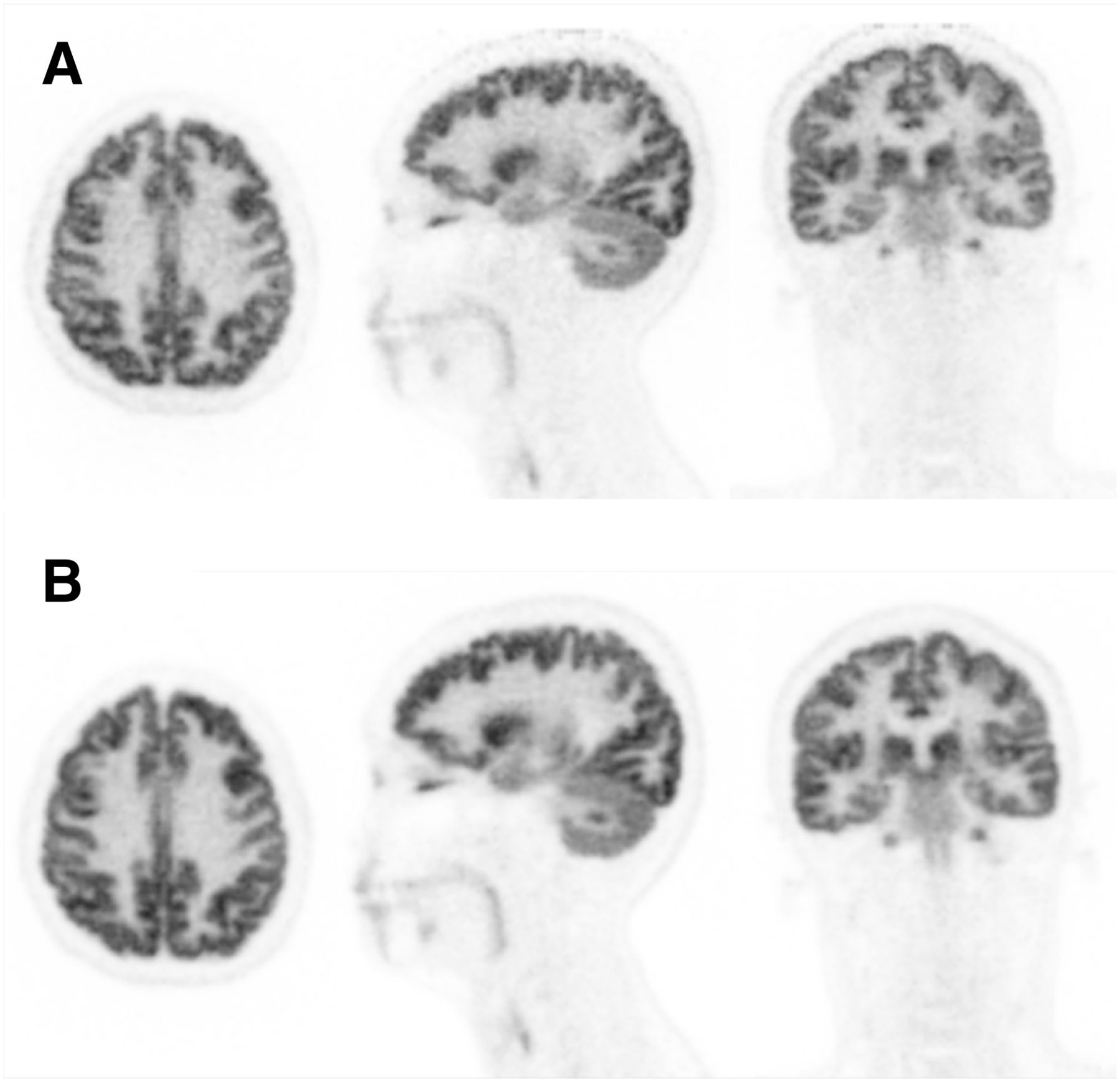
- FIGURE 5.
(A) Representative sagittal (left), axial (middle), and coronal (right) views of 20-min PennPET Explorer image of subject 1 at 1.5 h after injection of 555-MBq dose of 18F-FDG. All are 2-mm sections. (B) PennPET Explorer image, subsampled (⅛ data) to represent 2.5-min scan. (C) Clinical scan acquired with Philips Ingenuity TF PET/CT scanner at 1 h after injection, using clinical protocol with 10 bed positions for total of 20 min. These data were reconstructed off-line with same reconstruction method as for PennPET Explorer data.
- FIGURE 6.
Sagittal (left), axial (middle), and coronal (right) views of PennPET Explorer images of subject 2 positioned with head near edge of AFOV (A) and at center of AFOV (B). These scans were acquired starting at 1.5 h after injection of 18F-FDG for 10 min each. All are 1-mm sections.
- FIGURE 7.
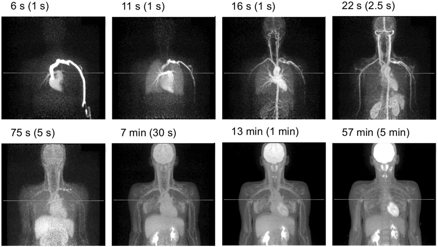
Dynamic 18F-FDG study of subject 7 acquired for 60 min after 555-MBq dose of 18F-FDG. Representative maximum-intensity projections are shown from 0 to 60 min after injection.


Tables
Parameter Radius (cm) Rings (n) Algorithm Radial (mm) Tangential (mm) Axial (mm) FWHM 1 1 Analytic 4.2 ± 0.3 3.9 ± 0.4 4.1 ± 0.2 1 1 Iterative 3.9 ± 0.3 3.8 ± 0.3 3.6 ± 0.2 1 3 Iterative 3.9 ± 0.2 3.9 ± 0.3 3.9 ± 0.3 10 3 Iterative 4.2 ± 0.2 3.9 ± 0.2 3.9 ± 0.3 20 3 Iterative 5.6 ± 0.2 3.9 ± 0.4 3.7 ± 0.3 FWTM 1 1 Analytic 8.5 ± 0.8 8.4 ± 0.9 7.9 ± 0.2 1 1 Iterative 7.2 ± 0.4 7.1 ± 0.2 6.8 ± 0.2 1 3 Iterative 7.4 ± 0.6 7.3 ± 0.2 7.8 ± 1.2 10 3 Iterative 8.1 ± 0.2 7.2 ± 0.2 7.6 ± 0.8 20 3 Iterative 10.4 ± 0.3 7.2 ± 0.2 7.3 ± 0.7 FWHM = full width at half maximum; FWTM = full width at tenth maximum.
Uncertainties are SD of replicate measurements.
Parameter Radius (cm) Rings (n) Algorithm Center (mm) Gap (mm) Edge (mm) FWHM 1–20 1 Iterative — — 3.5 ± 0.2 1–20 3 Iterative 4.0 ± 0.2 4.2 ± 0.1 3.6 ± 0.1 FWTM 1–20 1 Iterative — — 6.8 ± 0.1 1–20 3 Iterative 8.5 ± 0.6 8.2 ± 0.1 6.9 ± 0.2 FWHM = full width at half maximum; FWTM = full width at tenth maximum.
Uncertainties are SD of replicate measurements.
Supplemental Data
Files in this Data Supplement:


Jump to section
Related Articles
Cited By...
- Feasibility of an Ultra-Low-Dose PET Scan Protocol with CT-Based and LSO-TX-Based Attenuation Correction Using a Long-Axial-Field-of-View PET/CT Scanner
- Is Long-Axial-Field-of-View PET/CT Cost-Effective? An International Health-Economic Analysis
- [11C]Carfentanil PET Whole-Body Imaging of {mu}-Opioid Receptors: A First in-Human Study
- Total-Body PET System Designs with Axial and Transverse Gaps: A Study of Lesion Quantification and Detectability
- [11C]Carfentanil PET Whole-Body Imaging of Mu-Opioid Receptors: A First In-Human Study
- Performance Characteristics of a New Generation 148-cm Axial Field-of-View uMI Panorama GS PET/CT System with Extended NEMA NU 2-2018 and EARL Standards
- Dose Reduction in Pediatric Oncology Patients with Delayed Total-Body [18F]FDG PET/CT
- Whole-Body PET Imaging: A Catalyst for Whole-Person Research?
- Fully Automated, Semantic Segmentation of Whole-Body 18F-FDG PET/CT Images Based on Data-Centric Artificial Intelligence
- Total-Body 18F-FDG PET/CT in Autoimmune Inflammatory Arthritis at Ultra-Low Dose: Initial Observations
- Radioembolization Dosimetry with Total-Body 90Y PET
- 3D melanoma spheroid model for the development of positronium biomarker
- Principles of Tracer Kinetic Analysis in Oncology, Part II: Examples and Future Directions
- Performance Characteristics of the Biograph Vision Quadra PET/CT System with a Long Axial Field of View Using the NEMA NU 2-2018 Standard
- Developing a Novel Positronium Biomarker for Cardiac Myxoma Imaging
- Performance Evaluation of the uEXPLORER Total-Body PET/CT Scanner Based on NEMA NU 2-2018 with Additional Tests to Characterize PET Scanners with a Long Axial Field of View
- Investigating Low-Dose Image Quality in Whole-Body Pediatric 18F-FDG Scans Using Time-of-Flight PET/MRI
- Benefit of Improved Performance with State-of-the Art Digital PET/CT for Lesion Detection in Oncology