


Article Figures & Data
Figures
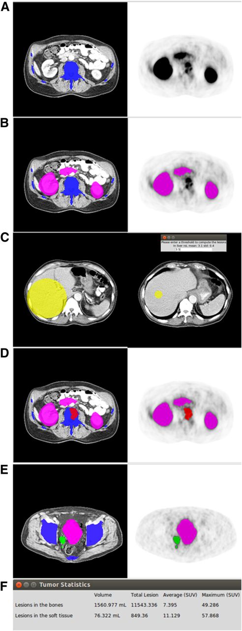
- FIGURE 1.
The 6-step workflow of qPSMA. First, bone mask (A) and normal-uptake mask (B) are automatically computed. Then, SUVthr_st is semiautomatically computed from liver background activity (C). Bone lesions are segmented using SUVthr_bone (D), whereas soft-tissue lesions are segmented using SUVthr_st, previously calculated at third step (E). Finally, output parameters are obtained by performing general statistics (F).
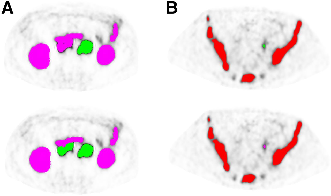
- FIGURE 2.
Examples of manual corrections in 2 metastatic castration-resistant prostate cancer patients. (A) Because of their large connections with intestine, retroperitoneal lymph nodes were wrongly classified as having normal uptake and not considered when SUVthr_st was applied. After correction of normal-uptake label, lymph nodes were segmented as soft-tissue lesions. (B) Ureter segmented as soft-tissue lesions and manually changed to normal-uptake label.
- FIGURE 3.
Bland–Altman plot of qPSMA and METAVOL agreement on semiautomatic computation of SUVthr_st. Solid line indicates average mean difference, and dotted lines delineate 95% limits of agreement (mean ± 1.96 × SD). No systematic difference between the 2 software programs was found.
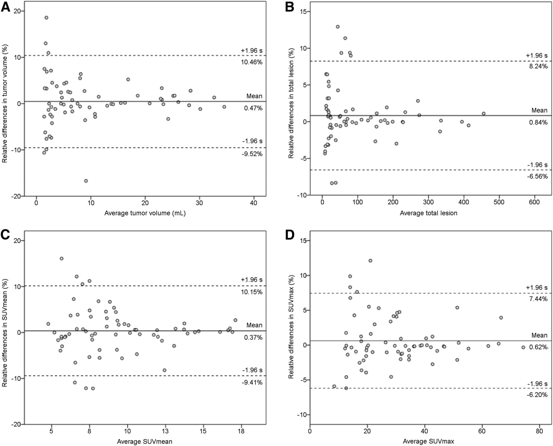
- FIGURE 4.
Bland–Altman plots for tumor volume (A), total lesion (B), SUVmean (C), and SUVmax (D) from 68 lesions segmented with qPSMA and Syngo.via software. Solid lines indicate average mean difference, and dotted lines delineate 95% limits of agreement (mean ± 1.96 × SD).

Tables
Characteristic Data Patients (n) 20 Age (y) Mean 73 Range 65–84 PSA (ng/mL) Mean 369 Range 1–2,222 Site of metastasis (n) Lymph node, overall 12 Lymph node only 1 Bone, overall 19 Bone only 1 Bone and lymph node 12 Local recurrence 4 Visceral, overall 3 Intraobserver analysis Interobserver analysis Output parameter Read 1 vs. 2 Difference (%) ICC User 1 vs. 2 Difference (%) ICC bPSMA-TV (mL) 801.9 vs. 800.4 −2.22 (−5.72;1.25) 1.000 (0.999;1.000) 801.9 vs. 800.1 2.53 (−2.60;7.68) 1.000 (1.000;1.000) bPSMA-TL 6397 vs. 6393 −2.94 (−7.75;1.86) 1.000 (0.999;1.000) 61397 vs. 6392 2.37 (−1.93;6.68) 1.000 (1.000;1.000) bPSMA SUVmean 7.34 vs. 7.39 −0.73 (−2.24;0.77) 0.998 (0.998;1.000) 7.34 vs. 7.33 −0.16 (−1.99;1.67) 0.998 (0.996;0.999) bPSMA SUVmax 38.43 vs. 38.45 −0.07 (−0.38;0.22) 1.000 (0.999;1.000) 38.43 vs. 38.43 0.05 (−0.20;0.31) 1.000 (1.000;1.000) stPSMA-TV (mL) 67.8 vs. 68.6 3.10 (−8.24;14.45) 1.000 (0.999;1.000) 67.8 vs. 67.1 9.05 (−1.49;19.61) 0.999 (0.998;1.000) stPSMA-TL 1026 vs. 1033 5.22 (−4.87;15.31) 1.000 (0.999;1.000) 1026 vs. 1016 8.70 (−1.37;18.77) 1.000 (0.999;1.000) stPSMA SUVmean 9.95 vs. 9.96 −0.43 (−1.86;0.98) 1.000 (0.999;1.000) 9.95 vs.9.92 −0.49 (−1.88;0.90) 0.999 (0.996;0.999) stPSMA SUVmax 31.23 vs. 31.20 0.18 (−0.15;0.53) 1.000 (1.000;1.000) 31.23 vs. 33.21 0.32 (−0.25;0.89) 1.000 (1.000;1.000) Data are mean; 95%CIs are in parentheses. P values on paired t testing are all >0.05
Supplemental Data
Files in this Data Supplement:


Jump to section
Related Articles
Cited By...
- RECIP 1.0: A Roadmap for Clinical Implementation
- RECIP 1.0 Predicts Progression-Free Survival After [177Lu]Lu-PSMA Radiopharmaceutical Therapy in Patients with Metastatic Castration-Resistant Prostate Cancer
- Prognostic Value of End-of-Treatment PSMA PET/CT in Patients Treated with 177Lu-PSMA Radioligand Therapy: A Retrospective, Single-Center Analysis
- Toward Single-Time-Point Image-Based Dosimetry of 177Lu-PSMA-617 Therapy
- Prognostic Value of Tumor Volume Assessment on PSMA PET After 177Lu-PSMA Radioligand Therapy Evaluated by PSMA PET/CT Consensus Statement and RECIP 1.0
- Using 68Ga-PSMA-11 PET/CT for Therapy Response Assessment in Patients with Metastatic Castration-Resistant Prostate Cancer: Application of EAU/EANM Recommendations in Clinical Practice
- Novel Framework for Treatment Response Evaluation Using PSMA PET/CT in Patients with Metastatic Castration-Resistant Prostate Cancer (RECIP 1.0): An International Multicenter Study
- The Influence of Specific Activity on the Biodistribution of 18F-rhPSMA-7.3: A Retrospective Analysis of Clinical PET Data
- Tumor Sink Effect in 68Ga-PSMA-11 PET: Myth or Reality?
- PSMA PET for the Assessment of Metastatic Hormone-Sensitive Prostate Cancer Volume of Disease
- Assessing Response to 177Lu-PSMA Radioligand Therapy Using Modified PSMA PET Progression Criteria
- Semiautomatically Quantified Tumor Volume Using 68Ga-PSMA-11 PET as a Biomarker for Survival in Patients with Advanced Prostate Cancer
- Comparison of 3 Interpretation Criteria for 68Ga-PSMA11 PET Based on Inter- and Intrareader Agreement